


Medically reviewed by Shaun Larsen, OD, Doctor of Optometry, Southern College of Optometry.
Last clinically reviewed: July 2026 | Next scheduled review: July 2027
What this article covers. “Vision” is not one ability but several, each handled by different cells and different parts of the visual system, and each capable of failing independently. That is why one person can read the bottom of the eye chart yet be unable to walk safely across a room, while another struggles to read a newspaper but has no difficulty driving. This guide explains the distinct kinds of vision, what each one does, how each is measured, and what it means when one of them is lost.
The kinds of vision at a glance
- Central vision. Fine detail, reading, faces. Handled by the cones of the macula, especially the fovea. Measured by visual acuity.
- Peripheral vision. Everything outside the point of gaze. Detects motion and supports orientation and mobility. Measured by perimetry.
- Binocular vision and depth perception. The two eyes combining into one three-dimensional image. Measured by stereoacuity testing.
- Color vision. Discriminating wavelength, via three cone types. Measured by Ishihara plates, D-15 or anomaloscope.
- Night vision and dark adaptation. Seeing in low light, handled by the rods. Measured by dark adaptometry.
- Contrast sensitivity. Distinguishing an object from a background of similar brightness. Measured by the Pelli-Robson chart.
Why this matters practically. A standard eye test measures the first of these and, sometimes, the third. Contrast sensitivity, dark adaptation and colour discrimination are rarely tested in a routine appointment, yet they are frequently what a patient is actually complaining about. If your difficulty does not match your chart result, these are the tests to ask for by name.
Central vision
The macula is the small central region of the retina, roughly 5.5 mm across, containing the highest density of cone photoreceptors in the eye. Its centre, the fovea, has no rods and no overlying inner retinal layers at all, an arrangement that maximises resolution.
Central vision does the work of reading, recognising faces, threading a needle and reading road signs. It occupies a tiny fraction of the retina but a disproportionate share of the visual cortex, which is why macular disease is so disabling relative to the area of tissue involved.
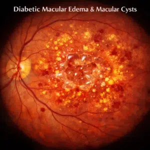
When it fails: age-related macular degeneration, diabetic macular oedema, macular hole, Stargardt disease and central serous chorioretinopathy all attack central vision while leaving peripheral vision intact. The characteristic experience is a grey or blank patch in the middle of the view with the surroundings still clear, often with distortion so that straight lines appear bent. New distortion or a new central dark patch warrants assessment within days, because in wet macular degeneration the damage happens over weeks and is not recoverable once scarring occurs.
Peripheral vision
Everything you can see while looking straight ahead without moving your eyes. A single healthy eye sees approximately 100 degrees temporally (outward), 60 degrees nasally (toward the nose), 60 degrees upward and 70 to 75 degrees downward. The limits are set partly by facial anatomy: the nose restricts the inner field, the brow the upper. Both eyes together span roughly 200 degrees horizontally, with a central overlap of about 120 degrees.
Peripheral retina is rod-dominated, so it excels at detecting motion and works well in dim light, but resolves very little detail and almost no color. This is why a movement caught “out of the corner of your eye” prompts you to turn and look directly at it: the periphery detects, the fovea identifies.
When it fails: glaucoma and retinitis pigmentosa are the classic causes, along with stroke, which produces a loss on the same side of the field in both eyes. Peripheral loss is disabling for mobility, driving and orientation while leaving reading intact, which is close to the opposite of macular disease. It is also insidious, because the fellow eye and the brain fill in missing areas seamlessly. A person with advanced glaucoma may read 20/20 and still meet the legal definition of blindness on field criteria alone.
Binocular vision
Normal vision involves two eyes working together to form one image, this process is known as binocular vision and essential for many visual tasks such as reading, driving and hand-eye coordination. Furthermore, binocular vision helps us perceive depth perception in three dimensions, enabling us to perceive objects from multiple dimensions. Unfortunately, some individuals experience problems with their binocular vision which interfere with daily activities, symptoms could include blurry images that wiggle or double up or blurredness around edges, these problems can often be treated by an eye doctor who specializes in vision therapy.
How depth perception actually works. Because the eyes are separated by roughly 6 to 6.5 cm, each receives a slightly different view of the world. The brain uses this small horizontal difference, called retinal disparity, to compute distance. This is stereopsis, and it is the most precise depth cue available, but it only operates out to a few meters. Beyond that, depth comes from monocular cues: relative size, overlap, perspective, texture gradient, shading and motion parallax. This is why someone with one eye can still judge distance reasonably well at a distance, and why the difficulty is concentrated on near tasks such as pouring a drink or threading a needle.
Squinting narrows the aperture the light passes through, which reduces the blur circle on the retina and temporarily sharpens blurred distance vision. It is the same optical principle behind the pinhole test used in clinic. What squinting does do is mask an uncorrected refractive error, so habitual squinting is worth mentioning at an eye examination, and sustained squinting can contribute to eye strain and headache.
Binocular rivalry is what happens when the two eyes are shown genuinely incompatible images, for example vertical stripes to one eye and horizontal stripes to the other. Rather than blending them, perception alternates, one image dominating for a few seconds before the other takes over. It has nothing to do with the wavelength of light. It is studied because it separates the physical stimulus from conscious perception: the input stays constant while what the person sees changes, which makes it a useful window onto how the visual system resolves conflict.
Single-binocular vision encompasses three related but separate phenomena: simultaneous perception, fusion and stereopsis. These are traditionally described as three grades, each building on the one before:
- Simultaneous perception, the ability to register images from both eyes at the same time without one being suppressed.
- Fusion, combining the two images into one.
- Stereopsis, extracting depth from the small difference between them. This is the highest grade, and it is lost first when binocular function breaks down.
Panum’s fusional area is the small zone around the point of fixation within which slightly different images from the two eyes are still fused into one. Objects falling outside it produce physiological diplopia, a normal and usually unnoticed doubling of objects much nearer or further than what you are looking at. You can demonstrate it by holding a finger up at arm’s length, focusing on it, and noticing that a more distant object appears doubled.
While some individuals with binocular vision can perform these tasks effortlessly, others require assistance such as prism glasses or vision therapy to strengthen weaker eyes and achieve enhanced clarity and depth perception.
A note on what vision therapy can and cannot do, since the term covers a wide range. There is reasonable evidence supporting supervised vision therapy for convergence insufficiency, a specific binocular disorder causing eye strain and difficulty sustaining near work, and for aspects of amblyopia treatment in children. There is no good evidence that eye exercises reduce refractive error, reverse eye disease, or improve vision in a structurally normal, fully corrected eye. Programmes marketed as improving general vision or eliminating the need for glasses are not supported by trial evidence.
Double vision (diplopia)
Diplopia means seeing two images of a single object. The first question a clinician asks is whether it disappears when one eye is covered, because the answer separates two entirely different categories.
- Binocular diplopia resolves when either eye is covered. It results from misalignment of the two eyes, and the causes range from benign to serious: cranial nerve palsies (third, fourth or sixth), thyroid eye disease, myasthenia gravis, orbital fracture, stroke, aneurysm and raised intracranial pressure.
- Monocular diplopia persists with one eye covered and is present in the affected eye alone. It is optical rather than neurological, and usual causes are cataract, uncorrected astigmatism, corneal irregularity including keratoconus, dry eye and dislocated lens.
New onset double vision needs prompt medical assessment, and the original statement that it “can often be improved through eye exercises or eye patches” is not adequate guidance. Sudden binocular diplopia, particularly with headache, pain, a drooping eyelid, a dilated pupil or any other neurological symptom, requires emergency evaluation, because a painful third nerve palsy with pupil involvement can indicate an aneurysm. Prisms, occlusion and surgery all have a role, but only once the cause has been identified.
Monocular vision
Monocular vision means functioning with one eye, either because only one eye is present or because one eye contributes little usable vision. Monocular vision may present challenges when it comes to judging distance and perceiving depth but our visual system is highly adaptable, people with limited binocular vision use various strategies such as head or eye movements to compensate for lack of depth perception.
Monocular vision may occur if one eye is impaired in any way, such as with amblyopia (lazy eye) or strabismus (crossed eyes). When this happens, the brain cannot properly integrate visual information from that particular eye, leading to distortion or blurring as well as difficulty performing tasks that require depth perception.
What functioning monocularly is actually like. Two things change. The visual field narrows on the side of the affected eye, by roughly 20 to 30 degrees, which is why people bump into door frames on that side initially. And stereopsis is lost, which affects near tasks most: pouring liquid, judging steps and kerbs, catching, parking. Adaptation is usually good within weeks to months as the brain leans harder on monocular depth cues and on small head movements that generate motion parallax deliberately.
Practical measures worth knowing: wear polycarbonate protective lenses for sport and workshop tasks, because protecting the remaining eye becomes a priority in a way it was not before; check the remaining eye regularly; expect an adjustment period for driving, and note that most states permit monocular driving subject to field requirements; and turn the head more deliberately rather than relying on peripheral awareness on the affected side.
There are steps available to improve monocular vision, including vision therapy. An experienced vision therapist can teach patients exercises designed to strengthen eye muscles and enhance coordination between eyes. Prism glasses may also be recommended as they shift images so the brain can better process information from affected eyes.
The comparison with animals is worth making, but the original version had it backwards. Eye position across species follows a consistent pattern: prey animals such as rabbits and horses have eyes on the sides of the head, giving a very wide field of view (a horse sees nearly 350 degrees) at the cost of binocular overlap, which is useful for detecting predators approaching from any direction. Predators such as cats, owls and humans have forward-facing eyes, giving a narrower field but a large binocular overlap and therefore good stereopsis, useful for judging the distance to prey. Humans sit firmly in the forward-facing, binocular group.
Color vision
Color is discriminated by three types of cone photoreceptor, sensitive to short (blue), medium (green) and long (red) wavelengths. The brain compares their relative activity to derive color, which is why colour discrimination is poor in dim light: rods handle low light and provide no colour information at all.
Inherited color vision deficiency affects roughly 8% of men and 0.5% of women of Northern European descent. Because the genes for red and green cone pigments sit on the X chromosome, it is far more common in males. Most cases are red-green deficiencies (protanomaly, deuteranomaly and their more complete forms), which are present from birth, stable and not a disease. Blue-yellow deficiency is much rarer, and complete absence of color vision (achromatopsia) is rarer still and accompanied by poor acuity and severe light sensitivity.
Acquired color vision loss is different and is clinically significant. A new change in color perception can signal disease, and the pattern helps localise it: red-green loss and red desaturation point toward the optic nerve, and are an early feature of optic neuritis and compressive optic neuropathy; blue-yellow loss points toward the retina, and is seen in glaucoma, diabetic retinopathy and macular disease. Nuclear cataract yellows the lens and selectively absorbs short wavelengths, which is why colours often look strikingly blue for a while after cataract surgery.
If one eye has started seeing colors as duller or washed out compared with the other, that asymmetry is worth reporting: it is a sensitive early sign and a quick thing to check by comparing a red object between your two eyes.
Night vision and dark adaptation
Rods outnumber cones by roughly twenty to one and handle vision in dim light. They are absent from the very center of the fovea, which is why a faint star is easier to see by looking slightly to one side of it than directly at it.
Dark adaptation is the process of the retina regaining sensitivity after bright light. Cones adapt within several minutes; rods take considerably longer, with full sensitivity reached after roughly 20 to 30 minutes. This is why stepping from bright sunlight into a dim room is briefly disabling, and why glare from oncoming headlights is followed by a period of reduced vision.
When it fails: retinitis pigmentosa classically presents with night blindness in childhood or adolescence, often years before peripheral field loss becomes obvious. Vitamin A deficiency is the leading cause of night blindness worldwide and is reversible if treated. Cataract impairs night vision mainly through glare and light scatter rather than reduced sensitivity, and age-related macular degeneration, particularly with reticular pseudodrusen, delays dark adaptation measurably, sometimes before other changes are apparent. Uncorrected myopia also worsens at night because the pupil dilates, increasing aberrations, a phenomenon known as night myopia.
Contrast sensitivity
The most under-appreciated kind of vision, and often the most functionally important. Visual acuity measures the smallest high-contrast black letter you can identify on a bright white chart. The real world is mostly low contrast: a kerb, a step edge, a face, a grey car in rain, a pale stair against a pale carpet.
Contrast sensitivity can be substantially reduced while acuity remains near normal, and it is degraded by cataract, macular degeneration, glaucoma, diabetic retinopathy and optic neuropathy. It is the strongest visual predictor of mobility difficulty, falls and driving problems, better than acuity in most studies, and it explains the common complaint of “I can read the chart fine but I cannot see properly”.
It is measured in a few minutes with a Pelli-Robson chart and is almost never included in a routine examination. If your day-to-day difficulty is worse than your chart result suggests, ask for it by name.
Refractive errors: how the eye focuses
The following are not kinds of vision but focusing errors, meaning the eye fails to bring light to a sharp point on the retina. They are grouped here to keep the distinction clear, and unlike everything above they are usually fully correctable with lenses.
Hyperopia
Farsightedness (also referred to as hypermetropia) is a common visual condition whereby individuals find it easier to see distant objects than closer ones. This occurs when the eye’s focusing power is diminished; either due to having too flat of lenses and corneas or too short eyes that redirect light onto areas behind rather than directly onto retina.
Farsightedness causes near objects to appear blurry, which is most commonly experienced by children and young adults. People with severe farsightedness often struggle with reading or performing tasks requiring close up vision; they may experience headaches, eyestrain or fatigue after close work and sore or burning eyes as a result of working close up, .
One point that surprises people: a young hyperope may see perfectly well at all distances, because the eye’s focusing muscle can compensate for several diopters of hyperopia. The error is masked, not absent, and the effort of maintaining it causes the eye strain and headache described above. This is why cycloplegic refraction, using drops that temporarily relax focusing, is used in children and young adults: without it, significant hyperopia can be missed entirely. As the focusing range declines with age, previously hidden hyperopia becomes apparent, which is why some people find their distance vision worsening in their forties and fifties despite never having needed glasses.
Uncorrected hyperopia in children matters more than in adults, because it can drive accommodative esotropia (an inward turning of the eye) and amblyopia, both of which are time-sensitive to treat.
Hyperopia can be effectively treated using eyeglasses or contact lenses designed to focus light directly onto the retina, and may also be treatable through refractive surgery in suitable candidates.
Optometrists offer comprehensive eye examinations to diagnose farsightedness accurately and efficiently. This exam should include testing visual acuity and refraction while also assessing lens and cornea health.
As one ages, some may develop the misperception that they are becoming farsighted as reading glasses are required for close up work. However, this condition called presbyopia should be distinguished from true farsightedness. As part of the natural aging process, eye focusing ability may decrease over time due to age-related changes to its crystalline lens within the eye, this change does not vary according to whether or not a person wears reading glasses.
Presbyopia is a loss of focusing range, not a refractive error. The crystalline lens stiffens progressively from childhood, and by the mid-forties it can no longer change shape enough to focus at reading distance. It affects everyone eventually, regardless of whether they are short-sighted, long-sighted or neither. Note one common misconception: using reading glasses does not accelerate it, and avoiding them does not slow it. The stiffening happens either way.
Astigmatism belongs here too, and it is a third type of refractive error rather than a variant of the others. The cornea, or occasionally the lens, is curved more steeply in one meridian than another, so light focuses at two lines rather than one point, blurring vision at all distances. Regular astigmatism is fully correctable with a cylindrical lens in spectacles or toric contact lenses. Irregular astigmatism, from keratoconus, corneal scarring or previous surgery, cannot be corrected with a spectacle lens because the surface is not a smooth toric shape, and requires rigid or scleral contact lenses instead.
Myopia
People with nearsighted vision, also known as myopia, can see close objects clearly while distant objects appear blurry. Nearsightedness occurs when light rays entering the eye do not properly focus onto the retina, usually as a result of having too long of an eye from front to back or having an excessively steeply curled cornea that causes light rays to converge too soon and focus front of rather than directly onto it.
Myopia is classified by which structure is responsible. Axial myopia, by far the most common form, means the eyeball is too long from front to back, so light focuses in front of the retina. Refractive myopia means the eye is of normal length but the focusing power is too strong, usually because the cornea is too steeply curved or, less often, because of changes in the lens. Nuclear cataract can produce an acquired myopic shift in later life, sometimes giving the temporary impression that reading vision has improved.
Myopia can often be linked to genetics. Research shows that an individual’s risk increases significantly if one or both parents are nearsighted, with risks increasing further as they get older; reaching its peak during early adulthood. Lifestyle factors also play a factor; people who spend long hours using digital devices without enough outdoor time tend to become myopic faster than others.
Time outdoors is the better-evidenced half of that. Randomised trials have found that increasing outdoor time in childhood reduces the incidence of myopia, with roughly two hours a day the commonly cited target. The evidence that near work and screen time independently drive myopia is weaker and harder to separate from reduced time outdoors.
Myopia control matters because high myopia is not just a strong prescription. Beyond about −6 diopters, lifetime risk rises substantially for retinal detachment, myopic maculopathy, glaucoma and cataract. Interventions with trial evidence for slowing progression in children include low-dose atropine drops, orthokeratology, and specially designed spectacle lenses and soft contact lenses using peripheral defocus. This is a genuine and fast-moving area, and worth raising if you have a myopic child.
Refraction exams can help detect myopia and identify corrective lenses necessary to improve vision. An autorefractor instrument measures how light bends as it passes through cornea and lens to give an ophthalmologist information on which prescription glasses or contact lenses may be necessary to improve your eyesight. An autorefractor gives a starting point rather than a final prescription: subjective refinement, where you compare lens options, produces the prescription actually issued.
Corrective lenses such as eyeglasses and contacts utilize various refractive technologies to bend light rays and focus them onto the retina, bringing distance vision into focus. While corrective lenses can be very successful at improving myopia, they do come with some drawbacks patients should be aware of. Among others are cosmetic, convenience and optical considerations when wearing lenses as well as potential minification of image which some patients may find unappealing; alternatively LASIK could offer more permanent solution that would eliminate lenses altogether compared with corrective lenses.
When to seek care
Same day, emergency: sudden loss of vision in one eye, whether or not it recovers; a curtain or shadow crossing the field; a sudden shower of floaters with flashing lights; sudden double vision, especially with headache, eyelid droop or a dilated pupil; severe eye pain with redness, haloes and nausea; any injury or chemical splash; new vision loss with headache, scalp tenderness or jaw pain in anyone over 50.
Within days: new distortion of straight lines; a new central grey or dark patch; new floaters without flashes; a new difference in colour perception between the eyes.
Routine: gradual blurring over months, increasing difficulty at night, difficulty with small print, eye strain with near work.
Frequently asked questions
What are the different types of vision?
Central vision (detail and reading), peripheral vision (motion and orientation), binocular vision and stereopsis (depth), color vision, night vision and dark adaptation, and contrast sensitivity. Each is handled by different parts of the visual system and each can fail independently, which is why one person can read the eye chart perfectly yet struggle to move around safely and another can navigate easily but cannot read.
What is the difference between binocular and monocular vision?
Binocular vision uses both eyes together, producing a single fused image and stereoscopic depth perception from the small difference between the two views. Monocular vision is functioning with one eye: the field narrows on that side by roughly 20 to 30 degrees and stereopsis is lost, though depth judgement at distance remains reasonably good because it relies on monocular cues such as relative size, overlap and motion parallax.
Can you have good visual acuity and still have poor vision?
Yes, and this is common. Acuity tests one narrow thing: the smallest high-contrast letter you can identify at distance under good lighting. Someone with advanced glaucoma may read 20/20 with a field so constricted they cannot cross a road safely. Someone with cataract may read 20/25 in a dim exam room and be unable to drive at night because of glare. Contrast sensitivity, visual field and glare testing capture what acuity misses.
Do eye exercises improve vision?
For specific binocular vision disorders, particularly convergence insufficiency, supervised vision therapy has reasonable trial evidence. In amblyopia treatment in children, some forms have a role alongside correction and patching. But no exercise program has been shown to reduce refractive error, eliminate the need for glasses, or improve vision in a structurally normal, fully corrected eye, and none treats eye disease. Programs marketed on those claims are not supported by evidence.
What is 20/20 vision, and is it perfect?
20/20 means you resolve at 20 feet what a reference observer resolves at 20 feet. It is a reference standard, not a ceiling and not a measure of overall visual health: many healthy young adults read 20/15 or better. And because it measures only central high-contrast acuity, 20/20 is entirely compatible with significant loss of field, color discrimination, contrast sensitivity or depth perception.
References
1. Clinical Methods: The History, Physical, and Laboratory Examinations. Chapter on Visual Fields. NCBI Bookshelf, NBK220.
2. Pelli DG, Robson JG, Wilkins AJ. The design of a new letter chart for measuring contrast sensitivity. Clin Vis Sci. 1988;2:187-199.
3. Owsley C, Sloane ME. Contrast sensitivity, acuity, and the perception of real-world targets. Br J Ophthalmol. 1987;71(10):791-796.
4. Blake R, Logothetis NK. Visual competition. Nat Rev Neurosci. 2002;3(1):13-21.
5. Convergence Insufficiency Treatment Trial Study Group. Randomized clinical trial of treatments for symptomatic convergence insufficiency in children. Arch Ophthalmol. 2008;126(10):1336-1349.
6. He M, Xiang F, Zeng Y, et al. Effect of time spent outdoors at school on the development of myopia among children in China: a randomized clinical trial. JAMA. 2015;314(11):1142-1148.
7. Chia A, Chua WH, Cheung YB, et al. Atropine for the treatment of childhood myopia: safety and efficacy of 0.5%, 0.1%, and 0.01% doses (ATOM2). Ophthalmology. 2012;119(2):347-354.
8. Flitcroft DI. The complex interactions of retinal, optical and environmental factors in myopia aetiology. Prog Retin Eye Res. 2012;31(6):622-660.
9. Owsley C. Aging and vision. Vision Res. 2011;51(13):1610-1622.
10. American Academy of Ophthalmology. Comprehensive Adult Medical Eye Evaluation Preferred Practice Pattern. aao.org.
11. American Academy of Ophthalmology. Amblyopia Preferred Practice Pattern. aao.org.



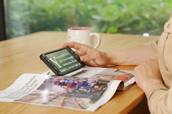
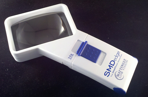
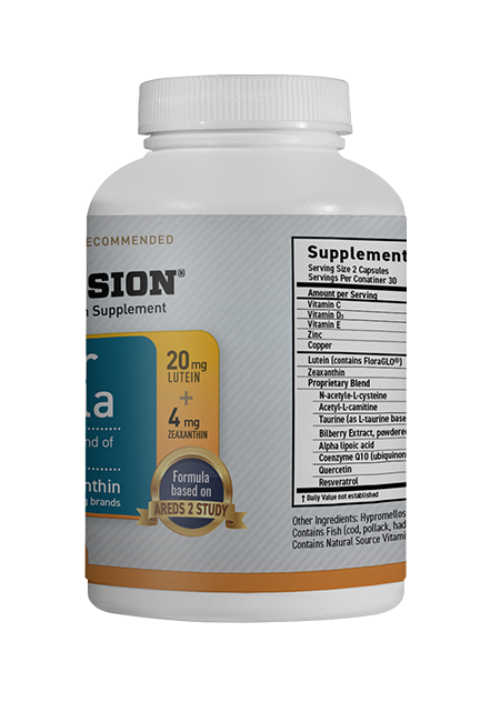

 Shop Best Low-Vision Aids with FREE Doctor Consultation.