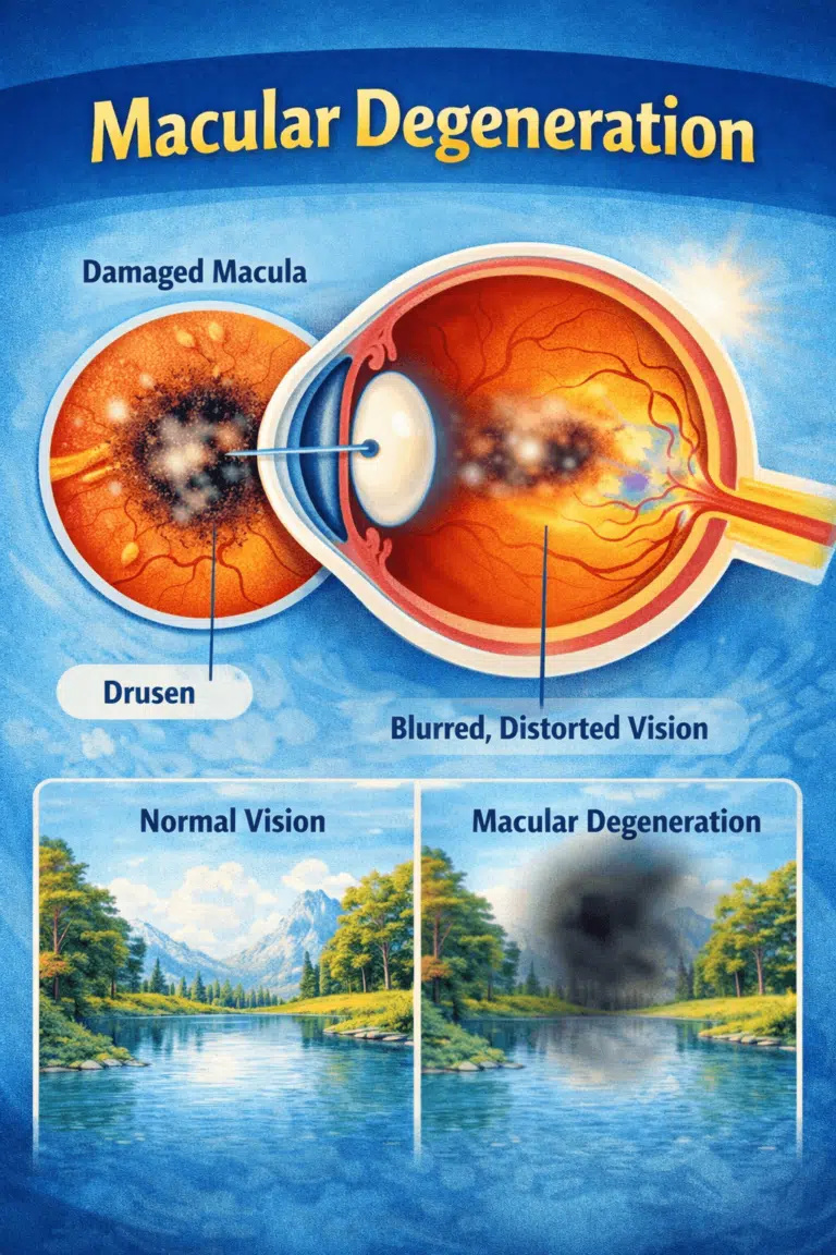
Stargardts Disease
Stargardt disease is the most common inherited macular dystrophy, affecting roughly 1 in 8,000 to 10,000 people [1][2]. It is caused, in about 95% of cases, by variants in the ABCA4 gene that allow toxic vitamin A byproducts called bisretinoids to build up in the retinal pigment epithelium, the support layer beneath the macula. Central vision blurs and fades, usually beginning in childhood or adolescence, while side vision is typically preserved. There is no approved treatment yet, but the first positive phase 3 drug trial in the disease’s history was reported in December 2025 and an FDA filing is under way [18][19].

Not Sure? Talk to a Doctor
We are offering Free Doctor Consultation
What Is Stargardt Disease?
Stargardt disease is a progressive, genetic macular dystrophy that most often begins in children, teenagers, and young adults [1][2]. It is an inherited illness, which means that it is handed down from parents to children.
Stargardt disease is also called Stargardt macular dystrophy, juvenile macular degeneration, or – when flecks are widespread with later onset – fundus flavimaculatus. It was first described by the German ophthalmologist Karl Stargardt in 1909 [1][3]. Photoreceptors, which are specific light-sensing cells in the macula, die out in persons with Stargardt disease. The central or detailed vision blurs or has dark regions. Color vision may also be compromised.
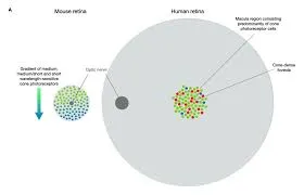
The macula is the small central zone of the retina responsible for reading, recognizing faces, and seeing fine detail. Because Stargardt disease attacks the macula while usually sparing the peripheral retina, most patients retain useful side vision for life even when central acuity falls to the level of legal blindness (20/200 or worse) [2][15].
How Stargardt Disease Damages the Retina (Pathophysiology)
Understanding the mechanism explains both the symptoms and the logic of every drug now in trials. The ABCA4 gene encodes a ‘flippase’ transport protein located in the outer segment discs of photoreceptor cells. Each time light bleaches the visual pigment, a reactive vitamin A aldehyde (all-trans-retinal) is released; some of it binds membrane lipid to form N-retinylidene-phosphatidylethanolamine. ABCA4’s job is to flip this compound back to the disc surface where it can be recycled through the visual cycle [5][6][7].
When both copies of ABCA4 are faulty, the retinaldehyde lingers and condenses with a second molecule of retinaldehyde to form bisretinoids, most notably A2E. As the retinal pigment epithelium (RPE) phagocytoses shed photoreceptor outer segments each day, these indigestible bisretinoids accumulate in RPE lysosomes as autofluorescent lipofuscin. Lipofuscin-laden RPE cells sicken and die; the photoreceptors they support die next; and the result is expanding atrophy of the central retina [6][7]. This sequence – RPE poisoning first, photoreceptor loss second – is why treatments aim to slow vitamin A dimerization rather than simply ‘feed’ the retina.
A clinically important variant of this picture is the foveal-sparing phenotype, in which a ring of atrophy surrounds a preserved foveal center and visual acuity remains good for years longer than expected. The mechanism of foveal survival is not fully understood, but foveal-sparing patients are recognized as a distinct subgroup and may be ideal candidates for early intervention trials [13].

What types of Stargardt disease exist?
STGD1: The recessive type of Stargardt disease induced by mutations in the ABCA4 gene is by far the most frequent, accounting for roughly 95% of cases [1][7].
STGD4 is a rare dominant form caused by mutations in the PROM1 gene, which disrupts photoreceptor disc formation [10].
STGD3: A rare dominant type of Stargardt disease caused by ELOVL4 gene mutations, first characterized in 2001 [9]. Because STGD3 and STGD4 are autosomal dominant, an affected parent has a 50% chance of passing the condition to each child – a different risk calculation from STGD1, which is why genetic confirmation matters (see Diagnosis below).
Late-onset Stargardt disease is linked to missense mutations in ABCA4 that occur outside of recognized functional domains; these patients often present after age 45, frequently with the foveal-sparing pattern and a slower course [7][13].
What Are the Symptoms of Stargardt Disease?
Someone may initially become aware of an issue with their center vision. It may be distorted, hazy, or have black regions. Side vision (peripheral vision) is frequently unaffected. Many patients also develop dyschromatopsia – acquired difficulty distinguishing colors, typically along the red-green axis – as cone photoreceptors are lost [1][3].
When moving between bright and dark environments, eyesight may take longer to adapt than normal. This delayed dark adaptation reflects the slowed visual cycle and is often one of the earliest symptoms, sometimes years before acuity drops [1].
For some patients, Stargardt disease advances slowly at first, then quickly accelerates and finally plateaus. Vision loss may accelerate at roughly 20/40 vision (meaning someone sees at 20 feet what a normal-seeing person sees at 40 feet). In the landmark ProgStar natural history studies of more than 350 genetically confirmed patients, average acuity loss was slow overall – about half a letter per year over two years – but individual trajectories varied widely with age of onset and genotype [14][15].
While most persons with Stargardt disease eventually lose their central vision, many may have strong side vision for the remainder of their lives.
Red flags that warrant prompt evaluation: a child who holds reading material unusually close, sits very near screens, struggles with the board at school, or fails vision screening despite ‘normal-looking’ eyes; a young person with unexplained central blur and a normal exam (early Stargardt maculae can look deceptively normal, and children are sometimes mislabeled as having functional vision loss); or any sudden, painless drop in central vision, which always deserves urgent eye care to exclude other causes [1][3].

What causes Stargardt disease?
Stargardt disease is often caused by mutations in the ABCA4 gene. This gene encodes a transport protein in the photoreceptors that clears reactive vitamin A byproducts after light exposure; when it fails, those byproducts dimerize into toxic bisretinoids [5][6][7].
The ABCA4 gene does not function in Stargardt disease, causing lipofuscin (bisretinoid) material to accumulate in yellowish clumps in the retinal pigment epithelium beneath the macula. This material, over time, damages the RPE and then the light-sensitive cells and degrades your central vision [6][7].
Stargardt disease is a hereditary genetic disorder that is handed on from parents to children. STGD1 is autosomal recessive: a child must inherit one non-working ABCA4 copy from each parent, and the parents – each carrying one normal copy – almost always have normal vision themselves. Roughly 1 in 20 people carries at least one pathogenic ABCA4 variant, which is why the disease can appear with no family history at all [7][8].
Carrier math. When both parents are carriers, each pregnancy has a 25% chance of an affected child, a 50% chance of an unaffected carrier, and a 25% chance of a child who inherits neither variant. Siblings of an affected person who themselves are unaffected still have a 2-in-3 chance of being carriers. An affected person’s children will all be carriers, but will develop STGD1 only if the other parent also contributes a pathogenic ABCA4 variant [2][7].
How Is Stargardt Disease Diagnosed?
Your doctor may order the following tests to diagnose Stargardt disease or to monitor your symptoms:
Color vision evaluation. Because Stargardt disease may result in acquired color vision loss, your eye doctor may also examine your color vision with tests such as the Farnsworth D-15 or Ishihara plates.
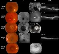
Photography of the fundus. To check for yellowish specks on your macula, your eye doctor may take a snapshot of your retina. The classic findings are yellow-white ‘pisciform’ (fish-tail-shaped) flecks at the level of the RPE and, in later stages, a ‘beaten-bronze’ or bull’s-eye appearance of the central macula [3][4].
Fundus autofluorescence (FAF). This quick, non-invasive photograph maps lipofuscin, which glows under blue light. Active flecks appear bright (hyperautofluorescent); dead RPE appears dark. FAF is now the standard tool for staging Stargardt disease and is the primary outcome measure in every current clinical trial, tracked as areas of ‘definitely decreased autofluorescence’ (DDAF) [14][16].
Fluorescein angiography (when needed). A dye study of retinal circulation classically shows a ‘dark’ or ‘silent’ choroid in the large majority of patients – reported in up to about 80% – because lipofuscin in the RPE masks the background choroidal glow. Modern imaging has made this invasive test less necessary, but the sign remains a useful diagnostic clue [3][4].
Electroretinography (ERG). Your eye doctor may use an ERG to determine how effectively your retina reacts to light. ERG findings define three prognostic groups (the Lois classification): Group 1, abnormality confined to the macula; Group 2, added generalized cone dysfunction; Group 3, both cone and rod dysfunction. Group 3 patients have the most severe, most widespread disease [12].
Optical coherence tomography (OCT). This test uses light waves to create a detailed image of your retina. In Stargardt disease, OCT shows thinning or loss of the ellipsoid zone – the band representing photoreceptor integrity – and outer retinal atrophy, often before acuity falls [3][16].
Genetic analysis. To confirm the diagnosis of Stargardt disease, your doctor may recommend a genetic test. Confirming two disease-causing ABCA4 variants (one on each chromosome, ‘in trans’) is now considered essential: it secures the diagnosis, separates recessive STGD1 from dominant mimics like STGD3 and STGD4, defines inheritance risk for the family, and is required for entry into most clinical trials. In the United States, no-cost sponsored panel testing with genetic counseling is available through the Foundation Fighting Blindness Open Access / My Retina Tracker program (testing performed by Blueprint Genetics) and through industry-sponsored programs such as Invitae’s inherited retinal disease panel [27]. Ask for formal genetic counseling alongside testing – variant interpretation in ABCA4 is genuinely difficult, with over 1,000 known disease-associated variants [7].
Clinical staging. Ophthalmologists also grade the fundus using the Fishman classification (stages I-IV, from a small foveal lesion with limited flecks through widespread atrophy) to describe severity and follow progression [11].
Differential diagnosis. Conditions that can mimic Stargardt disease include cone and cone-rod dystrophies, pattern dystrophies, Best disease, drug toxicity (notably hydroxychloroquine maculopathy), and – in older patients – dry AMD. Genetic testing plus multimodal imaging distinguishes these; the distinction matters because prognosis, family counseling, and trial eligibility differ for each [3][4].

Treatment: What Exists Today and What Is Coming (2024-2026)
Stargardt disease has no known cure. As of August 2026 there is still no FDA-approved treatment – but for the first time, a drug has succeeded in a phase 3 trial, and an approval decision is expected soon. Here is the verified state of the pipeline.
Tinlarebant (Belite Bio) – first positive phase 3, FDA filing under way. Tinlarebant is a once-daily oral tablet that partially blocks retinol-binding protein 4 (RBP4), the carrier that delivers vitamin A to the eye, thereby slowing bisretinoid formation. In December 2025, the pivotal phase 3 DRAGON trial (104 adolescents aged 12-20 with STGD1, randomized 2:1 to drug or placebo for 24 months) met its primary endpoint: a statistically significant 36% reduction in the growth rate of atrophic lesions (DDAF; p = 0.0033), with a 33.7% slowing on the key secondary lesion measure (p = 0.027) [18]. The most common side effects – a yellowish tint to vision (xanthopsia) and delayed dark adaptation – were expected from the mechanism and mostly mild [18][19]. Belite Bio completed a rolling New Drug Application to the FDA in 2026 under Breakthrough Therapy designation; a second adolescent study (DRAGON II) and a broader phase 3 (PHOENIX) continue [19].
Gildeuretinol / ALK-001 (Alkeus Pharmaceuticals) – deuterated vitamin A with Breakthrough Therapy designation. Gildeuretinol is chemically ‘hardened’ vitamin A: deuterium at the carbon-20 position makes the molecule roughly five times slower to dimerize into A2E, so the retina keeps its vitamin A supply while forming less toxin. In the 24-month, placebo-controlled TEASE-1 trial, it slowed atrophic lesion growth by 21.6% (square-root transformed; 29.5% untransformed) [20]. In the ongoing TEASE-3 study, patients treated at the earliest disease stage showed no progression and remained symptom-free over two to six years of therapy – a striking interim result, though from a small open-label cohort [21]. Gildeuretinol holds FDA Breakthrough Therapy, Fast Track, Orphan Drug, and Rare Pediatric Disease designations; an NDA has been in preparation since 2025, and the global phase 3 NORTHSTAR study is under way [21][30].
Emixustat (Kubota Vision) – phase 3 missed its endpoint. Emixustat, an oral visual-cycle modulator (RPE65 inhibitor), did not slow DDAF lesion growth versus placebo in its 24-month phase 3 trial of 194 patients reported in 2022. Post-hoc analyses hinted at benefit in eyes with smaller baseline lesions, but as of 2026 it is not an available therapy and should not be presented as one [22].
Gene therapy – promising but technically hard. ABCA4 is too large to fit in a single adeno-associated virus (AAV) vector, the workhorse of approved retinal gene therapy. The earlier lentiviral approach (SAR422459/StarGen, Sanofi) proved safe over three years in a phase 1/2a trial but showed no clear visual benefit, and the sponsor discontinued development in 2019 for portfolio reasons [23]. Current strategies split the gene across two AAV vectors that reassemble the protein inside photoreceptors; in 2025, dual AAV ‘intein’ vectors were reported safe and effective in large-animal models, supporting movement toward human trials [25].
In one promising research program, retinal cells are given a healthy form of the Stargardt disease gene, ABCA4, to restore normal protein synthesis [23][25].
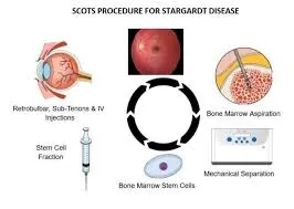
Another research avenue uses stem cells to replace the retinal pigment epithelial (RPE) cells. In phase 1/2 trials, human embryonic stem cell-derived RPE transplanted under the retina of Stargardt patients showed no tumor formation or rejection over follow-up – an important safety milestone – though visual gains were modest and unproven; this remains experimental [24].
Other treatments try to minimize the development of lipofuscin, which causes visual loss in Stargardt disease. Tinlarebant and gildeuretinol, above, are exactly this class [18][20].
A caution about the new ‘geographic atrophy’ injections. Pegcetacoplan (Syfovre) and avacincaptad pegol (Izervay) are FDA-approved only for geographic atrophy secondary to age-related macular degeneration. Stargardt atrophy is a different disease with a different mechanism, and these complement inhibitors are neither approved nor studied for it. Patients occasionally ask about them after seeing television advertising; they are not a Stargardt treatment [29].
Who Gets Stargardt Disease? (Epidemiology)
Stargardt disease is the most common inherited macular dystrophy, affecting roughly 1 in 8,000 to 10,000 people – about 30,000 or more Americans of all ages, not only children [1][2][3].
The condition affects both eyes and appears between the ages of five and early adulthood, with a median onset in the teenage years; a substantial late-onset group presents after 45 [1][7].
A genetic abnormality causes the photoreceptors in the eye to perish in Stargardt disease.
Vision loss starts slowly and eventually accelerates, impairing center vision so badly that an afflicted individual becomes legally blind while maintaining peripheral vision.
Genotype predicts course. Patients carrying two ‘null’ ABCA4 variants (no working protein at all) tend to have childhood onset and the fastest progression, while the common G1961E missense variant is associated on average with later onset, smaller lesions, and slower atrophy growth – though even siblings with identical variants can differ by decades, so no individual forecast is certain [7][12][16]. Childhood-onset disease generally carries the worse visual prognosis [1][14].

Is Stargardt Disease an Emergency?
Stargardt vision loss often manifests itself throughout the first 20 years of a person’s life, especially in late childhood and adolescence.
However, it is impossible to predict when or how quickly retinal damage may develop since variances can exist even among family members with comparable inherited tendencies.
In some circumstances, symptoms of Stargardt disease develop in childhood; however, a person with Stargardt disease (especially the fundus flavimaculatus type of the disease) may reach middle life before visual issues become apparent.
The general rule runs the other way: the later the onset, the better the outlook. Late-onset and foveal-sparing forms usually progress more slowly and retain reading vision longer, whereas childhood-onset disease is typically the most aggressive [1][13][14].
Stargardt disease itself is not a medical emergency – it progresses over years. But any sudden change (new distortion, a rapidly enlarging blind spot, flashes, or a curtain over vision) is not typical of Stargardt disease and warrants same-week ophthalmic assessment to rule out a treatable second problem such as retinal detachment or, rarely, choroidal neovascularization [3][4].
Low Vision Rehabilitation: Devices and Strategies That Actually Help
Because Stargardt disease spares peripheral vision, low vision rehabilitation is unusually effective – especially for young patients, whose adaptability is excellent. A structured program typically includes:
Eccentric viewing and preferred retinal locus (PRL) training. Patients learn to aim a healthy patch of off-center retina at what they want to see. Young Stargardt patients often develop a PRL spontaneously; formal training and magnification together support reading rehabilitation, though evidence for training beyond magnification alone remains limited in controlled studies [26].
Optical magnification. High-add reading spectacles, hand and stand magnifiers, and monocular telescopes for distance (board work, signs, sport). Classic clinical series in Stargardt patients found focusable monocular telescopes and high-add near corrections the most used devices, supporting education and employment [28].
Electronic and digital aids. Desktop and portable video magnifiers (CCTVs) with reversed contrast, screen magnification and text-to-speech built into phones and computers, OCR apps, and head-worn electronic visual aids. eSight combines a camera, display technology, and powerful computation to provide real-time video that allows persons with vision loss to see. Head-worn devices (eSight, OrCam, IrisVision and others) suit some patients and tasks but not all; a trial fitting with a low vision specialist beats any brand claim.
School and career support. Children qualify for an IEP or 504 plan: enlarged print, preferential seating, extended test time, accessible screens, and orientation training. Because intelligence and peripheral vision are untouched, educational and vocational outcomes with good support are strong – career counseling should aim high, not low [26][28].
Driving – honest counseling. Central acuity loss is the limiting factor. Some U.S. states license drivers using bioptic telescopes at intermediate acuity levels, but many Stargardt patients will not meet vision standards as the disease progresses. Planning early for driving alternatives (transit training, ride services) is part of responsible care, and a low vision specialist can assess realistic bioptic candidacy state by state [26].
Support and registries. The Foundation Fighting Blindness (My Retina Tracker registry) and macular disease charities offer peer support, trial matching, and up-to-date research news [27].

Daily Habits: What the Evidence Supports (and What It Does Not)

In the absence of a cure for Stargardt disease, physicians often propose several measures to help patients reduce the potential damage caused by the condition:
Dietary omega-3s (oily fish) are reasonable general nutrition, but evidence that they alter Stargardt progression is weak – no controlled trial supports a protective effect in STGD1. Do not present fish intake as disease-modifying [1][3].
Lowering the retina’s exposure to intense light. Because every photon bleaching rhodopsin generates the aldehyde that becomes A2E, light exposure plausibly fuels the disease. UV-blocking, and ideally blue-filtering, sunglasses plus a brimmed hat in bright conditions are a low-cost, guideline-endorsed precaution – though the supporting evidence is mechanistic and observational rather than from randomized trials [1][4][17].
Avoiding vitamin A SUPPLEMENTS – not normal food. In ABCA4-deficient mice, vitamin A supplementation accelerated lipofuscin bisretinoid accumulation (Radu et al., 2008), so most specialists advise Stargardt patients to avoid supplemental vitamin A, beta-carotene products, and AMD-formula ‘eye vitamins’ built around vitamin A precursors [17]. A normal, balanced diet does not require restriction; there is no evidence that avoiding vitamin A-containing foods helps, and outright deficiency harms the retina. And lipofuscin is a bisretinoid pigment, not ‘a fatty substance’ [6][17].
Maintaining a healthy diet and general health; smoking and being overweight may lead to faster macular degeneration, and smoking cessation in particular is advised: smoking adds oxidative stress to an RPE already burdened by bisretinoids, and it is the strongest modifiable risk factor in the related atrophic maculopathies [3][4].
What happens without any management? The disease progresses regardless – these measures aim to slow it and preserve function. Most patients stabilize in the 20/200 range (legal blindness) while keeping navigating peripheral vision; complete blackness is not the expected outcome. In ProgStar, atrophic lesions grew on average by about 0.76 mm2 per year, the benchmark against which the new drugs are measured [14][15][16]. Skipping low vision rehabilitation, school support, and trial screening is the real cost of ‘no treatment.’
Who Can Help With Treating Stargardt Disease?
Our Low Vision Aids (LVA) eye doctor can assist you in coping with the symptoms of vision loss. Stargardt disease is a hereditary eye condition that causes vision loss in children and adolescents, and sometimes first appears in adults. It affects over 30,000 persons in the United States [2][3].
As part of a dilated eye exam, your eye doctor will look for Stargardt disease. The exam is easy and painless: your doctor will dilate (widen) your pupils with eye drops and then look for evidence of Stargardt disease, such as yellowish flecks in your macula. Suspected cases should then be referred for multimodal imaging (FAF, OCT), electroretinography where indicated, and confirmatory genetic testing with counseling – and co-managed between a retina specialist (diagnosis, trial access) and a low vision optometrist (function, devices, school and work support) [3][4][27].

How Common Is Stargardt Disease – and Who Is at Risk?

Stargardt disease does not affect everyone. Vision loss linked with the disease normally starts in childhood or adolescence, although in certain cases, vision impairments may begin in adulthood. According to Orphanet, the gateway for rare diseases, Stargardt disease affects roughly one in every 8,000 to 10,000 births [1][2].
Stargardt disease is an inherited disorder, which means it is handed down from generation to generation. Approximately 95% of Stargardt cases are the recessive ABCA4 form (STGD1). In these families both parents are typically unaffected carriers of one variant each – they do not have the disease, and usually have no idea they carry it. When two carriers have children, each pregnancy carries a 25% chance of Stargardt disease [7][8]. The rare dominant forms (STGD3, STGD4) behave differently: one affected parent, 50% risk per child [9][10].
Frequently Asked Questions
If I have Stargardt disease, will my children have it? With recessive STGD1, all your children will be carriers, but they develop the disease only if your partner also contributes a pathogenic ABCA4 variant. Given the roughly 1-in-20 carrier rate, the risk to each child is on the order of 1-2%, and partner carrier testing can refine it further. Dominant forms (STGD3/STGD4) carry a 50% risk per child – which is why genetic confirmation of your subtype comes first [7][8][9].
Will my child go completely blind? Almost never. Stargardt disease destroys central vision – most patients eventually plateau around 20/200, meeting the definition of legal blindness – but peripheral vision, the vision used for orientation and mobility, is usually retained for life. Children adapt remarkably well with rehabilitation and school support [2][14][15].
Can adults develop Stargardt disease? Yes. Late-onset STGD1 typically appears after 45, often with foveal sparing and slower progression, and is regularly mistaken for early dry AMD. Genetic testing and the fleck pattern on autofluorescence distinguish the two [7][13].
Is there a treatment coming soon? Realistically, yes – the field’s first approval decision is pending. Tinlarebant’s phase 3 DRAGON trial met its endpoint in December 2025 (36% slowing of lesion growth in adolescents) and its FDA application was completed in 2026; gildeuretinol has Breakthrough Therapy designation with positive TEASE data. Neither restores lost vision – both slow progression, which is why early diagnosis matters [18][19][20][21].
What supplements should I avoid? Avoid vitamin A supplements, beta-carotene, and AMD ‘eye vitamin’ formulas containing vitamin A precursors, based on animal data showing accelerated toxin buildup. Eat a normal diet; do not restrict foods. Ask before starting any retinol-containing product, including some acne treatments and multivitamins [17].
Will I be able to drive? It depends on your acuity, your state’s law, and progression. Some patients with milder disease qualify, sometimes via bioptic telescope programs; many eventually will not. A low vision evaluation gives you a realistic, state-specific answer and a plan either way [26].
This article is for educational purposes and does not replace an examination by an eye care professional. If you notice changes in your vision, or if Stargardt disease runs in your family, schedule a comprehensive dilated eye examination and ask about genetic testing and counseling.
References
1. Tanna P, Strauss RW, Fujinami K, Michaelides M. Stargardt disease: clinical features, molecular genetics, animal models and therapeutic options. Br J Ophthalmol. 2017;101(1):25-30. doi:10.1136/bjophthalmol-2016-308823
2. National Eye Institute. Stargardt Disease. National Institutes of Health. https://www.nei.nih.gov/learn-about-eye-health/eye-conditions-and-diseases/stargardt-disease
3. Voigt AP, et al. Stargardt Disease. In: StatPearls Internet. Treasure Island (FL): StatPearls Publishing. https://www.ncbi.nlm.nih.gov/books/NBK587351/
4. American Academy of Ophthalmology. Stargardt Disease / Fundus Flavimaculatus. EyeWiki. https://eyewiki.org/Stargardt_Disease/Fundus_Flavimaculatus
5. Quazi F, Lenevich S, Molday RS. ABCA4 is an N-retinylidene-phosphatidylethanolamine and phosphatidylethanolamine importer. Nat Commun. 2012;3:925. doi:10.1038/ncomms1927
6. Lenis TL, Hu J, Ng SY, et al. Expression of ABCA4 in the retinal pigment epithelium and its implications for Stargardt macular degeneration. Proc Natl Acad Sci U S A. 2018;115(47):E11120-E11127. doi:10.1073/pnas.1802519115
7. Cremers FPM, Lee W, Collin RWJ, Allikmets R. Clinical spectrum, genetic complexity and therapeutic approaches for retinal disease caused by ABCA4 mutations. Prog Retin Eye Res. 2020;79:100861. doi:10.1016/j.preteyeres.2020.100861
8. Riveiro-Alvarez R, Aguirre-Lamban J, Lopez-Martinez MA, et al. Frequency of ABCA4 mutations in 278 Spanish controls: an insight into the prevalence of autosomal recessive Stargardt disease. Br J Ophthalmol. 2009;93(10):1359-1364. doi:10.1136/bjo.2008.148155
9. Zhang K, Kniazeva M, Han M, et al. A 5-bp deletion in ELOVL4 is associated with two related forms of autosomal dominant macular dystrophy. Nat Genet. 2001;27(1):89-93. doi:10.1038/83817
10. Yang Z, Chen Y, Lillo C, et al. Mutant prominin 1 found in patients with macular degeneration disrupts photoreceptor disk morphogenesis in mice. J Clin Invest. 2008;118(8):2908-2916. doi:10.1172/JCI35891
11. Fishman GA. Fundus flavimaculatus: a clinical classification. Arch Ophthalmol. 1976;94(12):2061-2067. doi:10.1001/archopht.1976.03910040721003
12. Lois N, Holder GE, Bunce C, Fitzke FW, Bird AC. Phenotypic subtypes of Stargardt macular dystrophy – fundus flavimaculatus. Arch Ophthalmol. 2001;119(3):359-369. doi:10.1001/archopht.119.3.359
13. Fujinami K, Sergouniotis PI, Davidson AE, et al. Clinical and molecular analysis of Stargardt disease with preserved foveal structure and function. Am J Ophthalmol. 2013;156(3):487-501. doi:10.1016/j.ajo.2013.05.003
14. Strauss RW, Ho A, Munoz B, et al. The Natural History of the Progression of Atrophy Secondary to Stargardt Disease (ProgStar) Studies: design and baseline characteristics. ProgStar Report No. 1. Ophthalmology. 2016;123(4):817-828. doi:10.1016/j.ophtha.2015.12.009
15. Kong X, Strauss RW, Cideciyan AV, et al. Visual acuity change over 24 months and its association with foveal phenotype and genotype in individuals with Stargardt disease: ProgStar Study Report No. 10. JAMA Ophthalmol. 2018;136(8):920-928. doi:10.1001/jamaophthalmol.2018.2198
16. Strauss RW, Munoz B, Ho A, et al. Progression of Stargardt disease as determined by fundus autofluorescence over a 12-month period: ProgStar Report No. 11. JAMA Ophthalmol. 2019;137(10):1134-1145. doi:10.1001/jamaophthalmol.2019.2885
17. Radu RA, Yuan Q, Hu J, et al. Accelerated accumulation of lipofuscin pigments in the RPE of a mouse model for ABCA4-mediated retinal dystrophies following vitamin A supplementation. Invest Ophthalmol Vis Sci. 2008;49(9):3821-3829. doi:10.1167/iovs.07-1470
18. Belite Bio, Inc. Belite Bio announces positive topline results from the pivotal global phase 3 DRAGON trial of tinlarebant in adolescents with Stargardt disease. Press release, December 1, 2025. https://investors.belitebio.com/news-releases/
19. Ophthalmology Times. Tinlarebant NDA submission completed for Stargardt disease type 1, targeting first approved therapy. 2026. https://www.ophthalmologytimes.com/view/tinlarebant-nda-submission-completed-stargardt-disease
20. Alkeus Pharmaceuticals. TEASE-1 randomized controlled trial results for gildeuretinol (ALK-001) in Stargardt disease, presented at the American Academy of Ophthalmology Annual Meeting, October 2024. https://alkeuspharma.com/
21. Alkeus Pharmaceuticals. Alkeus announces new positive interim TEASE-3 study results showing gildeuretinol prevented disease progression in early-stage Stargardt patients. 2024. https://alkeuspharma.com/alkeus-pharmaceuticals-announces-new-positive-interim-tease-3-study-results-showing-gildeuretinol-prevented-disease-progression-in-early-stage-stargardt-patients/
22. Kubota Vision Inc. Kubota Vision announced end of phase 3 clinical trial of emixustat in patients with Stargardt disease. Business Wire, June 24, 2022. https://www.businesswire.com/news/home/20220624005070/en/
23. Parker MA, Erker LR, Audo I, et al. Three-year safety results of SAR422459 (EIAV-ABCA4) gene therapy in patients with ABCA4-associated Stargardt disease: an open-label dose-escalation phase I/IIa clinical trial, cohorts 1-5. Am J Ophthalmol. 2022;240:285-301. doi:10.1016/j.ajo.2022.02.013
24. Schwartz SD, Regillo CD, Lam BL, et al. Human embryonic stem cell-derived retinal pigment epithelium in patients with age-related macular degeneration and Stargardt’s macular dystrophy: follow-up of two open-label phase 1/2 studies. Lancet. 2015;385(9967):509-516. doi:10.1016/S0140-6736(14)61376-3
25. Retinal gene therapy for Stargardt disease with dual AAV intein vectors is both safe and effective in large animal models. Sci Adv. 2025;11:eadt9354. doi:10.1126/sciadv.adt9354
26. American Academy of Ophthalmology. Vision Rehabilitation Preferred Practice Pattern. Ophthalmology. 2023;130(3):P271-P335. doi:10.1016/j.ophtha.2022.10.031
27. Foundation Fighting Blindness. My Retina Tracker Genetic Testing Program (Open Access, with Blueprint Genetics). https://www.fightingblindness.org/open-access-genetic-testing-program
28. Fonda G, Thomas H, Gore GV. Educational and vocational placement, and low-vision corrections in albinism; characteristics and low vision corrections in Stargardt’s disease. Ophthalmology / low vision clinical series. PMID: 4047602. https://pubmed.ncbi.nlm.nih.gov/4047602/
29. Complement inhibition in geographic atrophy: clinical evidence for pegcetacoplan and avacincaptad pegol. Cureus. 2025. PMID: 42326286. https://pubmed.ncbi.nlm.nih.gov/42326286/
30. Retinal Physician. Novel therapies for Stargardt disease. October 2025. https://www.retinalphysician.com/issues/2025/october/novel-therapies-for-stargardt-disease/

Find a nearby low vision professional
Visit a low vision specialist to learn more about low vision aids and choose the proper ones. Find one in your region.
Latest Posts
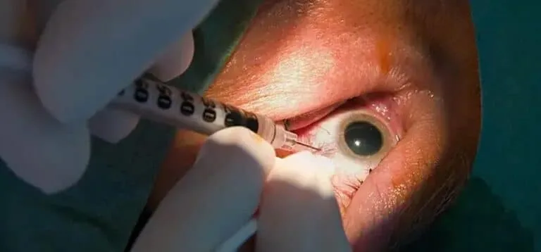
Avastin Eye Injection: Complete Guide to Cost, Side Effects & Effectiveness
If your doctor has recommended an eye injection for a retinal condition, it’s normal to feel worried or scared.
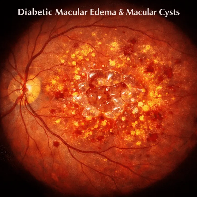
Diabetic Macular Edema and Macular Cysts: Causes, Symptoms & Treatment
Receiving a diagnosis related to your vision can feel overwhelming, especially when terms like macular cysts or cystoid macular edema are mentioned alongside diabetes.
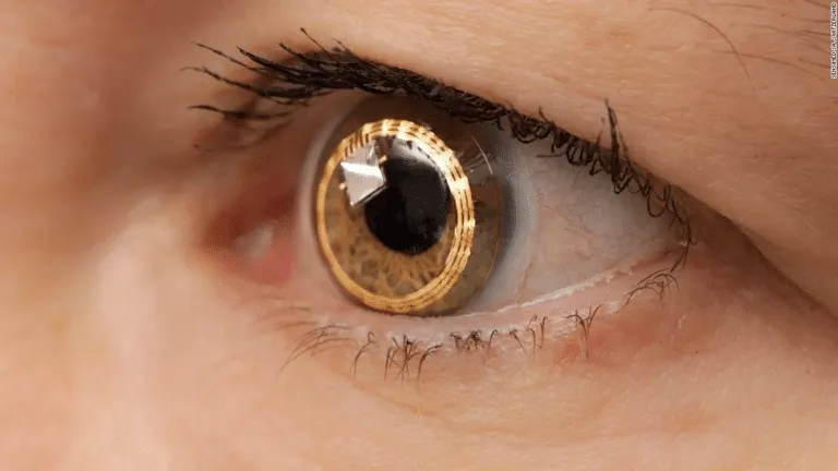
Low Vision Aids: Complete Guide to Visual Aids for Independence
Living with changes in vision can feel overwhelming, especially when regular glasses no longer help.
Macular Degeneration Self-Care: Complete Guide to Managing AMD Daily
If you’ve been diagnosed with macular degeneration (AMD), I understand that fear.