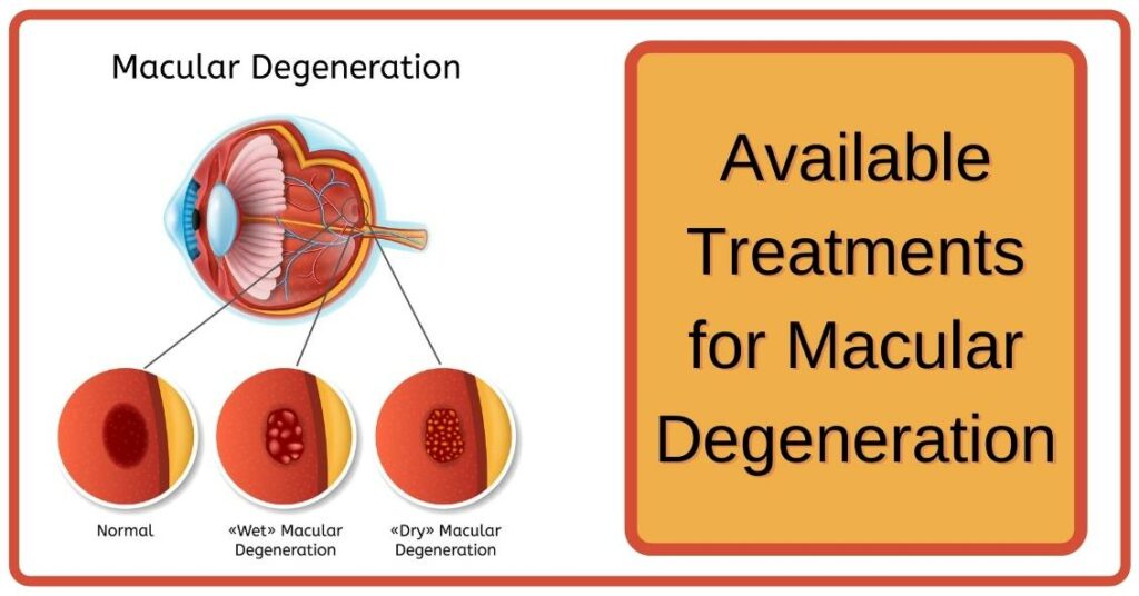
Left untreated, wet age-related macular degeneration can quickly progress and lead to vision loss. Learn the early warning signs of this condition as well as treatments that have been shown to effectively slow its progress.
A 76-year-old woman suffering from Atrophic AMD visited our clinic with an increased central scotoma on home Amsler grid testing. Her previous medical history included Atrophic Macular Degeneration, High Cholesterolemia and Colon Cancer being surgically treated 20 years earlier.
Dry Macular Degeneration
Under dry macular degeneration, the macula gradually breaks down due to thinning retinal pigment epithelium (RPE) cells in the eye. This leads to blurry, distorted or fuzzy central vision and difficulty seeing fine details, such as straight lines appearing wavy or crooked. Luckily, most people do not lose all their central vision as AMD worsens; rather it becomes increasingly blurry over time without ever leading to blindness.
Drusen are waste deposits found beneath the retina that form in dry macular degeneration, believed to be caused by build-ups of fats, protein and cholesterol in between layers of retina and layers more immediately underneath it. These deposits disrupt light-sensitive photoreceptor cells responsible for translating light into vision.
As macular degeneration progresses, small areas of central vision become blurry or distorted when trying to focus on near objects. These central regions tend to appear darker than their surrounding area and may give an illusion that there is an empty spot at the center of your vision. If these symptoms appear in your life, it is wise to visit an eye care professional as they can determine its severity as well as suggest possible treatments options.
About 85 to 90% of cases of macular degeneration fall under the category of dry macular degeneration, meaning those affected can still enjoy good peripheral vision but it may no longer be as sharp. Sometimes dry AMD can advance to wet AMD where abnormal blood vessels grow beneath the retina and leak fluid and blood, damaging retinal tissue quickly causing rapid vision loss.
There is no cure for wet macular degeneration, but its progression can be reduced with regular eye examinations and medications that block an enzyme responsible for abnormal blood vessel growth and leakage – such as those available through prescription. Eye exams should detect early symptoms such as central blurriness or distortion in your visual field and treat accordingly with medications provided only through valid medical prescription.
Wet Macular Degeneration
Wet macular degeneration occurs when abnormal blood vessels form under the retina and leak blood or fluid into it, damaging the macula and leading to rapid central vision loss. Symptoms may include blurriness, distortion of straight lines and blind spots – these typically begin in one eye but may spread. While wet AMD is much less common than its dry counterpart, prompt treatment must be sought to prevent permanent vision loss.
The macula is a small area at the back of your eye that provides central and color vision, including central-depth perception. It contains light-sensitive cells that transmit signals from light sources back to your brain for translation into images of objects and people present before you. With wet macular degeneration, this signaling process becomes damaged due to blood vessels growing underneath the retina that leak fluid, damaging retina cells while scarring scar tissue covers its surface.
15-20% of people living with age-related macular degeneration suffer from its wet subtype, the faster-growing form that can lead to permanent vision loss if left untreated. Wet macular degeneration occurs when tiny new blood vessels form under the retina and leak blood or fluids into it quickly degrading vision fast; this process is known as choroidal neovascularization or CNV.
If you have wet type macular degeneration, regular testing with an Amsler grid can help detect distorted or fuzzy vision as an early indicator of fluid accumulation under the retina. Without treatment, CNV may damage retinal pigment epithelium resulting in blind spots on retina.
If you suffer from wet macular degeneration, your doctor may suggest photodynamic therapy as a solution to damage leaking blood vessels and slow vision loss. The procedure involves injecting a drug into an arm vein; when examined by laser light energy directed at leaky vessels it attaches itself to low-density lipoprotein (LDL) molecules attached to low-density lipoprotein, which then absorbs light energy which causes them to clot shut and close off completely.
Early Symptoms
Macular degeneration often progresses gradually over time and may go undetected, as its initial stages do not cause noticeable visual changes. The macula, located within the retina and responsible for sharp and clear central vision, allows a person to see objects, read, and recognize faces without difficulty. When affected, this area allows people to recognize faces quickly.
As the macula thins, central vision blurs and dark spots appear at the center of a person’s field of view. This can make everyday tasks such as driving or reading challenging; colors no longer appear vibrantly; low light visibility may become difficult.
In 90% of AMD cases, eye doctors can typically detect its early symptoms through a standard dilated exam. This involves placing eye drops into each eye to dilate them, enabling your health professional to observe your retina and macula more closely. They’ll check for clumps of protein called drusen that form on retinal layers; as well as performing an Amsler grid test: this square pattern features straight and wavy lines which they ask you to focus on; any deviation could signal macula thinning and disease advancement.
While many do not experience vision loss during the initial stage of macular degeneration, others progress to wet AMD. With wet AMD, abnormal blood vessels begin growing underneath the retina and leak fluid into it causing damage to macula that results in rapid and severe vision loss.
Macular degeneration risks should have an eye exam annually in order to catch it early and treat it more successfully, or stop further progression of their disease. An exam is particularly recommended if any sudden changes occur in vision.
Late Symptoms
The macula is the central portion of retina, the light-sensitive tissue located at the back of your eye that enables you to see fine details and straight ahead. Light hits this light-sensitive tissue and converts into electrical impulses for interpretation and recognition in your brain. Macular degeneration damages this macula, leading to central vision loss; it is one of the leading causes of blindness among adults aged 60 or above and progresses from early through intermediate and finally advanced stages.
Dry macular degeneration often manifests itself through tiny deposits called drusen under the retina. While not visible to the naked eye, they may be visible during dilated examination of your eyes. Drusen are harmless; it is part of normal aging; however if your doctor notices them it would be wise to schedule regular appointments to evaluate your eye health thoroughly.
Some individuals with dry AMD do not progress to wet macular degeneration. If they do, symptoms may include distortion of straight lines such as wavy ones and blurring of vision in their center field of vision. If abnormal blood vessels leaking fluid cause by wet macular degeneration aren’t stopped from leaking, central vision will continue to deteriorate over time.
Doctors struggle to accurately predict who will experience central vision loss and who won’t. Researchers are actively searching for early indicators that indicate faster disease progression among certain patients – these could include structural or functional changes, genetic markers or proteins as well as imaging tests like optical coherence tomography (OCT).
Maintaining regular eye exams can help slow macular degeneration progression by keeping track of reduced or distorted vision, testing your ability to read letters on an Amsler grid, discussing family history of macular degeneration and any risk factors with eye care professionals, as well as discussing family histories or potential risk factors with them. Schedule an appointment online or over the phone today –


