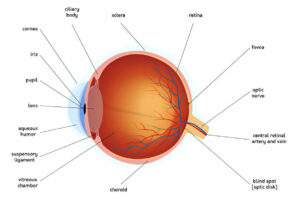
High blood pressure damages the small blood vessels within the retina, disrupting their function and potentially leading to reduced blood supply for it. This may cause swelling within it.
Untreated, the dilated blood vessels can block blood from reaching the eye, potentially leading to sight loss and necessitating regular checks on your blood pressure. Therefore, it’s crucial that you get yours checked regularly in order to maintain good eye health.
Grade 1
High blood pressure (hypertension) can damage the delicate blood vessels in your eyes, eventually leading to them bleeding and leading to symptoms like blurred vision or loss of sight. Furthermore, elevated blood pressure increases stroke risks which further compromise vision; so taking regular readings of your blood pressure and seeking medical assistance if sudden vision changes or headaches arise are both essential measures.
Hypertension can lead to hypertensive retinopathy, in which blood vessels in the retina at the back of the eye change and leak. Swollen blood vessels reduce oxygen delivery to the retina resulting in visual acuity loss or stroke affecting 50-80% of people with high blood pressure.
Hypertensive retinopathy‘s early signs include a generalized narrowing or widening of retinal blood vessels known as arteriolar narrowing or vascular congestion. Furthermore, blood vessels within the retina may become twisted or dilate significantly as well.
Research has established that the progression of retinopathy correlates with blood pressure severity. The Beaver Dam Eye Study concluded that patients with hypertension exhibited a direct relationship between their systolic blood pressure levels and retinal vascular changes; those with the highest systolic blood pressure had more severe cases.
Retinopathy signs also correlate to arterial stiffness and aortic root diameter in the aorta, with S stage of retinopathy more closely correlating with both than H stage; suggesting it represents early stages of atherosclerosis.
Good news is that even early signs of hypertensive vascular disease can be reversed with healthy lifestyle choices and by cutting salt intake. Regular visits to your optometrist can assist in the detection of these early warning signs, so your GP can intervene and prescribe treatments or lifestyle modifications that can protect blood vessels and limit potential damage.
Grade 2
As blood pressure rises, retinal blood vessels become more vulnerable to becoming blocked and not allowing adequate blood flow through their walls, depriving areas of retina from receiving adequate oxygen supply and potentially leading to sight loss if it becomes severe enough.
Retinal blood vessels are controlled through an autoregulation process in which their diameter automatically narrows or widens in response to changes in blood flow to and from retina. High blood pressure may alter this mechanism and cause vessels to thicken up further and narrow more drastically, restricting how much blood reaches retina.
Hypertensive retinopathy refers to any condition which disrupts how retinal blood vessels operate, and as it progresses may lead to microaneurysms, hemorrhages, hard exudates and cotton wool spots developing on retinal vessels. Sometimes blood can also leak through between layers of eyeball, known as vitreous haemorrhage.
Studies demonstrate a direct relationship between hypertension and severity of retinopathy. Individuals who had uncontrolled high blood pressure were 50-70% more likely to have retinal hemorrhages or microaneurysms, as well as 30-40% more likely to suffer AV nicking, than normotensive people.
Researchers have also demonstrated that mild to moderate retinopathy is an independent predictor of cardiovascular disease in young patients with preclinical hypertension. However, its grade should not be used solely as a basis for judging an individual; its influence does not determine their lifespan or the degree to which they will develop cardiovascular complications such as heart disease and stroke.
Controlling blood pressure through diet, exercise and lifestyle modifications is one of the best ways to lower the risk of hypertensive retinopathy. Reducing salt and alcohol intake, quitting smoking and managing weight can all help maintain healthy levels of blood pressure.
Grade 3
Hypertension damages small blood vessels which supply the retina (a light-sensitive tissue at the back of the eye) with blood. When these blood vessels expand and leak fluid into it, this causes swelling which prevents transmission of visual signals to the brain as well as damage to optic nerve and white spots appearing on retina; over time this condition may lead to permanent blindness.
Hypertensive retinopathy can be diagnosed through a dilated exam that allows doctors to see inside of your eye. An ophthalmologist may detect narrowed blood vessels, areas of poor circulation (whitening of areas corresponding with poor circulation), hemorrhages or cotton wool spots as well as swelling in your optic nerve.
Research has demonstrated that hypertensive retinopathy can serve as an accurate predictor of cardiovascular diseases. Moderate signs are strongly correlated to subclinical and clinical stroke and other cerebrovascular outcomes, congestive heart failure and cardiovascular death despite no traditional risk factors present.
An assessment tool has been created to assist physicians in classifying the severity of hypertensive retinopathy. It takes into account disease progression in relation to blood pressure levels and provides a predictive score of cardiovascular events, providing accurate predictions of cardiovascular events both among individuals with normal blood pressure levels as well as those treated with antihypertensive drugs.
Hypertensive retinopathy can be broken down into four stages with increasing severity: grade 1 is marked by mild generalized retinal arteriolar narrowing or focal areas of narrowing, arteriovenous nicking or arteriovenous nicking; grade 2 shows more severe focal or generalized narrowing as well as microaneurysms and hard exudates; while grade 3, commonly known as accelerated hypertensive retinopathy, has symptoms seen in grades 1-3 plus macular edema and optic disc swelling.
Treatment for hypertensive retinopathy follows the same course as treating high blood pressure elsewhere. Medication to maintain a blood pressure below 140/90 mmHg can reduce risks for complications. Beta blockers, diuretics, calcium channel blockers and angiotensin converting enzyme inhibitors may all be prescribed to control this issue.
Grade 4
Hypertension damages blood vessels in the retina (light-sensitive tissue at the back of the eye). Thickening and narrowing of these vessels reduces blood flow, potentially resulting in swelling of optic nerve and macula (known as macular edema) as well as signs of severe retinopathy such as dilations twisted vessels that leak fluid into retina – signs that could potentially lead to sudden vision loss as well as serious risks such as cardiovascular disease (heart attack and stroke).
High blood pressure damage to retinal vessels often comes as a result of atherosclerosis, the condition which affects all the vessels throughout the body and leads to plaque build-up and narrowed vessel linings; blood vessels may clot and rupture allowing blood leakage into retina.
As retinal blood vessels deteriorate, retinal pigment epithelium (the thin layer at the back of your eye) may thin and lighten in color, as well as developing small white spots known as drusen that appear either circularly or linearly on its surface. Drusen are often associated with vitreous hemorrhage or choroidal haemorrhage.
Condition may progress into severe retinopathy and sclerosing choroidopathy, or macular scarring (macular fibrovascular dystrophy). At this stage, symptoms include microaneurysms, blot haemorrhages, flame-shaped haemorrhages, hard exudates and cotton wool spots; signs include an increased level of vascular endothelial growth factor (VEGF) in vitreous fluid; changed ratio between A/V size ratio; tortuosity of vascular arcades etc.
Hypertensive Retinopathy can quickly worsen without treatment, potentially leading to permanent vision loss. But by managing blood pressure with lifestyle modifications and medication, most patients can have their retina healed over time. Regular check-up with both an eye doctor and primary care provider is recommended in order to protect the health of both.
Control of blood pressure is of primary importance in treating this condition. Drugs from various classes, including beta blockers, diuretics, calcium channel blockers and angiotensin converting enzyme inhibitors may be prescribed; laser surgery may be performed for serious cases involving retinal edema that threaten vision loss.


