sudden, painless vision loss should always be taken seriously and evaluated promptly, with a comprehensive history providing insight into possible causes.
Patients over 60 presenting with unilateral painless loss of vision are at an increased risk for Giant Cell Arteritis (GCA). Aside from visual symptoms, headaches and systemic constitutional symptoms should also prompt GCA screening.
Central retinal artery occlusion (CRAO)
This condition occurs when a blood clot blocks the central retinal artery of one eye. As oxygen supply to the retina decreases, which could potentially result in permanent vision loss. It should be treated immediately. It constitutes an urgent medical situation requiring immediate care.
The central retinal artery begins at its source – the ophthalmic artery, a branch of the internal carotid artery – before traveling intracranially for some distance before entering orbit and joining up with optic nerve. Pressure changes throughout this journey; both within the eye itself and from blood vessels that support it.
Blood clots that form within the arteries can quickly cause sudden, painless vision loss – an ocular stroke. Ocular strokes can occur for various reasons including arterial disease such as atherosclerosis, trauma or inflammation conditions; emboli (fragments of plaque that break off from other vessels and enter eye); embolisms from another blood vessel entering eye; embolisms (pieces of plaque from one vessel entering another one), embolisms containing plaque fragments breaking off another blood vessel entering another, blocking central retinal artery or outer retinal arteries within it all causing sudden vision loss – an anocular stroke in other words!).
People at risk for chronic renal allograft (CRAO) include those with high blood pressure, age over 50 and diabetes; additionally those who use birth control pills are more at risk than others for this condition.
CRAO can cause sudden, painless vision loss in one eye. Other symptoms may include reduced brightness of red reflex (or lack thereof) and floaters. Left untreated, this condition often leads to complete blindness.
Health care providers will review your history and conduct a physical exam that focuses on what condition is causing vision loss and the severity of symptoms. An eye doctor may suggest other tests in order to get an idea of the source of discomfort; such tests might include blood tests to check blood sugar or vessel issues; measurement of blood pressure and cholesterol levels; as well as fluoroscopy to inspect inside your eyes for damage signs.
Retinal vein occlusion (RVO)
Retinal Vein Occlusion (RVO) is one of the leading causes of vision loss among older people worldwide, due to a blockage of blood flow in retinal veins which results in elevated pressure causing hemorrhages, swelling and oxygen-deprivation in the retina resulting in hemorrhages, swelling and even ischemia (lack of oxygen supply to retina). RVO may occur either centrally or branch vains and lead to sudden painless loss of vision.
Loss of vision that occurs without pain requires immediate medical intervention and should be evaluated by a retinal specialist immediately. A dilated eye exam and additional testing such as digital fundus photography, OCT and fluorescein angiography should be conducted to ascertain the severity of RVO, along with checking history of cardiovascular disease, inflammation conditions or systemic diseases that might complicate things further.
RVO occurs when an embolism forms from plaque that breaks away from main arteries in the heart or carotid arteries and occludes central retinal artery from feeding retina, leading to rapid and usually painless loss of vision in one eye. Central retinal vein occlusion differs from branch retinal vein occlusion by being more likely to involve more of retina. As a result, its prognosis tends to be worse.
No effective treatment exists to unclog retinal veins occluded by RVO; however, treatments implemented within 1.5 hours of its onset have shown some success in terms of improving vision. Such therapies include ocular massage to dislodge any emboli that might clog retinal veins; anterior chamber paracentesis; intraocular pressure-lowering medication and intraocular massage as potential remedies. It’s also important to optimize modifiable risk factors such as blood pressure regulation, smoking cessation, cholesterol control and diabetes control to reduce future instances.
Patients suffering from moderate or severe RVO should receive anti-VEGF injections, which will decrease vascular leakage, lessen cystoid macular oedema, and enhance visual acuity. A recent study also concluded that these injections improve tortuosity of affected retinal veins – possibly explaining why the oedema subsides over time.
Retinal detachment
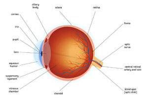
The retina lines the inner wall of your back eye, turning light that enters into nerve signals sent directly to your brain that form images you perceive. Unfortunately, due to age-related and other medical conditions, your retina can tear or detach itself, creating vision loss across any or all visual fields, potentially leading to permanent blindness if untreated quickly.
Retinal detachments often begin suddenly with floating black or white flecks or strings moving before your eyes, followed by dark curtains or shadows moving across your vision, like veiling. If the detachment extends all the way into the macular region (macula), central vision may become lost; most retinal detachments require surgery in order to return it back into its proper position.
Retinal detachments typically result from retinal holes or tears that allow liquid vitreous to seep under the retina, accumulating under it, causing it to separate from the back wall of the eye and eventually detach itself from it. A surgical procedure called pneumatic retinopexy or scleral buckle surgery may be performed in order to repair this tear and return the retina back into its proper place; using a silicone band indented onto the back of eye to press against retinal detachment create a bump seal against retinal tears while keeping fluid away from entering space underneath it.
Retinal detachments can often be treated by using a gas bubble and laser treatment in an office (pneumatic retinopexy). More complex retinal detachments may require surgery at a hospital operating room involving either scleral buckle or vitrectomy; nearly all procedures are successful at restoring retina to its correct place, though vision may take time to improve after treatment has taken effect.
If you experience sudden and painless vision loss, reach out to Clinton Warren, MD and the THIRDCOAST Retina team immediately. A comprehensive examination and ultrasounds will determine the extent of detachment as well as which surgical procedure would best repair it.
Glaucoma
Glaucoma is characterized by damage to the optic nerve, which sends information about your surroundings directly into your brain. It is the second leading cause of blindness globally and often caused by elevated eye pressure which damages optic nerve and leads to vision loss. Glaucoma can lead to total blindness if left untreated, increasing with age and family history of disease. Eye doctors are equipped to diagnose it through regular eye exams that include measurements of eye pressure, pachymetry (the thickness of cornea) and computerized testing of peripheral visual fields and nerve fiber layers.
Your eyes produce aqueous humor to stay moisturized and healthy, which is produced in a region behind the colored part of the eye (iris) and drains out via channels near the cornea. If more fluid is produced than can be drained off quickly enough, eye pressure rises, potentially damaging optic nerves in turn.
There are two primary types of glaucoma. Primary open-angle glaucoma (POAG), for instance, may slowly and painlessly reduce peripheral vision without your knowing until tunnel vision arises. Closed angle or acute angle-closure glaucoma should be treated as a medical emergency and can lead to severe eye pain, blurred vision, halos around lights and redness of the eye if untreated immediately.
Glaucoma can be slowing its progress by using medication to lower your eye pressure. Some medicines may interact with others you take, so be sure to inform your ophthalmologist of everything you’re taking.
Glaucoma treatments include surgery or implants that aid drainage of fluid from your eyeball, while implants may help control eye pressure. An ophthalmologist may also create an iridotomy, creating a small hole in your iris that allows more effective drainage of fluid from your eyeballs. It is vital that those living with glaucoma adhere to their treatment plans and attend regularly scheduled follow-up appointments.


