Age related macular degeneration (AMD) is a prevalent eye disease that leads to central vision loss. Affected areas include your retina’s macula.
Drusen formation under the retina doesn’t cause symptoms and is easily detectable with a comprehensive eye exam.
Dry Age-Related Macular Degeneration (DAMD)
Macular Degeneration, commonly referred to as AMD or ARMD, occurs when damage to the macula, the part of the retina responsible for central vision, occurs and results in its damage deterioration and the loss of sharp, straight-ahead (central) vision over time. This process usually occurs gradually due to light sensing cells becoming less effective over time – and is one of the main causes of legal blindness among Americans aged over 65 today.
Macular degeneration comes in two main varieties – dry and wet. Dry macular degeneration occurs when tiny deposits known as drusen form under the retina and begin thinning out the macula, leading to blurry central vision over time. Unfortunately, this form of macular degeneration cannot be reversed so regular visits to your central Louisiana retina specialist for regular dilated eye exams should be undertaken in order to protect vision in this way.
Wet macular degeneration occurs when abnormal blood vessels form under the retina and leak fluid, leading to macular degeneration and ultimately leading to central blind spots. Unfortunately, this form of macular degeneration is difficult to treat but new medications are being researched that target abnormal blood vessel growth; Faricimab-svoa or vabysmo is one such promising drug which targets and inhibits two disease pathways that drive diabetic macular edema as well as wet age related macular degeneration – such as faricimab-svoa or vabysmo.
Late-Stage Dry Age-Related Macular Degeneration (LASIUM)
A specific region of our retina called the macula is responsible for central vision – essential for reading, driving, recognising faces and colors, as well as seeing fine details. Over time however, light-sensing cells within this specialized region begin to break down, leading to vision loss in what is known as age related macular degeneration (ARMD) affecting more than 13 million North Americans and being the leading cause of blindness among those aged 75 or above.
Eighty-five percent of patients suffering macular degeneration experience its dry form, where deposits known as drusen form under the retina. While progressive, it often progresses slowly enough that most patients still retain reading vision. However, approximately fifteen percent of cases also feature abnormal blood vessels known as wet form macular degeneration that begin leaking fluid or bleeding beneath the retina, potentially leading to rapid loss of vision.
Early diagnosis and treatment of wet macular degeneration is crucial to protecting vision. One effective strategy involves injecting anti-vascular endothelial growth factor (VEGF) agents like Lucentis (ranibizumab) or Avastin (bevacizumab) directly into the eye to stop new blood vessels forming under the retina, as well as any leakage under it. Lucentis (ranibizumab) or Avastin (bevacizumab) have shown significant promise in stopping wet macular degeneration’s progress – typically stopping further central vision deterioration while some patients even experience some improvement!
Early-Stage Dry Age-Related Macular Degeneration (E-SAMD)
Macular degeneration, also known as macular atrophy, occurs when the central portion of the retina (which provides light-sensitive inner lining in the back of the eye) breaks down and leads to irreversible vision loss in Americans over 60. It’s a progressive condition.
Macular degeneration comes in two varieties, dry and wet. Dry macular degeneration accounts for 85-90% of cases. It results from macula thinning, and yellow deposits called drusen appear under the retina causing gradual central vision loss while maintaining peripheral vision.
10-20% of macular degeneration cases progress to wet macular degeneration (neovascular). With wet AMD, blood vessels form underneath the retina and leak blood and fluid into it; vision is lost due to central retinal cells being destroyed and becoming blind spots.
Photodynamic therapy may provide some patients with relief from wet macular degeneration, providing better vision. Under this procedure, a doctor will administer drops to numb your eyes before shining a special laser light into each eye. The laser light interacts with verteporfin drug which enters abnormal blood vessels and closes them off preventing them from leaking blood and fluid, slowing further progression of wet macular degeneration into more serious stages.
Late-Stage Atrophic Age-Related Macular Degeneration (A-SAMD)
At this stage, patients may notice blurry vision that gradually worsens over time. Central areas of their visual field tend to be first affected, and straight lines may appear crooked or bent; patients may no longer be able to read, drive or recognize faces; it is crucial that any such patients visit an eye doctor immediately as treatment can help protect against further loss of vision.
Fovea: the central portion of the macula that contains retinal pigment epithelial (RPE) cell layer in AMD. If dry AMD progresses further, RPE layers thin out and no longer function normally; eventually becoming so thin as to make light detection difficult preventing you from seeing fine detail. Underlying structures may change and small deposits known as drusen may form on your retina.
These drusen can grow into larger masses known as geographic atrophy. If these enlarged drusen are allowed to continue growing, retinal fluid leakage into the vitreous cavity may occur and cause distortion of macula.
Regular examinations, OCT imaging and fundus fluorescein angiography tests as well as injection therapy are key in managing wet macular degeneration. Anti-VEGF medication can often help stop fluid leakage within the macula; additional vitamins and supplements may also be added into treatments to provide relief from symptoms.
Late-Stage Exudative Age-Related Macular Degeneration (EXUDA)
The macula is responsible for central vision. We rely on it for reading, driving a car, using computers or smartphones, recognising faces, seeing fine detail and seeing in low light conditions. Age related macular degeneration (ARMD), however, causes progressive loss of central vision until eventually only your peripheral vision remains functional (the sides of your field of vision). ARMD is one of the primary causes of blindness among people aged 50 or above.
At first, this disease manifests itself with gradual vision loss that occurs without pain or discomfort in either eye. At the onset, you may notice straight lines appear wavy or blurry and require brighter lighting for reading and detail viewing; later on this condition could interfere with driving ability and the recognition of faces.
Studies have demonstrated the efficacy of certain foods, supplements and vitamins for slowing the progression of ARMD, such as foods high in lutein and zeaxanthin; vitamins C and E; B6, and folate acid. Recently approved treatment options such as faricimab-svoa (Vabysmo) have also proven useful against this form of macular degeneration as it works by blocking two disease pathways that lead to its progression while offering much faster relief than traditional therapies with less frequent injections/treatment sessions than other therapies can.
Late-Stage Neovascular Age-Related Macular Degeneration (NVE-AMD)
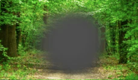
Advanced AMD (NVE-AMD) typically occurs without discomfort, with symptoms that include a dark black spot in the center of your visual field and straight lines being bent or crooked. Other signs include loss of fine detail and difficulty seeing faces, lines or colors.
NVE-AMD occurs when abnormal blood vessels form beneath the retina and leak fluid or blood into the macula, leading to severe and permanent vision loss if left untreated. Treatment options for NVE-AMD include injections of medication directly into the eye.
These medications, called anti-vascular endothelial growth factor agents, work by blocking a specific molecule that stimulates new blood vessel formation. When combined with other therapies such as laser therapy or photodynamic therapy with verteporfin injections, injections may significantly slow progression of neovascular macular degeneration while significantly improving visual acuity.
NVE-AMD is the leading cause of legal blindness in North America and its prevalence is expected to increase dramatically with baby boomers’ aging population. Current therapies, including focal/grid laser photocoagulation and panretinal laser photocoagulation with ranibizumab treatment are proven effective in maintaining visual acuity when administered according to established protocols in randomized phase 3 clinical trials; moreover, faricimab – the first bispecific antibody approved specifically for treating neovascular AMD – provides another treatment option with promising potential.