Medically reviewed by Shaun Larsen, OD – Doctor of Optometry, Southern College of Optometry.
Last clinically reviewed: July 2026 | Next scheduled review: July 2027
In brief: the pinhole test is a fast way of separating blur caused by the eye’s focusing optics from blur caused by disease behind them. Looking through a small aperture, if your vision improves, the problem is largely refractive – the eye needs a different lens prescription. If it does not improve, the blur is coming from somewhere a lens cannot fix: a cloudy media, the retina, or the optic nerve. In macular degeneration, vision characteristically does not improve through a pinhole, which is exactly why the test is useful – but a pinhole result on its own never diagnoses AMD, and it is not a screening test.
What the pinhole test actually is
A pinhole occluder is an opaque disc with one or more small holes, typically 1.0 to 1.5 mm in diameter, with around 1.2 mm the usual working size. The patient reads the acuity chart through it, one eye at a time, with their spectacles on if they normally wear them.
Why the aperture size matters. Below about 1.0 mm, diffraction at the edges of the hole starts to blur the image again and retinal illumination falls too far; above about 1.5 mm the aperture no longer narrows the light cone enough to neutralise refractive error. The 1.0–1.5 mm range is the compromise between those two failure modes.
How it works
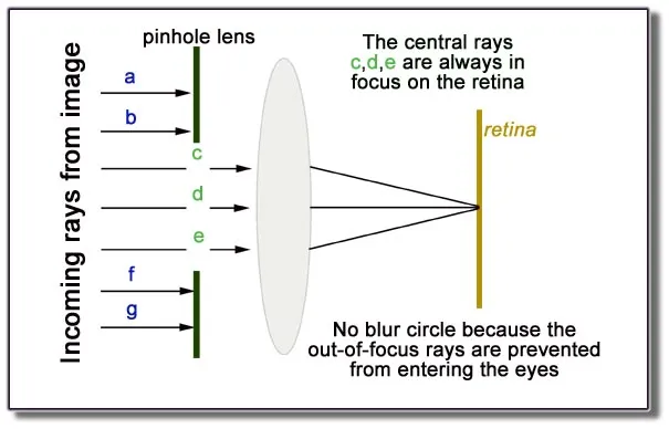
In an eye with refractive error, light from a point in the scene does not converge to a point on the retina – it lands as a blur circle. The size of that blur circle is what makes vision look soft.
A pinhole restricts the eye to a narrow bundle of near-axial rays. Two things follow. First, the blur circle shrinks, because the cone of light reaching the retina is narrower. Second, depth of focus increases – the range of distances over which the image stays acceptably sharp widens. The same optical principle explains why a camera stopped down to a small aperture has greater depth of field, and why squinting sharpens distance vision.
A pinhole also bypasses peripheral corneal and lens irregularities, and reduces the effect of higher-order aberrations. That is why it can partially improve vision in irregular astigmatism – keratoconus, corneal scarring, post-surgical ectasia – even though a spectacle lens cannot.
The critical limitation: this only helps when the blur originates in front of the retina. If the light is already being focused correctly and the problem is the tissue receiving it – a damaged macula, an unhealthy optic nerve – then narrowing the beam changes nothing about the image quality the brain receives. A pinhole cannot improve a signal that is being lost after it lands.
Practically, the pinhole neutralises roughly 3–5 dioptres of refractive error. Very large refractive errors may improve only partially, which is one reason a pinhole result is a pointer rather than a verdict.
What each result means
Vision improves through the pinhole. The reduction is largely refractive. The eye needs a new or updated prescription, or – in irregular astigmatism – a rigid or scleral contact lens. In a pseudophakic eye, improvement can point to a residual refractive surprise after cataract surgery.
Vision does not change. The blur is not refractive. Attention moves to media clarity, the retina and the optic nerve. This is the typical finding in macular degeneration, and also in amblyopia, optic neuropathy, and many retinal conditions. It is also what you would expect in someone already wearing an accurate, up-to-date prescription – which is why the test is interpreted alongside the refraction, not instead of it.
Vision gets worse through the pinhole. This is a real and informative result, and the original version of this page treated it as an alarming anomaly. It occurs most often with dense media opacity – typically advanced cataract – where the patient is relying on scattered light across a wide pupil, and restricting the aperture drops retinal illumination below what they need. It can also happen with a central corneal or lens opacity sitting directly on the axis, or simply when the pinhole is not aligned with the pupil.
A note on technique, because misalignment is the most common source of a misleading result: the pinhole must sit centred over the pupil, the room should not be dark (the test needs a reasonably bright chart), and the patient should be encouraged to move the occluder slightly to find the best position rather than assume the first placement is optimal.
What the pinhole test cannot do
This section matters more than any other on this page, because the title pairs the test with a disease it cannot diagnose.
It does not diagnose macular degeneration. It tells you the blur is not refractive. Cataract, retinal disease, optic neuropathy and amblyopia all produce the same non-improving result. Diagnosis requires a dilated examination and OCT.
It does not detect early AMD. Early and intermediate AMD often leave acuity normal. A patient with large drusen can read 20/20 and improve normally through a pinhole. A normal pinhole result excludes nothing.
It does not detect distortion. Metamorphopsia – straight lines appearing bent – is the symptom most specific to macular disease and the one most likely to signal conversion to wet AMD. The pinhole test does not assess it at all. That is what an Amsler grid is for.
It does not measure contrast sensitivity, visual field or reading speed – the measures that often track function in macular disease better than acuity does.
It is not a home test for AMD. Improvised pinholes tell you very little on their own, and a reassuring result may delay presentation. If you have new distortion, a new central dark patch, or sudden central blurring, the correct response is a prompt appointment, not a self-test.
Where the pinhole test is genuinely useful
Triage in a busy clinic. It takes seconds and immediately separates “needs a refraction” from “needs a dilated look”.
Estimating potential acuity before cataract surgery. In eyes whose vision is limited purely by cataract, a near-pinhole test helps predict the acuity achievable after surgery – useful for setting expectations, particularly where coexisting macular disease may cap the result.
Predicting benefit from a rigid contact lens in corneal disease. Pinhole acuity correlates with the acuity achievable with rigid or hybrid lenses in keratoconus, so it helps identify who is worth fitting.
Checking a suspicious refraction. If acuity is poor with a fresh prescription but improves through a pinhole, the refraction warrants repeating before anything else is investigated.
Tests that actually assess the macula
Since this page is found by people searching for AMD testing, here is what is genuinely used, and what each one contributes.
Dilated fundus examination. Direct assessment of drusen, pigmentary change, atrophy, haemorrhage and fluid.
Optical coherence tomography (OCT). The workhorse of retinal practice: micron-resolution cross-sections of the retinal layers, detecting intraretinal and subretinal fluid invisible on examination, quantifying central subfield thickness, and driving most retreatment decisions in wet AMD.
OCT angiography (OCTA). Maps macular neovascularisation without dye injection.
Fundus autofluorescence (FAF). Images retinal pigment epithelium lipofuscin; the reference standard for delineating and measuring geographic atrophy.
Fluorescein angiography. Dye-based; still the standard for characterising leakage where OCT and OCTA leave the picture unclear.
Amsler grid. Assesses central distortion and scotoma – see the corrected description below.
Microperimetry. Maps retinal sensitivity point-by-point and locates the preferred retinal locus – particularly useful in geographic atrophy, where acuity can look reasonable while reading is severely impaired.
Contrast sensitivity testing (Pelli–Robson) and reading speed (MNREAD, Radner), which frequently reflect real-world function better than Snellen acuity.
How the Amsler grid actually works
The Amsler grid is a square grid of straight horizontal and vertical lines with a single dot at the centre, held at normal reading distance under good light.
How to use it: wear your reading glasses; cover one eye; fix your gaze steadily on the central dot and keep it there; then, without moving your eyes, notice whether any of the lines look bent, wavy, broken, blurred, or whether any area is missing or dark. Repeat with the other eye. Steady central fixation is the whole point of the test – scanning your eyes around the grid defeats it, because you will look directly at each area in turn and may fixate past a scotoma without noticing it.
Report any new distortion or missing area promptly. And know its limits: the Amsler grid is insensitive, perceptual completion can fill in small scotomas, and a normal grid does not exclude progression. It supplements scheduled examinations rather than replacing them.
Macular degeneration patients will typically not show an improvement in visual acuity when looking through a pinhole, which is what makes the test useful: the absence of improvement tells the clinician that uncorrected refractive error is not the explanation, and directs attention to the retina and optic nerve instead.
Photostress recovery testing is a genuine and relevant adjunct. A bright light is shone into the eye for a set period, bleaching the photoreceptor pigments, and the time taken to return to within one line of baseline acuity is measured – normally under about a minute. Recovery is characteristically prolonged in macular disease, because photopigment regeneration depends on the retinal pigment epithelium, and comparatively normal in optic neuropathy, where the photoreceptors and RPE are intact. That contrast is what makes the test useful: it helps distinguish a macular cause from an optic nerve cause when both are plausible.
How the test is performed
Pinhole occluders (small circular holes cut in opaque panels with small circular openings) restrict incoming light to a narrow path that bypasses refractive irregularities and presents one focused image directly to the retinal fovea. They can be used to assess visual acuity in patients suffering from dry macular degeneration; they work well with all types of eyewear (bifocals, progressive lenses or contact lenses).
Procedure: the patient wears their current distance correction. One eye is occluded. Baseline acuity is recorded on the chart at the standard testing distance. The pinhole is then placed over the tested eye, aligned with the pupil, and acuity is measured again. The process is repeated for the fellow eye, and both results are recorded alongside the unaided and corrected acuities.
Interpretation – the point on which the original text was inverted: if acuity improves through the pinhole, the cause is refractive. If it does not improve, the cause lies elsewhere – media opacity, retina, or optic nerve. Improvement points toward the lens prescription; no improvement points away from it.
Where acuity does not improve, the next steps are a dilated fundus examination and OCT. Worsening through the pinhole most often reflects dense media opacity reducing retinal illumination, or simple misalignment of the aperture, and should prompt re-testing before it is treated as a clinical finding.
When to seek care, by urgency.
Same day – emergency: sudden painless loss of vision in one eye; a curtain or shadow moving across the field; a sudden shower of floaters with flashing lights; severe eye pain with redness, haloes, nausea and vomiting; sudden double vision; any eye injury or chemical splash; new vision loss with headache, scalp tenderness or jaw pain in anyone over 50 (possible giant cell arteritis – sight- and life-threatening).
Within days: new distortion of straight lines; a new central grey or dark patch; sudden central blurring. In suspected wet AMD these symptoms determine the outcome, because untreated neovascular disease progresses over weeks and vision lost to scarring does not return.
Routine: gradual blurring over months, increasing glare at night, difficulty with small print, changes in colour perception.
Diagnosis
Additionally, other tests can be undertaken to fully evaluate the health of both eyes and visual systems, including direct and indirect ophthalmoscopy, slit-lamp biomicroscopy, tonometry, contrast sensitivity testing, color vision tests and motility checks. Some of these involve a bright light directed into the eye, which causes temporary glare and afterimages lasting a few minutes. That is expected and harmless – it is not “temporary blindness”, and describing it that way is needlessly alarming to someone reading before an appointment. Dilating drops blur near vision and increase light sensitivity for several hours, so patients should arrange not to drive immediately afterwards and may want sunglasses.
Pinhole apertures are opaque panels containing one or more holes 1.0 to 1.5 mm in diameter that restrict light rays entering through refractive irregularities into one narrow path, providing a single image aimed at the retina. If vision improves through the pinhole, the reduction is refractive in origin. Media problems such as cataract, vitreous haemorrhage or retinal detachment characteristically do not improve through a pinhole – and dense cataract may look worse through one, because the aperture cuts the light the patient was relying on.
Alternatively, if the patient’s best-corrected acuity does not improve with pinhole testing, this suggests that their visual acuity issues are caused by something other than refractive error (for instance Salzmann nodular degeneration or epithelial basement membrane dystrophy). Those two examples are corneal surface disorders and are worth naming, but the far more common explanations in this age group are cataract, macular disease and optic neuropathy. Careful retinoscopy and trial frame refraction will need to be conducted in order to ascertain whether an RGP lens would help correct their visual acuity.
Where the pinhole produces no improvement and macular disease is suspected, an Amsler grid is a reasonable next step – it assesses central distortion and scotoma, not motion perception. See the corrected description of how the grid works earlier in this article.
Treatment
Pinhole lenses enable light rays to pass more efficiently through irregular or partially opaque media (i.e. cataract) with less scattering than would be possible with lenses of equal power, thus potentially improving visual acuity without correcting refractive errors; rather, they reduce the effect of irregularities in the cornea and lens – the eye’s refracting surfaces. A pinhole has no effect on irregularities of the retinal surface, which sit behind the optics, and “irregularities on Amsler grids” describes a test result rather than a structure. It may be noticeable that those suffering ocular diseases experience improvements when viewing through pinhole lenses compared with looking through conventional lenses; this result shouldn’t be used as the basis for spectacle correction; professional retinoscopy/trial frame refraction should be undertaken prior to considering wearing glasses or not.
On pinhole glasses sold commercially. Perforated “pinhole glasses” marketed as vision trainers exploit the same optical effect, and they will make blurred distance vision look temporarily sharper. They do not treat refractive error, they do not strengthen the eye, and there is no evidence they produce lasting change in acuity. They also cut a large amount of light and severely fragment the visual field, which makes them unsafe for driving or moving about. The small-aperture intraocular lens used in some cataract surgery is a legitimate clinical application of the same principle – that is a prescribed implant, not the same thing as a novelty product.
Home monitoring for conversion to wet AMD. Patients with intermediate or advanced AMD in one eye are at meaningful risk of developing neovascular disease, and early detection changes the outcome. Options include the Amsler grid, described above, and home monitoring devices that test visual function – for example by asking the patient to identify distortions in a pattern of dots – and alert the clinic when performance changes. These are functional tests performed by the patient; they do not photograph or image the retina, and they do not replace scheduled examinations. Discuss suitability with your retina specialist.
Your eye care specialist will conduct an eye exam that includes inspecting both the front of your eye and its back retina through its pupil using special drops to dilate your pupils. He or she may then ask you to read letters on a Snellen chart in various sizes and lighting conditions; the smaller letters that can be read, the better your vision will be.
Wet AMD is detected by imaging, not by charts. An Amsler grid can raise suspicion, but macular oedema and subretinal fluid are identified on OCT, and neovascular membranes on OCT angiography or fluorescein angiography. No eye chart can distinguish macular oedema from central serous chorioretinopathy.
Hard exudates and cotton wool spots are different findings and should not be equated. Hard exudates are well-defined yellow lipid and protein deposits left behind where fluid has resorbed, typically in diabetic retinopathy and vein occlusion. Cotton wool spots are soft, fluffy, pale grey-white patches caused by ischaemic infarction of the retinal nerve fibre layer, seen in diabetes, hypertension and vascular occlusion. They look different, they arise from different mechanisms, and they carry different implications.
Vitrectomy surgery may be recommended in cases of exudative macular degeneration with persistent fluid accumulation or retinal detachment, combined with subretinal injection of t-PA, intraocular gas tamponade or anti-VEGF injections to address associated macular edema or neovascularization.
Prevention
The macula is the central region of retina that provides sharp and clear central vision necessary for activities like reading and driving. Age-related macular degeneration (AMD), a progressive disease that affects people over 50, causes blurry central vision as well as distortion or blank spots in your field of view and difficulty with distinguishing colors – not to mention making straight lines appear wavy or crooked!
Macular degeneration cannot be fully prevented, but lifestyle changes may help slow its progress. These include not smoking, eating a healthy diet including plenty of green leafy vegetables and other antioxidants, wearing sunglasses when outdoors or wearing protective eyewear such as sunnies when needed outside, maintaining a healthy weight and exercising regularly. Smoking cessation is by a wide margin the highest-value action: smoking is associated with roughly two- to four-fold increased AMD risk and earlier onset. For those with intermediate AMD or advanced AMD in one eye, AREDS2 supplementation (500 mg vitamin C, 400 IU vitamin E, 10 mg lutein, 2 mg zeaxanthin, 80 mg zinc, 2 mg copper) reduces progression risk to advanced disease by approximately 25% – note that it does not prevent AMD in people who do not have it, and does not restore lost vision. Regular eye exams also play a key role in the detection of macular degeneration; an Amsler grid test involves looking at vertical and horizontal lines that form perfect squares; any distortion in them could indicate the start of macular degeneration.
There are two primary forms of macular degeneration, dry and wet. The former occurs when the macula thins over time while its opposite, less frequent but faster progressing variant (wet form) forms abnormal blood vessels underneath the retina that leak fluid and blood, leading to loss of central vision and potentially permanent blindness. Intravitreal anti-VEGF injection is the standard treatment for wet AMD and has been for well over a decade – agents include bevacizumab, ranibizumab, aflibercept, brolucizumab and faricimab, given on fixed, as-needed or treat-and-extend schedules. Photodynamic therapy is now reserved for narrow indications such as polypoidal choroidal vasculopathy and is no longer front-line therapy. Presenting PDT as the primary treatment substantially misrepresents current care.
It is also worth stating that AMD does not cause total blindness: peripheral vision is characteristically preserved, and patients typically retain independent mobility even when central vision is severely affected.
Frequently asked questions
If my vision improved with the pinhole, does that mean I don’t have macular degeneration?
It makes uncorrected refractive error the likely explanation for that particular blur, but it does not exclude AMD. Early and intermediate AMD frequently leave acuity normal, and a person can have both a refractive error and macular drusen. Only a dilated examination with OCT can answer the AMD question.
My vision didn’t improve with the pinhole. Does that mean I have macular degeneration?
No. It means the blur is not refractive. Cataract, macular disease, optic nerve disease and amblyopia all produce the same result. The pinhole narrows the possibilities; it does not identify which one.
Can I do a pinhole test at home?
You can reproduce the optical effect – looking through a small hole punched in card will sharpen blurred distance vision if you have an uncorrected refractive error. But the result is not reliable enough to act on, and a reassuring result may delay presentation for something that needs prompt attention. If you have new distortion, a new central dark patch, or sudden blurring, book an appointment rather than testing yourself.
Do pinhole glasses improve eyesight?
They temporarily sharpen blurred distance vision through the same optical effect, but there is no evidence they produce any lasting improvement in acuity or treat refractive error. They also reduce light substantially and fragment the visual field, so they should not be worn for driving or moving around.
What is the best test for macular degeneration?
There is no single test. A dilated fundus examination establishes the diagnosis; OCT is the key imaging study for detecting fluid and staging disease; fundus autofluorescence measures geographic atrophy; and OCT angiography or fluorescein angiography characterises neovascular membranes. The Amsler grid supports monitoring between visits. The pinhole test is not a macular test – it is a way of ruling refractive error in or out.
References
1. Lin H-Y, Lee C-Y, et al. Applications of the pinhole effect in clinical vision science. *Taiwan J Ophthalmol*. 2023;13(4):427–435.
2. Kanclerz P, et al. Results of the pinhole test correlate with hybrid contact lens visual acuity in patients with visual impairment due to corneal diseases. *J Ophthalmol*. 2022;2022:4932856.
3. Melki SA, Safar A, Martin J, et al. Potential acuity pinhole: a simple method to measure potential visual acuity in patients with cataracts, comparison to potential acuity meter. *Ophthalmology*. 1999;106(7):1262–1267.
4. Glaser JS, Savino PJ, Sumers KD, et al. The photostress recovery test in the clinical assessment of visual function. *Am J Ophthalmol*. 1977;83(2):255–260.
5. Amsler M. Earliest symptoms of diseases of the macula. *Br J Ophthalmol*. 1953;37(9):521–537.
6. Ferris FL, Wilkinson CP, Bird A, et al. Clinical classification of age-related macular degeneration. *Ophthalmology*. 2013;120(4):844–851.
7. Age-Related Eye Disease Study 2 Research Group. Lutein + zeaxanthin and omega-3 fatty acids for age-related macular degeneration: the AREDS2 randomized clinical trial. *JAMA*. 2013;309(19):2005–2015.
8. American Academy of Ophthalmology Retina/Vitreous Panel. Preferred Practice Pattern: Age-Related Macular Degeneration. aao.org.
9. Rosenfeld PJ, Brown DM, Heier JS, et al. Ranibizumab for neovascular age-related macular degeneration. *N Engl J Med*. 2006;355(14):1419–1431.
Related Posts
-
Eye Floaters vs. Macular Degeneration: What’s the Difference?
When you notice changes in your vision, it’s natural to feel concerned.
-
What is Age-Related Macular Degeneration?
Your retina at the back of your eye converts light rays into signals recognized by your brain as images.
-
Margarine Causes Macular Degeneration
Margarine may get a bad rap these days, but when used sparingly it can actually be beneficial in terms of health benefits.
All articles on macular degeneration signs, causes and tests