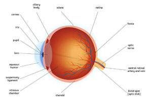
Your retina at the back of your eye converts light rays into signals recognized by your brain as images. If these cells become damaged, your central vision may blur or appear wavy.
Wet age-related macular degeneration (AMD) occurs when abnormal blood vessels form underneath your macula in your retina and leak blood and fluid, damaging your vision and potentially leading to further vision problems.
Macular pucker
The retina is a layer of light-sensing cells lining the back of your eye that converts light entering to signals for transmission to your brain, which are then recognized as images you see. When damaged, retinal damage may cause blurred or distorted central vision due to damage to macula which provides clear central vision essential for reading, driving and other activities requiring fine detail; with macular pucker, scar tissue forms on this area resulting in wrinkles or bulges which distort your view.
Macular pucker occurs when the vitreous gel inside your eye shrinks and pulls away from your retina – the thin film at the back that captures visual images – as a natural part of aging, yet in certain instances this change may lead to macular pucker. If too much vitreous gel pulls too hard on your retina, a membrane may form that wrinkles and bulges it, impairing central vision and disrupting its functionality.
An eye care specialist can diagnose macular pucker during a comprehensive exam. They will look out for symptoms such as wavy straight lines or missing or blank spots in your central vision, along with looking at an Amsler grid resembling a checkerboard pattern of straight lines; when looking at this grid they ask you to focus on one small dot in the center and report whether any lines appear wavy or missing.
Depending on the severity of macular pucker, an ophthalmologist may prescribe eyeglasses or contact lenses to assist in reading fine details. If it worsens further, surgery to flatten out the macular surface (called vitrectomy ) could restore central vision.
If you are experiencing wavy straight lines or missing or blank areas in your central vision, make an appointment at THIRDCOAST RETINA to speak with one of our ophthalmologists about treatment options. Our wide range of surgical and non-surgical treatments includes macular pucker, retinal holes and macular detachments as well as other eye disorders.
Age-related macular degeneration (AMD)
Age-related macular degeneration (AMD) is an eye disease that gradually erodes central vision – the sharpest and clearest part of your retina used for reading, driving and recognising faces and colors – over time. AMD is one of the primary causes of severe vision loss among older adults.
This condition affects the macula, located at the back of each eye and composed of millions of light-sensing cells. Over time, it can lead to gradual and progressive loss of central vision as well as distortion or wavy lines in your vision – the primary symptoms being blurry vision in dim light conditions – one or both eyes may experience vision changes which worsen over time.
AMD comes in two forms, wet and dry. Around 85% to 90% of those diagnosed with AMD fall under the dry form, typically diagnosed by yellow spots called drusen appearing around their macula, believed to be deposits or debris from retinal tissue decaying over time. Small drusen may not cause symptoms at first; as they grow larger they could eventually damage central vision-forming cells of their macula, leading to vision loss in some instances.
Macular degeneration may progress from its dry form into wet AMD, leading to faster and more severe vision loss. Wet AMD occurs when abnormal blood vessels grow underneath or near the retina and macula resulting in bleeding, fluid leakage and severe vision loss. The more advanced a case of dry macular degeneration becomes, the higher its chances of progressing into wet macular degeneration are.
Treatment for wet macular degeneration focuses on halting further damage. Treatment typically includes injecting anti-vascular endothelial growth factor (anti-VEGF), an eye medication that interferes with chemicals found in your body that stimulate the formation of new blood vessels, into your eye. Anti-VEGF can prevent abnormal vessels from growing and leaking further within your eyeball, helping slow down progression of wet macular degeneration while possibly saving some central vision.
Epiretinal membranes
Epidermal membrane (ERM), is a semitranslucent fibrous tissue cover which may form on the inner surface of your retina – the light-sensitive area at the back of your eye. ERM is more prevalent with age and can lead to blurred or distorted vision as well as loss of central vision which impacts reading or driving abilities. ERM symptoms range from mild to severe severity depending on its location in your retina.
Your eye doctor will examine your eye and use special diagnostic tools, including a fundus camera which takes rapid-sequence photographs of retinal vasculature following injection of fluorescein sodium dye, and an imaging device known as Optical Coherence Tomography that creates cross sectional images of your retina, to asses the severity of ERM.
If the ERM is causing significant visual distortion or loss of function, your doctor may suggest vitrectomy surgery to remove it. This procedure is performed under local anesthesia using a small instrument to lift and peel the epiretinal membrane.
This procedure has been proven to dramatically increase vision quality for most patients; however, it cannot treat dry macular degeneration permanently; instead recurrences of this issue remain quite prevalent. Furthermore, active cases of wet macular degeneration should avoid this surgery, since its risks include creating new blood vessels which leak fluid into your macula which could quickly damage or destroy it and your vision quickly.
An epiretinal membrane may be caused by several conditions, including diabetic macular edema, proliferative retinopathy from diabetes and choroidal neovascularization related to high blood pressure or age related factors. Individuals diagnosed with Ehlers-Danlos syndrome may also be more at risk for epiretinal membrane development.
Epiretinal membrane symptoms often manifest themselves with wavy or distorted lines appearing in your central vision. If this is something you detect, make an appointment immediately with your ophthalmologist who may suggest looking through a grid with one eye covered while looking through another without covering either eye. Look at its center dot to check if any areas appear blurred, darkened or otherwise abnormal.


