

Pinhole Test for Macular Degeneration
Macular degeneration patients will typically not show an improvement in visual acuity when looking through a pinhole, making this test useful in identifying whether refractive error or other media opacities may be the source of their visual impairment.
Photostress recovery testing should also be administered (see Chapter 3). This tests the time it takes a patient’s vision to recover following exposure to bright lights; any prolongations could indicate macular disease or optic nerve pathology.
Symptoms
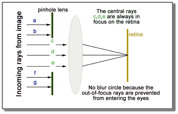
Pinhole occluders (small circular holes cut in opaque panels with small circular openings) restrict incoming light to a narrow path that bypasses refractive irregularities and presents one focused image directly to the retinal fovea. They can be used to assess visual acuity in patients suffering from dry macular degeneration; they work well with all types of eyewear (bifocals, progressive lenses or contact lenses). An easy test for visual acuity would involve covering one eye while asking someone else read off Snellen chart at 20 feet then covering both eyes before repeating this test; if visual acuity improves with pinhole, it suggests more likely cause lies within optic nerve or macula rather than lenses or vitreous.
If the acuity does not improve with treatment, seek an explanation such as an eye disease, systemic disorder (eg sarcoidosis or collagen vascular diseases) or other pathology. If it worsens with use of an occluder, ask about any sudden symptoms that have developed recently or any recent worsening; consider referral for more immediate assessment in cases involving monocular patients or those who have a history of Stargardt disease.
Clinicians from the Tennent Institute of Ophthalmology detailed, in a case report published by BMJ, their experience treating an 80 year-old who presented to an accident and emergency department with sudden, deteriorating vision in his left eye that limited him to counting fingers in that eye. His medical history included diabetes, hypertension, dementia and glaucoma and an neurological examination and CT scan were conducted, all which proved normal.
The author of the case report advised all patients with painless, gradual visual loss for an evaluation by an ophthalmologist. Furthermore, rapid or dramatic visual changes, distorting vision and loss of vision in one eye should all prompt immediate referral for further evaluation by ophthalmology. In cases involving pain or additional symptoms such as flashes that could indicate more serious pathologies immediate referral should occur immediately.
Diagnosis
Additionally, other tests can be undertaken to fully evaluate the health of both eyes and visual systems, including direct and indirect ophthalmoscopy, slit-lamp biomicroscopy, tonometry, contrast sensitivity testing, color vision tests and motility checks. While additional tests may take longer and require bright light being shined directly into a patient’s eyes – sometimes leading to temporary blindness – these additional procedures must always be explained to patients prior to beginning them.
Pinhole apertures are opaque panels containing one or more holes 1.0 to 1.5 mm in diameter that restrict light rays entering through refractive irregularities into one narrow path, providing a single image aimed at the retina. If vision improves upon inserting this pinhole aperture, this indicates not an issue with refractive error but instead something media-related like cataract, vitreous haemorrhage, or retinal detachment is to blame.
Alternatively, if the patient’s best-corrected acuity does not improve with pinhole treatment, this suggests that their visual acuity issues are caused by something other than refractive error (for instance Salzmann nodular degeneration or epithelial basement membrane dystrophy). Careful retinoscopy and trial frame refraction will need to be conducted in order to ascertain whether an RGP lens would help correct their visual acuity.
When the pinhole test results remain unchanged, it may be worthwhile to perform an Amsler grid test to assess their perception of motion. An Amsler grid is a geometric chart with rows and columns of dots which allow patients to move their eyes around to identify individual dots; an ideal result would be being able to read the central “E” on the top row as well as counting fingers at 3 feet distance.
Treatment
Pinhole lenses enable light rays to pass more efficiently through irregular or partially opaque media (i.e. cataract) with less scattering than would be possible with lenses of equal power, thus potentially improving visual acuity without correcting refractive errors; rather they reduce distortion caused by retinal surface irregularities and irregularities on Amsler grids. It may be noticeable that those suffering ocular diseases experience improvements when viewing through pinhole lenses compared with looking through conventional lenses; this result shouldn’t be used as the basis for spectacle correction; professional retinoscopy/trial frame refraction should be undertaken prior to considering wearing glasses or not.
Monitor changes in vision closely for signs of wet macular degeneration. An at home monitoring program known as Foresee can detect vision changes earlier than traditional eye exams by taking digital pictures of the back of each eye through its pupil. It then creates a computerized record of each patient’s vision.
Your eye care specialist will conduct an eye exam that includes inspecting both the front of your eye and its back retina through its pupil using special drops to dilate your pupils. He or she may then ask you to read letters on a Snellen chart in various sizes and lighting conditions; the smaller letters that can be read, the better your vision will be.
Other tests used to detect wet macular degeneration include the Amsler grid and eye chart that displays lines of various thicknesses, enabling your eye care specialist to observe whether you have macular edema or central serous retinopathy. They’ll also check for any evidence of vascular leakage or hard exudates – yellow spots within your retina that look like cotton wool spots on the macula that indicate either leakage or swelling on its own.
Vitrectomy surgery may be recommended in cases of exudative macular degeneration with persistent fluid accumulation or retinal detachment, combined with subretinal injection of t-PA, intraocular gas tamponade or anti-VEGF injections to address associated macular edema or neovascularization.
Prevention
The macula is the central region of retina that provides sharp and clear central vision necessary for activities like reading and driving. Aged-related macular degeneration (AMD), a progressive disease of AMD that affects people over 50, causes blurry central vision as well as distortion or blank spots in your field of view and difficulty with distinguishing colors – not to mention making straight lines appear wavy or crooked!
Macular degeneration cannot be fully prevented, but lifestyle changes may help slow its progress. These include not smoking, eating a healthy diet including plenty of green leafy vegetables and other antioxidants, wearing sunglasses when outdoors or wearing protective eyewear such as sunnies when needed outside, maintaining a healthy weight and exercising regularly. Regular eye exams also play a key role in the detection of macular degeneration; an Amsler grid test involves looking at vertical and horizontal lines that form perfect squares; any distortion in them could indicate the start of macular degeneration.
There are two primary forms of macular degeneration, dry and wet. The former occurs when the macula thins over time while its opposite, less frequent but faster progressing variant (wet form) forms abnormal blood vessels underneath the retina that leak fluid and blood, leading to loss of central vision and potentially permanent blindness. Photodynamic therapy uses light-activated lasers to destroy these abnormal blood vessels, thus protecting vision while stopping further damage to it – this treatment may need to be repeated every three months for maximum effectiveness.


