

What is Phacolytic Glaucoma?
A mature cataract’s capsule leaks lens material, which sets off an inflammatory process that leads to phacolytic glaucoma. The altered lens protein, macrophages, and other inflammatory cells found in the discharged lens material cause trabecular meshwork blockage and glaucoma.
Phacolytic glaucoma is a condition that affects cataractous lenses with intact lens capsules, in contrast to several types of lens-induced glaucomas (such as lens particle glaucoma and phacoanaphylactic glaucoma). The research that is now available suggests that lens protein produced from minute flaws in the clinically healthy lens capsule directly obstructs outflow channels. In experimental perfusion investigations, the high molecular weight proteins present in cataractous lenses cause outflow blockage comparable to that seen in phacolytic glaucoma. Although there is frequently a macrophagic response, it is thought that macrophages are a normal reaction to lens protein in the anterior chamber rather than the root of the outflow blockage.
In a recent report, it was suggested that there may be two types of phacolytic glaucoma: (1) a more acute presentation brought on by the rapid leakage of lens proteins that obstruct the trabecular meshwork, and (2) a more slower presentation with secondary macrophages brought on by an immune reaction to lens proteins in the anterior chamber.
What is Glaucoma?
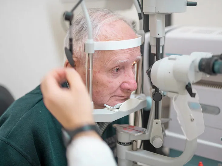
A collection of eye conditions known as glaucoma can result in vision loss and blindness by causing atrophy of the optic nerve, a nerve located in the back of the eye.
You may not notice the symptoms at first since they might appear gradually. A thorough dilated eye exam is the only method to determine if you have glaucoma.
Although there is no known cure for glaucoma, early detection and treatment can frequently reverse visual loss.
What kinds of glaucoma are there?
There are several distinct varieties of glaucoma, but open-angle glaucoma is the most prevalent one in the US and is what most people refer to when they discuss glaucoma. Angle-closure glaucoma and congenital glaucoma are two less typical varieties.
American Epidemiology/Frequency
Because wealthy nations like the United States have easier access to healthcare and perform cataract surgery early, phacolytic glaucoma is less common there.
International
More cases of phacolytic glaucoma are seen in developing nations.
Mortality/Morbidity
After cataract surgery, the majority of patients result in great visual improvement.
Race
There is no racial preference.
Sex
There is no sexual preference.
Age
Older persons are often affected by phacolytic glaucoma. The reported youngest patient was 35 years old.
Presentation/Diagnosis
A painful eye, photophobia (light sensitivity), impaired vision, and significant conjunctival hyperemia (redness) are the characteristic symptoms of phacolytic glaucoma. These symptoms are frequently present in elderly patients who have a history of visual problems. The typical diagnostic criteria for phacoantigenic glaucoma include corneal edema, increased intraocular pressure, flare reaction in the anterior chamber with no keratic precipitates (distinguishing this entity from phacoantigenic glaucoma), prominent cell or white material/particles in the anterior chamber, and signs of a mature cataract. There may also be pseudohypopyon (lens protein deposits layering in the inferior angle). The anterior chamber angle is open, according to gonioscopy.
Phacolytic Glaucoma Treatment
Medication
Glaucoma and inflammation are immediately controlled by medical therapy. Hyperosmotic agents, aqueous suppressants, anti-inflammatory medicines, and cycloplegics make up the first course of therapy.
Surgery
The lens must be removed through extracapsular cataract extraction, either with or without an IOL, as the only effective therapy. Although some ophthalmologists wait until the inflammation has subsided before implanting an IOL, there is no discernible difference in the patients’ ultimate visual acuity between those who received an IOL and those who did not.
A simultaneous trabeculectomy may be required if the phacolytic glaucoma has persisted for a long time (greater than seven days) in order to avoid postoperative IOP rises.
One must be extremely careful while handling eyes with hypermature Morgagnian cataracts since the capsule is delicate, the zonules are weak, and the vision is challenging because of the white, milky cortex. The removal of a cataract is not prohibited by a vision that is only capable of seeing light at presentation.
Management\sMedication
Glaucoma and inflammation are momentarily controlled by medical therapy. Hyperosmotic agents, aqueous suppressants, anti-inflammatory medicines, and cycloplegics make up the first course of therapy.
Prognosis
The prognosis is favorable, with the majority of patients seeing a significant improvement in vision after cataract removal; nevertheless, delaying surgery may result in a subpar result.
Those who suffer from phacolytic glaucoma (PG) may not fare as well as those who suffer from phacomorphic glaucoma.
After cataract surgery, therapy to reduce intraocular pressure is typically no longer necessary. A small percentage of individuals with persistently elevated intraocular pressure may require ongoing medical treatment or a filtration operation to regulate intraocular pressure.
The patient should be treated postoperatively with topical steroids, aqueous suppressants, and/or hyperosmotics, as indicated. Occasionally, eyes develop vitreous opacification behind the posterior capsule. The majority of these vitreous opacities disappear one to two weeks after surgery.
Once the cataract has been removed, IOP is often under control without the need of glaucoma meds. To determine the full amount of glaucomatous damage, a thorough glaucoma assessment (including a second gonioscopy to check for peripheral anterior synechiae, the visual field, and the condition of the optic nerve) should be performed.
The length of increased IOP, PAS, and optic nerve injury all affect the outcome. In one research, patients with glaucoma that had been present for more than five days and who were older than 60 performed considerably worse than a reference group of younger people with shorter illness duration.
Complications
The following are examples of phacolytic glaucoma complications:
Visual loss brought on by glaucoma that is not under control or by prolonged corneal edema
Suprachoroidal hemorrhage, capsular rupture with lens material loss into the posterior region, corneal damage, and vitreous prolapse are a few surgical side effects.
Differential Diagnosis
Pathophysiology of Lens-Particle Glaucoma
In contrast to phacolytic glaucoma, lens particle glaucoma develops as a result of “disruption of the lens capsule,” which can happen during cataract surgery, penetrating lens damage, or laser posterior capsulotomy. The damaged lens discharges material that contains lens particles into the anterior chamber, obstructing the aqueous outflow.
Diagnosis
The presentation normally happens a few weeks after the first trigger, although it can sometimes happen for months or even years afterward. Making an appropriate diagnosis requires taking into account the patient’s surgical or trauma history. Elevated intraocular pressure and indications of cortical lens material in the anterior chamber are examples of clinical findings. Synechiae, cell/flare response in the anterior chamber, and ocular edema are other potential symptoms.
Management
Initial medical treatment goals include controlling intraocular pressure, reducing inflammation with topical steroids, and avoiding the development of synechiae using mydriatics. The lens is surgically removed if the lens particle or material does not resorb, there is a significant amount of lens material in the anterior chamber, and the intraocular pressure cannot be regulated.
Glaucoma that is phacoantigenic (formerly known as Phacoanaphylaxis)
Pathophysiology
After surgery or penetrating trauma, the trabecular meshwork becomes obstructed, increasing intraocular pressure. This condition is known as phacoantigenic glaucoma. It is characterized by a granulomatous inflammatory reaction directed against the patient’s own lens antigens. It is crucial to note that, because this disorder is not an allergy, phacoanaphylaxis is not the proper name for it. The immune complex reaction of the Arthus type, which is mediated by IgG and the complement system, appears to be the mechanism triggering the reaction.
Diagnosis
Typically, phacoantigenic glaucoma develops one to fourteen days following cataract surgery or eye injury. Keratic precipitates, anterior chamber cell/flare response, synechiae, and leftover lens material are examples of clinical findings. The occurrence of glaucomatous optic neuropathy is less frequent.
Management
IOP-lowering medicines and topical steroids are used as the first line of treatment to lessen inflammation and regulate intraocular pressure. Once inflammation is under control and medicinal therapy is ineffective, surgical removal of any leftover lens material is advised.
PG may potentially be mistaken for traumatic glaucomas including angle-recession glaucoma and ghost cell glaucoma. Evidence of previous trauma may be discovered with a thorough history and examination of the anterior chamber angle. Long-term vitreous bleeding in the past should raise concerns about ghost cell glaucoma.
Last but not least, the posterior segment of seemingly aphakic eyes needs to be carefully examined for a displaced lens that, when hypermature, may start to leak and produce PG even though a cataract is not immediately obvious.
Summary
Gonioscopy at an open angle is critical to effective diagnosis and therapy. Prior to cataract extraction, the patient is stabilized with topical steroids and IOP-lowering medicines. Optimal visual prognosis if detected and treated quickly.
FAQ’s
What distinguishes phacolytic vs phacomorphic glaucoma?
Glaucoma that is phacomorphic: The primary distinguishing characteristic of phacomorphic glaucoma is gonioscopically closed angle and a shallow anterior chamber, both of which are related to hypermature cataract production. There is a significant anterior chamber inflammatory component to phacolytic glaucoma.
Treatment options for phacolytic glaucoma?
Cataract removal is the only effective therapy for phacolytic glaucoma (PG). The preferred method for removing cataracts has mostly changed from intracapsular cataract extraction to extracapsular cataract extraction (e.g., phacoemulsification with an intraocular lens implant).
What glaucoma phacolytic symptoms are there?
Phacolytic Glaucoma Symptoms: Ocular discomfort and decreased vision may appear suddenly or gradually.
Signs: unilaterally occurs (majority of cases) hyperemia of the conjunctiva (associated) increased intraocular pressure suddenly (may reach as high as 80 mmHg) widespread corneal edema
Is phacolytic glaucoma open or closed angle?
The abrupt development of open-angle glaucoma known as phacolytic glaucoma (PG) is brought on by a leaking mature or hypermature cataract. Cataract removal is the treatment.
Why does phacolytic glaucoma develop?
A mature cataract capsule leaks lens material, which sets off an inflammatory process that leads to phacolytic glaucoma. The abnormal lens protein, macrophages, and other inflammatory cells found in the discharged lens material cause trabecular meshwork blockage and glaucoma.
Is glaucoma be cured by cataract surgery?
Even though cataract surgery by itself may help keep glaucoma under control, it is typically ineffective for treating active glaucomatous development, which necessitates a large IOP reduction. However phacolytic glaucoma, which is rare can be cured by removing the cataract.
Which glaucoma kind is the most severe?
Secondary glaucoma is a disease kind that can be brought on by specific medications and eye conditions. Closed-angle glaucoma, however, is most likely the disease’s most severe kind. It happens when the angle is suddenly blocked, which causes a quick increase in ocular pressure.
Which glaucomas require urgent care?
Acute angle-closure glaucoma may be the cause of abrupt onset of symptoms. Severe headache and eye discomfort are two symptoms. You must receive treatment immediately. Call an ophthalmologist’s office right away or go to the emergency room.
What is phacomorphic glaucoma’s course of treatment?
Cataract removal is the only effective treatment for phacomorphic glaucoma, however it is simpler and safer to do so after decreasing IOP with medication. Usually, glaucoma drugs that lessen aqueous production are used initially.


