

Macular Degeneration Treatment
Macular degeneration is a prevalent eye disease among those over 50. It affects your central vision and may result in blurry or distorted straight ahead sight.
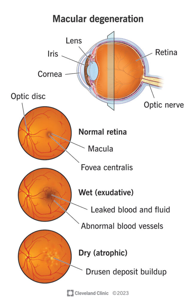
The macula is an area of retina that stores detailed images for transmission to your brain. When its function becomes compromised, symptoms include gradual loss of straight-ahead vision as well as distortion of objects and shapes beyond straight lines.
Dilated Eye Exams
Macular degeneration is one of the leading causes of central vision loss among adults over 50, as it leads to gradual breakdown or degradation of an area in the retina called macula that’s responsible for fine details, straight lines and colors. Over time this condition progresses into blurred, dark or distorted areas that make simple tasks like threading needles, driving or reading difficult or impossible for its victims. There’s currently no cure but treatments may slow its progress or help protect from its progression further down the line.
Macular degeneration usually develops slowly, with symptoms only becoming noticeable as it progresses further. At first, symptoms may only affect one eye and be limited to stray lines or patches of darkness in the center of vision; most times however, central vision will still remain clear but patients may notice straight lines seeming crooked or wavy or less vibrant colors; or in more serious forms the condition could suddenly bring on sudden loss of sharp central vision due to abnormal blood vessel growth which leaks fluid beneath retina.
An annual dilated eye exam is essential in detecting macular degeneration and other eye conditions, including diabetic retinopathy, retinal tears and tumors. A dilated exam typically includes administering drops that enlarge pupil diameter so your doctor has an improved view of what’s behind your eye, helping diagnose problems such as glaucoma, diabetic retinopathy, retinal tears or tumors more accurately.
Patients living with diabetes must undergo an annual dilated eye exam in order to monitor the progression of their disease and detect any retinal bleeding or hemorrhage. High blood pressure patients are encouraged to have this same examination performed, as hypertension can lead to complications known as retinopathy which damage small blood vessels within the retina, increasing your chances of retinopathy.
Fluorescein Angiography
Fluorescein angiography uses fluorescent dye to visualize blood vessels at the back of your eye. After injecting the dye into a vein in your arm, it travels quickly to reach its destination: retinal blood vessels at the back. A special camera then takes photographs without using X-rays and interprets these photographs; abnormal blood vessels or leakage under retina will show up as bright spots or distortion in photographs while new blood vessel growth can also be monitored effectively; in addition, angiogram helps identify where drusen are present which can help in treatment plans being devised.
Your doctor can use an angiogram to determine if laser treatment might help, which has been found to slow vision loss in some cases of wet macular degeneration. Laser treatments tend to work best when localized neovascularization does not spread across large portions of retina. An ophthalmologist can use the angiogram as well to monitor its success.
Regular eye exams, testing, and injection therapy may help protect patients with wet macular degeneration from suffering severe vision loss. This may involve viewing an Amsler grid with black lines arranged in a grid pattern to measure both central and peripheral vision; and performing Fluorescein angiography tests on retinal blood vessels using Fluorescein dye injection.
People suffering from wet macular degeneration are at an increased risk of abnormal blood vessels that leak and bleed, eventually deteriorating the macula. Symptoms may include gradual haziness or distortion in straight objects like telephone poles or venetian blinds; difficulty seeing faces and details, sudden vision loss; as well as gradual or sudden vision loss altogether. People living with wet macular degeneration usually lose central vision but retain peripheral vision so they can continue working, driving, living independently without the assistance of anyone.
Anticipating wet macular degeneration requires regular eye exams and treatment with anti-VEGF (a class of drugs such as Avastin, Lucentis and Macugen) or light-activated PDT therapies; such therapies target the protein responsible for new blood vessel formation in wet macular degeneration and have proven successful at slowing its progress – sometimes even restoring it completely.
Photocoagulation
The retina is a thin layer of cells at the back of your eye that converts light into electrical signals that travel to your brain. At its center lies a macula that helps you see fine details clearly along your direct line of vision. Macular degeneration causes macular thickness to reduce, leading to deposits being formed underneath its surface; additionally, abnormal blood vessels may form beneath retina that leak fluid and alter or distort vision; in these instances an Amsler grid chart can help identify changes and determine if they have had an impactful or significant influence over your vision; your doctor can use such charts in order to identify changes and identify any significant affect upon it as soon as they arise.
If you suffer from dry age-related macular degeneration (AMD), symptoms include gradual blurring in your central vision that makes reading or driving difficult, difficulty with recognising colors or facial features and blind spots in vision. If any change occurs to your central vision or there is an increase in eye pain, seek professional medical assistance immediately.
Wet macular degeneration is a more severe form of macular degeneration that results when abnormal and leaky blood vessels grow under the retina, breaking or bleeding, leaking fluids such as blood into the macula, distorting vision and eventually leading to permanent loss of straight-ahead vision. Your doctor can treat wet macular degeneration by performing pan-retinal laser photocoagulation; this involves using a laser beam to create multiple burns on the retina which seal off new blood vessel growth which might bleed or leak again later on.
Research indicates that you can lower your risk for macular degeneration by eating a diet rich in antioxidants – vitamins or minerals that combat damage from oxygen-charged molecules (free radicals). Two key antioxidants found naturally in fruits and vegetables that could potentially help lower macular degeneration risk are carotenoids such as lutein and zeaxanthin, both found in abundance within macula cells.
PDT
Photodynamic therapy (PDT) is being employed in some cases of wet age-related macular degeneration (AMD). This procedure uses light-sensitive dye and laser technology to seal leaky blood vessels, potentially helping stop further vision loss from wet AMD. Before the procedure, your eye doctor will administer drops to numb your eye before activating a non-thermal laser that targets abnormal blood vessels that leak fluid or are bleeding under the retina – these abnormal vessels contain blood which reacts with oxygen present in blood, leading to destruction while sparing normal blood vessels or retinal damage.
This procedure is an office-based treatment and should only take approximately 20 minutes, without being painful.
Age-related macular degeneration (ARMD) is a progressive condition that can lead to significant visual loss, most noticeably in the center of your visual field. Affected areas include the macula, which provides central vision. People living with this disease experience gradual loss of straight-ahead vision while typically maintaining color and peripheral vision. Macular degeneration comes in two varieties; dry (characterized by deposits under the retina) and wet (when abnormal blood vessels grow under retina and leak fluid into its area of impact).
Smoking, high blood pressure, and elevated cholesterol are known risk factors of wet macular degeneration; it may also be related to genetic changes. Regular dilated eye exams with your eye doctor are necessary in order to detect early signs of macular degeneration; testing includes distance and near vision acuity tests, fundus examination, optical coherence tomography angiography to assess how well your macula functions as well as fluorescein angiography which identifies any abnormal blood vessels under the retina.
Macular degeneration cannot be entirely prevented, but diet rich in green leafy vegetables, orange and yellow fruits and vegetables as well as foods containing lutein and zeaxanthin may lower your risk. Furthermore, taking a vitamin C, E and zinc supplement may reduce the likelihood of transition from dry macular degeneration to wet macular degeneration.


