

What is the Macular Degeneration Symptoms Test?
When a person has macular degeneration or another macular disorder, the Amsler grid test is a quick and easy way to evaluate and track their central vision. It aids in the detection of any alterations or anomalies in the macula, the region of the retina in the center of the eye that is important for fine, detailed vision.
A dot sits in the center of the Amsler grid, which is a pattern of horizontal and vertical lines. The grid may be any dimension, although commonly it is a square with sides of 10 cm. The test is carried out as follows:
Setup
Position the Amsler grid at eye level in a well-lit environment. Make sure to put on your corrective glasses if you wear them throughout the test.
Position yourself between 12 and 16 inches (30 to 40 centimeters) away from the grid.
One eye at a time
Cover one eye with your palm or an eye patch, and focus the eye that isn’t covered to the dot in the middle of the grid.
Looking at the grid
While focusing on the dot, take note of the lines and the general grid layout. Make a note of any alterations or anomalies you spot, such as missing regions, fuzzy lines, or distortion.
After evaluating one eye, switch to the other and repeat the process for that eye.
Any changes in the grid pattern should be noted since they could be symptoms of macular degeneration or other visual illnesses. It is advised to visit an eye care professional for a thorough examination if you detect any inconsistencies during the test, such as wavy lines, dark or blank regions, or areas of distortion. They can carry out additional tests, assess your condition further, and, if necessary, offer you the right advice and care.
The changes seen on the Amsler grid test in people with age-related macular degeneration (AMD) can provide important details about the condition and its impact on the macula, the area of the retina in the center of the eye that is responsible for fine, detailed vision. Typically, the abnormalities affecting the macula are responsible for the specific changes shown on the Amsler grid test in AMD.
Causes of the Changes Experienced on the Amsler grid test
AMD comes in two primary varieties: dry AMD and wet AMD. The Amsler grid can show several variations for each type:
Dry AMD
Making up between 85 and 90% of cases, dry AMD is the more prevalent type of illness. Under the macula, there is an accumulation of tiny yellow deposits known as drusen, which is how it is identified. The Amsler grid may vary in the early stages of dry AMD in the following ways:
central vision that is hazy or distorted: The presence of macular distortion can be seen as wavy, twisted, or uneven lines on the grid.
Dark or blank spots may be present in the center of your field of vision, signifying the existence of scotomas (blind spots).
Wet AMD is a less prevalent but more serious variation of the disease. It happens when abnormal blood vessels develop behind the macula, which has the potential to leak fluid or blood and seriously harm central vision. The following modifications could be seen on the Amsler grid in wet AMD:
Greater and more obvious distortions The grid’s overall distortion may be more pronounced, and wavy or bent lines may be more noticeable.
Bigger scotomas
In the center of vision, dark or blank spots may be more pronounced and greater in size.
It’s crucial to remember that the Amsler grid test is a subjective evaluation, and any alterations should be verified by a thorough eye exam performed by an eye care professional. The Amsler grid test is a screening method for identifying visual abnormalities, but it does not offer a conclusive assessment of AMD. It’s critical to see an eye care specialist for a proper evaluation and diagnosis if you detect any changes on the Amsler grid or suffer other symptoms.
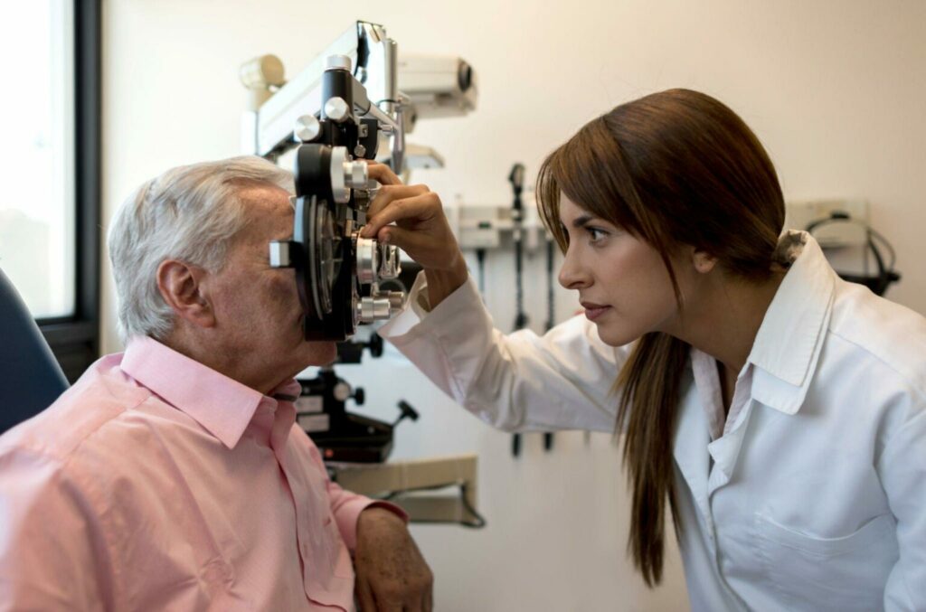
Additional Tests for Macular Degeneration Symptoms
Acuity test: Using an eye chart, the visual acuity test evaluates your vision at different distances. It aids in identifying whether your central vision has changed at all.
Dilated eye exam
During this examination, eye drops are used to enlarge your pupils so the eye doctor can inspect the macula at the rear of your eye.
Retinal imaging
The retina, particularly the macula, can be visualized in great detail using imaging techniques like optical coherence tomography (OCT). This aids in determining the macular tissue’s integrity and thickness.
Fluorescein Angiography
This test involves injecting a fluorescent dye into an arm vein, and as it travels through the blood vessels in your eyes, pictures are taken to assess blood flow and spot aberrant blood vessel growth.
It’s crucial to keep in mind that these examinations are normally carried out by medical specialists who have a focus on eye care. It is advised to make an appointment with an eye care professional who can conduct a thorough examination and make a diagnosis if you are having symptoms or have concerns about macular degeneration.
What are macular degeneration’s early signs and symptoms?
Symptoms of macular degeneration might vary based on the condition’s stage and type. You might not have any obvious symptoms in the beginning or the changes might be slight. However, when the illness worsens, the earliest signs and symptoms below may appear:
Central vision that is hazy or blurry is one of the early symptoms of macular degeneration that is most frequently experienced. As a result, it could be challenging to read, identify people, or carry out other tasks that call on precise eyesight.
Straight lines may appear wavy, crooked, or warped due to impaired vision. You might observe that normally straight objects or lines, like telephone poles or door frames, appear bent or crooked.
Reading challenges or difficulty performing close-up tasks
Because macular degeneration impairs central vision, you can find it challenging to sew, read the small print, or perform other close-up jobs.
Reduced color perception
Some macular degeneration sufferers may have a harder time accurately identifying colors or noticing changes in their color vision.
You might detect dark or void patches in the middle of your field of vision. Your vision may be obstructed by these blind spots, which could also grow with time.
Who is most susceptible to AMD?
The main risk factor for age-related macular degeneration (AMD) is advancing age. The risk rises with age, and people over 50 are the group most likely to experience it. It is crucial to remember that AMD can, albeit less frequently, affect people who are younger.
In addition to age, a number of additional factors can increase the risk of having AMD:
Family history
The risk is higher if AMD runs in the family. Your risk may be higher if you have close relatives who have been diagnosed with AMD, such as parents or siblings.
Smoking
Smoking is a major AMD risk factor that can be modified. Smokers may experience a faster rate of illness progression and are more likely to develop the disorder.
AMD is more prevalent in people who are Caucasian or European in origin. But it can strike persons of all racial and ethnic backgrounds.
Genetics
A higher risk of getting AMD has been linked to specific genetic abnormalities. Specific genes that may contribute to the onset and course of the disease have been identified by researchers.
Cardiovascular Health
AMD risk can be raised by factors that affect cardiovascular health, such as high blood pressure, high cholesterol, and obesity.
It’s crucial to remember that although these factors can make AMD more likely to occur, they do not ensure that someone will have the illness. On the other hand, AMD can still occur in people without these risk factors. Regular eye exams can help detect and monitor AMD early, enabling appropriate management and intervention, especially for people over 50 or with other risk factors.
Treatment for macular degeneration
The type, stage, and severity of macular degeneration determine the available treatments. Macular degeneration cannot be cured, but there are ways to manage the symptoms, slow down the condition’s progression, and maximize vision that still exists. Here are a few typical methods:
Nutritional Supplements as a Treatment for Dry AMD
Research has suggested that some high-dose antioxidant vitamins and minerals, such as vitamins C and E, zinc, copper, and lutein/zeaxanthin, may in some circumstances help halt the progression of dry AMD. However, it’s imperative to speak with an expert in eye care before using any supplements.
Lifestyle changes
Making healthy lifestyle choices, such as eating a balanced diet, exercising frequently, quitting smoking, and maintaining normal blood pressure and cholesterol levels, may help lower the risk of AMD progression or development.
Treatment for Wet AMD
Anti-VEGF Therapy: The main form of treatment for wet AMD, which is characterized by the development of abnormal blood vessels, is anti-vascular endothelial growth factor (anti-VEGF) therapy. To stop the development of abnormal blood vessels and lessen leakage, drugs such as ranibizumab, aflibercept, or bevacizumab are injected into the eye. This preserves vision and stops further damage.
Low Vision Aids and Rehabilitation
Low-Vision aids: Magnifiers, telescopic lenses, and specialized lighting are examples of low vision aids that can help improve remaining vision and help with daily activities.
In order to maximize functional vision and enhance quality of life, vision rehabilitation programs—which may also include vision therapy, occupational therapy, and counseling—can offer assistance, instruction, and adaptive techniques.
What is the prognosis for macular degeneration over the long term?
The type of macular degeneration (AMD), the stage at which it is discovered, the patient’s general health, and the success of treatment are some of the variables that can affect the long-term prognosis of AMD. Regarding the long-term prognosis of AMD, the following are some general considerations:
Compared to wet AMD, dry AMD advances more slowly. It may however eventually result in a gradual and progressive loss of central vision. In some circumstances, dry AMD can move to a more severe stage known as geographic atrophy, where the macular tissue is noticeably thinned and lost, severely impairing vision.
Wet AMD
When compared to dry AMD, wet AMD has the potential to lead to a faster and more severe loss of eyesight. However, vision loss can be reduced or stabilized with early detection and the right treatment, like anti-VEGF therapy, and some people may even see an improvement in their eyesight.
Loss of Vision
Macular degeneration primarily affects the center vision, which can make it difficult to read, drive, recognize faces, or do intricate jobs. Peripheral vision, on the other hand, is typically unaffected, allowing people to retain some degree of functional vision.
Individual Variability
Everybody’s experience with AMD is different, both in terms of its course and effects. While some people may endure a gradual decline in vision with little loss, others may experience a more aggressive course with severe visual impairment.
Summary of Macular Degeneration Symptoms Test
Despite the fact that there is presently no cure for macular degeneration, it is crucial to remember that continuous research and improvements in available treatments offer promise for better management and results. Optimizing the long-term prognosis of AMD requires regular monitoring, early discovery, and prompt action. It is advised to collaborate closely with an eye care specialist to create a customized management strategy based on unique situations and requirements.
FAQ’s for Macular Degeneration Symptoms Test
How can I perform a macular degeneration home test?
Amsler grid, distance vision, and close vision testing are the three types of vision exams that can be performed at home. This examination can identify macular degeneration. Vision distortion, blank areas, or blurriness are symptoms of this illness. If you often wear reading glasses, put them on for this exam.
What might be misdiagnosed as macular degeneration?
Cone dystrophy, adult vitelliform dystrophy, pattern dystrophy, North Carolina macular dystrophy, Doyne honeycomb dystrophy, and Sorsby macular dystrophy are autosomal dominant retinal dystrophies that can mimic AMD.
What is the typical age at which macular degeneration begins?
Before the age of 55, the syndrome is uncommon. Most adults 75 years of age or older experience it. Risk elements for AMD include AMD in the family history.
Can macular degeneration be cured with glasses?
8 types of glasses for macular degeneration, additional vision aids: Although macular degeneration cannot be fully cured with glasses, they can help you get the most out of your existing eyesight. You may find it simpler to carry out daily tasks that call for close attention to detail as a result. Sunglasses are also beneficial since they might stop your eyes from suffering more harm.


