

Macular degeneration deteriorates the central region of your retina (thin transparent tissue that lines the back wall of your eye). This area provides fine details, such as straight lines in your vision; however, it cannot detect faces or objects beyond these straight lines.
Macular degeneration can cause retinal detachment, leading to permanent vision loss if untreated. A dilated examination, ultrasound imaging and high-definition photographs can be helpful tools in detecting changes within the retina.
Causes
The retina – located on the back wall of the eye – contains millions of light-sensing cells that convert light entering your eye into electrical impulses that travel down your optic nerve and ultimately into sight. When something damages or detaches the retina, these light-sensing cells cannot receive signals to create clear vision; this results in blurry or distorted vision as a result; retinal detachments should be treated immediately for their immediate treatment and to avoid future vision loss.
Age related macular degeneration occurs when the macula becomes degraded, leading to two forms of macular detachment: dry and wet. Eighty-five to ninety percent of people diagnosed with macular degeneration exhibit dry form symptoms. Drusen deposits appear beneath the retina and typically do not cause vision loss, only becoming detectable through a thorough dilated eye exam. However, 10-15 percent of cases of macular degeneration present as wet form which develops when abnormal blood vessels grow under the retina and leak fluid into the macula. This form is one of the leading causes of legal blindness among those over fifty and typically leads to faster rates of central vision loss than dry ARMD.
Macular degeneration can result in either tractional or exudative retinal detachments. When vitreous gel contracting causes strain on retina and in some instances tears it, liquid from within it flows out through tear to detach it causing detachments that often lead to blind spots. This type of retinal detachment is most frequently experienced during macular degeneration due to age, family history of macular degeneration, trauma, high nearsightedness (myopia), certain diseases/infections such as diabetic retinopathy/glaucoma as well as inflammation/autoimmune eye diseases like Vogt-Koyanagi-Harada syndrome/uveal Melanoma/choroidal Hemangiomas etc causing these types of retinal detachments affecting these parts of the eyeball as a whole affecting this part of it from being attached.
Exudative detachments occur when scar tissue or abnormal membranes form on or within the retina and lead to its contraction, such as in cases such as choroidal hemangiomas, diabetic retinopathy, uveal melanomas, macular holes, and various miscellaneous conditions such as Vogt-Koyanagi-Harada disease and scleritis.
Symptoms
The retina is the layer of nerve cells at the back of your eye that transmits light signals from light-sensing cells to your brain, creating images you see. Comprised of millions of light-sensitive cells, it plays an integral part in central vision. People suffering from advanced macular degeneration can experience abnormal blood vessel growth beneath their retina that leads to retinal detachment resulting in blurry spots in central vision that gradually grow larger and darker as time progresses, or straight objects like telephone poles or venetian blinds appearing crookedly.
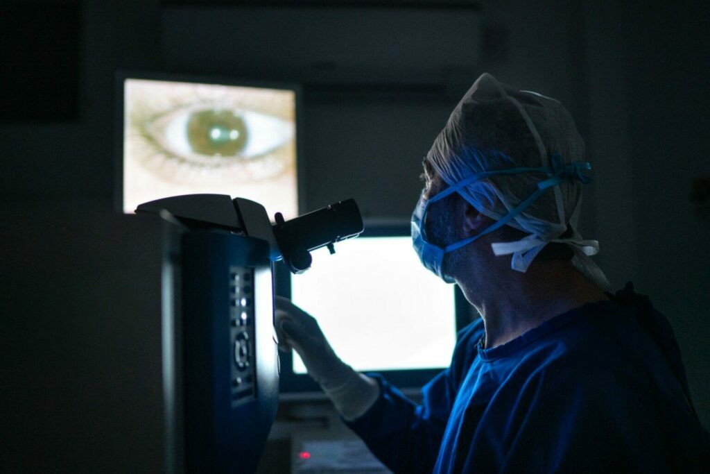
Retinal detachments can be serious medical conditions that need immediate medical treatment, or they could lead to permanent vision loss if left untreated. If any symptoms appear, seek medical advice immediately from your ophthalmologist as soon as possible for an examination using both a slit lamp and an ophthalmoscopy examination – these two tools allow doctors to dilate your eyes in order to detect tears or retinal detachment more accurately.
There are three categories of retinal detachments: rhegmatogenous, tractional and exudative. Of these detachments, rhegmatogenous is the most prevalent and occurs when a hole or tear forms in the retina allowing fluid to seep through and cause detachment. Tetheral detachments also arise without physical breaks in the retina – instead scar tissue caused by trauma to the eye can pull apart its connections with surrounding tissues causing detachments to form within eye tissues causing it detachments as well. Exudative retinal detachments can result from surgery procedures as well as trauma to eye trauma from injuries caused during cataract surgery procedures or procedures for which procedures for patients will cause detachments as well.
Wet macular detachments are relatively rare conditions in which retinal cells begin to degenerate, leading to retinal detachment. Although its cause remains unknown, Ehlers-Danlos Syndrome appears to play a part. With EDS, joints and skin become overextended and flexible which increases risk of retinal detachment due to pulling by muscles in the eye. Wet detachments may be treated by injecting gas or silicone oil into one eye to push back the retina into place before using photocoagulation or cryopexy to reattach it permanently.
Diagnosis
Macular degeneration is a slowly progressive condition in which central vision fades over time, making it more challenging to read, drive or recognize faces. While peripheral (side) vision remains unaffected, macular degeneration should never lead to total blindness as peripheral vision remains unchanged. If you detect any changes in vision it is important to visit your doctor immediately as macular degeneration often doesn’t exhibit symptoms in its early stages and goes undetected; regular eye exams could detect the presence of drusen deposits under the retina associated with dry macular degeneration if present – an eye exam could potentially detect these as early signs as early detection could reveal macular degeneration causing total blindness would result from side (peripheral) vision remaining unaffected.
Wet macular degeneration occurs when abnormal blood vessels grow underneath the retina and leak fluid or blood, blurring or distorting central vision and leading to faster vision loss than its dry counterpart. Furthermore, there is also an increased risk that it will spread into another eye; your doctor should therefore monitor both eyes closely.
With wet macular degeneration, there are treatments available that can either slow or halt its progression. Medication may reduce abnormal blood vessel growth that leaks fluid and blood. Alternatively, laser light surgery can be used to permanently destroy these vessels and stop their leakage.
Vitamins C and E, lutein and zeaxanthin may help protect or delay macular degeneration’s progression; your doctor can recommend the best oral supplement or injection treatment plan for you.
For wet macular degeneration, one of the most effective treatments is injections of medication which inhibits abnormal blood vessel formation and leakage. This injection can be given every four weeks at Beraja Medical Institute’s offices and should be taken in office. With regard to dry macular degeneration medications can only slow the rate of vision loss; unfortunately they cannot restore lost vision. If you are experiencing vision loss contact Beraja Medical Institute by phone or online today to arrange an appointment with one of our doctors!
Treatment
An retinal detachment is a medical emergency and should be addressed as quickly as possible. When diagnosed and treated quickly enough, patients often regain most of their lost vision. Treatment options range from in-office laser therapy to outpatient surgery; initially though, the focus should be on stopping fluid leakage under the retina – this can be accomplished by injecting special medicine that causes an eye bubble to form at its center which then pushes against it, helping seal off its attachment back to its wall of the eye and sealing up holes or tears with vitrectomy/membrane peel surgery procedures; repairs on damaged retinal tears/holes/ruptures can then repair holes/tears etc with these surgical procedures or techniques as needed.
Next comes the removal of any leaking blood vessels that caused retinal detachment, performed in an operating room using laser technology to destroy abnormal vessels quickly and painlessly.
Laser surgery cannot restore severe retinal cell damage; however, macular degeneration cases that involve small yellow protein deposits called “drusen”, but haven’t resulted in retinal detachments can often be helped with injections to slow or stop their progression and preserve vision.
People living with lattice degeneration should schedule an annual dilated exam to check for small retinal breaks or tears that could lead to retinal detachments, which may be sealed with laser. Sometimes they can even be sealed prior to any detachment occurring if photodynamic therapy (using medication that causes retinal blood vessels to light up when viewed through special cameras) can help – followed by laser cauterizing of abnormal vessels which causes macular detachments (safe and effective in most cases; not a cure);


