

Healthy eyes produce and circulate an eyewash known as aqueous humor that must drain properly or risk creating pressure within the eye that could result in damage to its optic nerve.
Everyone, especially those at risk for glaucoma, should get their eye pressure checked regularly using a device called a tonometer after having their eye numbed.
Causes
Your eyes produce aqueous humor to maintain healthy and moist conditions in your eye, which must then drain freely out to avoid vision loss. If this balance between production and drainage becomes upset, pressure in the eye can rise rapidly resulting in Glaucoma; untreated, it can even lead to blindness by damaging optic nerves (parts of your eye which transmit information from sight back into brain).
Open-angle glaucoma is one of the most prevalent forms of glaucoma and typically develops when drainage canals become blocked and cannot drain away fluid properly, leading to an accumulation of fluid pressure inside your eye and an increase in pressure that damages optic nerves and leads to vision loss.
Closed-angle glaucoma is less prevalent and occurs when drainage canals become completely clogged or closed off due to injury, cataract surgery or long-term use of certain drugs such as steroids. This condition could also result from diabetes or blood vessel problems known as ocular hypertension affecting one or both eyes.
Normal-tension glaucoma is another subtype of glaucoma that can develop even though eye pressure levels appear normal. While its exact cause remains unknown, researchers believe it may be down to abnormally sensitive optic nerves or reduced blood supply to the eyes that cause damage leading to glaucoma.
Glaucoma affects everyone, though it’s more prevalent among adults over 40 and ranks second as a cause of blindness. Anyone at higher risk, or with family histories of the condition should make regular visits to an eye doctor between 35-40 for eye pressure measurements to help detect glaucoma early and begin treatment in order to protect their vision and preserve your eyesight.
Symptoms
Healthy eyes typically maintain fluid pressure below 21 millimeters of mercury (mmHg). This means the passages that allow fluid to drain are working as intended and that glaucoma – a progressive disease which can lead to permanent vision loss – is being managed properly. Most commonly found among those over 40 and can be detected through an eye exam, even without symptoms present such as blurry vision, pain or redness in the eye, blind spots in peripheral (side) vision or colored rings around lights.
Open-angle glaucoma is the most prevalent form of glaucoma. It often develops slowly without producing symptoms until it’s too late to reverse any damage caused by high intraocular pressure, eventually damaging optic nerve fibers and leading to vision loss.
Narrow-angle glaucoma is less prevalent and advances faster than open-angle glaucoma, often as a result of blocked drainage canals by the iris which leads to an increase in pressure rapidly, potentially leading to blindness within two days if left untreated; it requires immediate medical intervention in order to avoid blindness. This medical emergency must be addressed promptly in order to save sight.
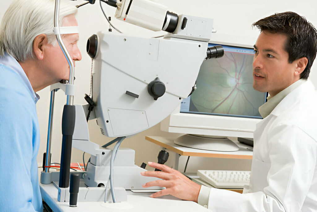
Your doctor will use a tonometer to take an accurate reading of eye pressure during an eye exam, after numbing your eye with drops. As part of the examination, the tonometer is gently pressed against the front of your eye – some models use puffs of air while others utilize small probes that rest against your cornea – with recorded data being sent directly back to them and diagnosis being given based on this reading.
If your eye pressure is elevated but no other signs of glaucoma such as optic nerve damage or blank spots in your side vision have surfaced, this condition is known as normal-tension glaucoma or low-tension glaucoma and needs to be closely monitored by an ophthalmologist as some individuals with this condition do develop full-blown glaucoma in due course.
Diagnosis
For diagnosis of low eye pressure, your doctor will use a device known as a tonometer after placing numbing drops into each eye. The tonometer measures intraocular pressure (IOP). Normal intraocular pressures range between 10-21mm of mercury. Your optic nerve may also be examined for signs of glaucoma, including blank spots in peripheral vision or blank spots along its path; additional tests could include visual field tests that measure blind spots, optical coherence tomography to examine nerve fiber layer thickness as well as gonioscopy to examine where eye fluid drains away.
Some individuals can have glaucoma with eye pressure lower than 21 mm of mercury – known as normal-tension glaucoma – which requires close monitoring as these individuals may still develop optic nerve damage and lose vision.
Individuals at risk for narrow-angle glaucoma, including those over 40 and particularly those at high risk, should have their eye pressure regularly checked from age 35 onward. This will allow early detection before it causes symptoms that threaten vision loss; and allows detection before damage to optic nerves becomes too severe to treat effectively. People who have had prior history of narrow-angle glaucoma should visit an eye doctor every year for this type of exam.
Treatment
Eye pressure increases when fluid in the eye doesn’t circulate as intended, a condition known as intraocular pressure or IOP. Increased IOP can damage optic nerves that connect images from eyes to brain. Once damaged, vision blurs and blindness may result; untreated high IOP could even lead to complete blindness within years.
Open-angle glaucoma is the most prevalent form of glaucoma, in which eye structures appear normal but fluids do not drain from an angle where cornea and iris meet properly – this fluid known as “aqueous humor” does not drain properly and causes pressure in the eye to increase, ultimately damaging optic nerve.
Tonometers are used to assess eye pressure. A doctor administers numbing drops before pressing the tonometer against the surface of the eye; then presses down with force applied against resistance from eye cells, measuring IOP in real-time.
Glaucoma treatment entails taking various medications that aim to lower eye pressure, with beta blockers, alpha adrenergic agonists, cholinergic agonists, carbonic anhydrase inhibitors and carbonic anhydrase inhibitors being the most frequently prescribed types. They may be taken alone or together and side effects include bitter taste in mouth or dryness and red or itchy eyes or lids.
If these treatments fail to reduce eye pressure, if necessary your ophthalmologist may employ laser surgery – known as laser trabeculoplasty and performed in-office. This procedure creates a flap in the sclera of the eye to create a bubble called filtration bleb in the conjunctiva which absorbs aqueous humor, thus helping lower eye pressure.
Laser Iridotomy, another form of laser surgery for glaucoma, works by creating a hole in the iris to drain away aqueous humor and lower eye pressure. If narrow-angle glaucoma persists after treatment with laser surgery or with other means, your eye surgeon can perform an additional procedure known as Trabeculectomy; this surgery can be completed in-office using eye drops for anesthesia before beginning.


