

What is IntraOcular Pressure?
The fluid pressure inside your eyes is called “intraocular pressure” in medicine. Eye pressure, or IOP, is another name for it. Your normal intraocular pressure is a big part of how well you see and how healthy your eyes are.
There’s fluid in your eyes. Vitreous humor, a thick, gel-like substance, fills the space at the back of your eye. The space in front of your eye (between your lens and iris) is filled with aqueous humor, a liquid. The aqueous humor is less thick and more watery than the vitreous humor.
Your body instantly controls the pressure inside your eyes. The same amount of old aqueous humor leaves your eye every time it makes new aqueous humor. The drainage angle is where the iris (the colored part of your eye) meets the sclera (the white part of your eye). This is where old watery fluid leaves your eye.
Having ocular hypertension means that your intraocular pressure measurement is too high. This extra stress on the parts inside your eye can hurt your optic nerve if you don’t fix it. If you don’t address ocular hypertension, it can turn into glaucoma and take away your sight for good.
How Is Intraocular Pressure Measured?
Tonometers are a group of tools that have been suggested since the 1800s as a way to measure IOP. Depending on how they work, these instruments can be divided into two main groups: Applanation tonometers and indentation tonometers.
Indentation Tonometry
Schiotz tonometers, which were first made many years ago but aren’t used anymore, are the model for indention tonometers.
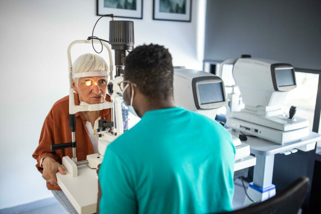
Applanation Tonometry
Eye pressure can be found in a lot of different ways. An easy way to do this is with a test called “applanation tonometry.” To applanate something means to make it flat. The “gold standard” measurement of intraocular pressure is with “Goldmann applanation tonometry,” which is used in most ophthalmologist’s offices.
For this test, numbing drops are used to make the eyes feel numb. A small amount of a non-toxic dye is also put in the eye. The person who treats your eyes will tell you how to put your head in a machine called a slit lamp. After that, a small tip is used to gently touch the eye’s surface and read the pressure inside the eye. How hard it is to gently flatten a set part of the cornea is used to measure eye pressure.
Stationary Applanation Tonometry
You can use a fixed tonometer or a Goldmann tonometer for applanation tonometry. The tonometer probe touches the cornea after numbing the surface of the eye and adding a dye. The device then counts how much pressure is needed to flatten the cornea. People have to be very still for any eye test, but it can be especially hard to sit still in front of a Goldmann tonometer. Because Goldmann tonometry is rigid and hard to get to, there are other ways to measure eye pressure besides it being the gold standard for IOP.
Tonometry with a Handheld Applanation
Handheld applanation tonometry is based on the idea that doctors can find out the intraocular pressure by figuring out how resistant the cornea is to pressure. This idea is then turned into a device that can measure eye pressure for almost anyone, not just people who can be examined on a Goldmann tonometer. The Reichert Tono-Pen takes the tools and strips them down to make a gadget that can be used by patients wherever they are. The Tono-Pen is a good option as long as the patient doesn’t have a rubber allergy which would make it impossible to use the tip covers that keep the probe clean. The Tono-Pen is also very flexible and easy to use.
Several things can change the results of this test, such as whether the cornea is too thick or too thin or whether there is too much or too little dye in the eye (in the case of goldmann). For instance, we know that this device can give an incorrectly low reading when the cornea is thin. This can happen naturally or because of laser surgery for vision correction. Also, naturally thin corneas are a risk factor for glaucoma on their own, so measuring corneal thickness is an important part of a full eye exam and should be done at your first visit and any others that follow.
A few years ago, other applanation devices were made that work in the same way. The Tono-Pen and the newer Tono-Pen Avia are small, light, battery-powered devices that work by pressing and pulling on the cornea. A small screen shows the standard deviation of the average of 10 readings to show how reliable each number is. Each patient is given a single-use rubber cap, which helps lower the risk of getting an infection from one patient to the next.
Tonometry with Air-Puff
Non-contact tonometry, also called air-puff tonometry, is a way to check your eye pressure by pressing down on the lens with a quick burst of air. Modern systems have been shown to connect with Goldmann tonometry, even though there is some disagreement about how accurate it is.
Rebound Tonometry
Another way to measure eye pressure is with rebound tonometry. Bounces a small tool with a plastic tip against the eye. There’s no need for numbing drops for this test, and you can use gadgets like iCare at home instead. The device is small and movable, and it’s easy to use with kids or people who can’t do what’s needed to take their eye pressure. Tonopen, a type of electronic indentation tonometry, is another small and simple device that you can take with you. On the other hand, you need numbing drops to use the Tonopen instead of iCare.
What Is A Normal Intraocular Pressure Measurement?
Millimeters of mercury are used to measure eye pressure, just like mercury is used to measure temperature in a thermometer. Most people think eye pressure between 10 and 20 millimeters of mercury (mmHg) is normal. It can hurt your eyes to have eye pressure that is too low or too high.
Ocular hypertension is high eye pressure that doesn’t occur with any other signs. Some people can have high eye pressure and not be hurt by it. Some people may lose their sight even if their eye pressure is normal.
Eye pressure hurts the optic nerve in people with glaucoma. Unfortunately, this damage makes it impossible to recover that lost vision. Glaucoma can make you totally blind if you don’t treat it.
What Causes High IOP?
When the clear fluid that fills the front area of the eye can’t flow properly out of the eye, this is called high IOP. The aqueous humor’s job is to keep the eye full of fluid so it doesn’t collapse and to feed certain parts of the front part of the eye, like the lens and some of the iris and cornea.
To keep IOP normal, the drainage system that lets the watery humor leave the eye must stay clear and work properly. A buildup of fluid can happen when the fluid can’t drain properly, which leads to an increase in eye pressure.
Are Intraocular Pressure Measurements Constant?
No. IOP can change every hour, every day, and every week. So, taking your eye pressure during a test can only give you a rough idea of your normal IOP. Large changes in IOP levels during the day can be bad for your eye health, especially if you have glaucoma.
Even though small changes in eye pressure are usual, you should talk to your eye doctor about how to keep an eye on your IOP, either at home or during more frequent eye tests. Eye doctors usually say that you should take your reading in the morning, so keep that in mind when you make your next appointment.
What are the symptoms of High IOP?
One tricky thing about ocular hypertension is that it doesn’t always show any signs. You should get your eyes checked regularly because of this. If the pressure inside your eye is high, your doctor will be able to tell during the exam.
You might feel eye pain when you move your eyes or touch your eyes.
Some eye problems can give you headaches or make you feel dizzy, but this doesn’t usually happen with ocular hypertension.
Risk Factors for Ocular Hypertension
A risk factor is something that makes it more likely that you will get a disease. Some things that put you at risk, like your age, race, or having another health problem, can’t be changed. When it comes to eye hypertension and getting glaucoma, having other diseases like:
- Hypotension and Hypertention
- Having diabetes.
- To be very nearsighted (myopia).
- Thinner Corneas
- The head of the optic nerve is bleeding.
When you have pigment dispersion syndrome, the colored part of your iris falls off. The colored part of your eye is called the iris. The pigment can get stuck in your drainage system.
People who have pseudoexfoliation syndrome (PXF) may notice that protein flakes start to build up in their organs, such as their eyes.
Other things that can put you at risk for ocular hypertension are:
- Being more than 40 years old.
- There is a history of glaucoma or eye hypertension in your family.
- Being Hispanic or Black.
- Taking medications containing steroids for a very long time.
- Having had eye surgeries or damage in the past.
What Causes Ocular Hypertension
Too much or too little fluid production or flow in the eye can lead to high pressure inside the eye. The pathways that should drain the fluid from inside the eye aren’t working right. It’s making more fluid, but it can’t drain. Because of this, there is more fluid inside the eye, which raises the pressure.
Thinking of a water balloon is another way to picture high pressure in the eye. If you add more water to the balloon, the pressure inside it will rise. The same thing happens when there is too much fluid in the eye: the more fluid there is, the higher the pressure. If you put too much water into a water balloon, it can pop. The same thing can happen to the optic nerve in the eye if the pressure is too high, it doesn’t pop but can be damaged.
People whose corneas are normal but very thick often have high ocular pressure in their eyes, even a little higher than usual. Their pressures may be lower and average, but because their corneas are so thick, the measurements give a falsely high number.
How often should your intraocular pressure be measured?
The American Academy of Ophthalmology (AAO) says that everyone younger than 40 who doesn’t have any known risk factors for glaucoma should get a full eye test every 5 to 10 years. Some of these tests look for glaucoma.
The AAO recommends that people who are at risk for glaucoma get comprehensive eye examinations on the following dates:
- Every two to five years for people under 40
- Every one to three years for people 40 to 54 years old
- Every one to two years for people 55 to 64 years old
- Every one to two years for people 65 and up
Depending on how at-risk you are and how healthy your eyes are in general, your eye doctor may tell you to get eye tests more often.
Glaucoma is more likely to happen to people who:
Are between middle-aged and old. Glaucoma is more likely to happen as you age, especially after age 40.
- Have a history of cataracts in your family.
- High intraocular pressure means you have high eye pressure.
- Have open-angle glaucoma and are African American.
- East Asians and people with East Asian ancestry are more likely to have closed-angle glaucoma.
- More likely to get closed-angle glaucoma if they are farsighted.
- Have hurt their eyes or had surgery on them, like fixing cataracts.
- You have diabetes.
- Have hypertension,
- Have been taking steroidal medications
Conclusion of Intraocular Pressure Measurement
The measurement of intraocular pressure is vitally important to the overall health of your eyes. Since it mostly is asymptomatic, it must be done by an eye doctor to know if you are at risk of developing glaucoma. If you haven’t had your eyes checked in a while you better get in to “see” what you’re missing.


