

A collection of tests known as glaucoma tests are used to identify glaucoma, an eye condition that can lead to blindness and visual loss. When fluid accumulates in the front of the eye, glaucoma results. The additional fluid raises ocular pressure. The optic nerve can be harmed by elevated eye pressure. Information is sent from the eye to the brain via the optic nerve. Serious vision issues may result from injury to the optic nerve.
Various forms of glaucoma exist: The three common types are:
Primary open-angle glaucoma is another name for open-angle glaucoma. The most typical form of glaucoma is this one. It occurs when the drainage channels of the eye’s fluid do not function properly. Like a clogged sink drain that backs up with water, the fluid accumulates in the canals. The pressure in the eyes rises as a result. Over the course of several months or years, open-angle glaucoma slowly worsens. Most people initially don’t have any symptoms or changes in vision. Both eyes are typically affected simultaneously by open-angle glaucoma.
Angle-closure or narrow-angle glaucoma, also known as closed-angle glaucoma. In the US, this kind of glaucoma is uncommon. One eye is typically affected at a time. The drainage channels in the eyes get blocked off in this form of glaucoma as if a stopper had been placed over a drain. Acute or chronic closed-angle glaucoma is both possible.
When acute closed-angle glaucoma develops, ocular pressure quickly rises. A medical emergency has arisen. If acute closed-angle glaucoma is not treated right away, patients could lose their eyesight in a matter of hours.
The onset of chronic closed-angle glaucoma is gradual. Many times, damage doesn’t show signs until it’s quite severe.
Who has a higher chance of getting glaucoma?
Your lifetime chance of acquiring glaucoma is greatly increased if you have a family history of the disease. You could be at greater risk if you:
- You’re Black (open-angle glaucoma or POAG).
- Have East Asian ancestors (closed-angle glaucoma).
- You’re farsighted (closed-angle glaucoma)
- Underwent a difficult eye surgery or sustained an eye injury.
- You’re a diabetic.
- Experiencing hypertension.
- You’ve taken corticosteroid medications in the past (like prednisone).
- You’re over 40 years old.
When is a glaucoma screening exam necessary?
If you are having vision issues like those listed below, you may require an eye exam that includes glaucoma tests.
- Blind areas
- Fuzzy vision
- Eye pressure or discomfort
- Headaches.
- When you gaze at lights, you see rainbow circles.
- Eyes are red.
- A narrowing of your visual field or tunnel vision
Additionally, the American Academy of Ophthalmology (AAO) advises that those who are glaucoma-risk individuals undergo thorough eye exams in accordance with the following schedule:
- Every one to three years, for people 40 to 54.
- Every one to two years, for people 55 to 64.
- Every six to twelve months for people 65 and older.
What occurs throughout glaucoma testing?
A glaucoma evaluation is a collection of examinations used to identify glaucoma. An ophthalmologist or optometrist conducts these examinations the majority of the time. These are physicians with a focus on treating and preventing eye diseases.
Eye tests for glaucoma include:
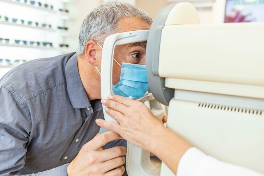
Tonometry
You will sit in an exam chair next to a specialized microscope known as a slit lamp during a tonometry test. To numb your eyes, your doctor or other healthcare professional will apply drops. You will then place your chin and forehead toward the slit lamp. Your medical professional will place a tonometer on your eye as you lean into the slit lamp. The instrument gauges ocular pressure. There will be a tiny air puff, but it won’t harm.
Pachymetry
You’ll first receive drops to numb your eye, just like in a tonometry exam. Your healthcare professional will then place a pachymeter, a little instrument, on your eye. This tool measures the corneal thickness. The iris, the colored portion of the eye, and the pupil are protected by the cornea, which is the eye’s outer covering. Your chance of developing glaucoma may be higher if you have a thin cornea.
The thickness of your cornea can interfere with a reliable reading of your IOP and lead to an incorrect perception of the internal pressure. There is a chance of misdiagnosis if your eye doctor does not consider your corneal thickness. You can get a precise diagnosis by having your cornea’s thickness measured and your eye pressure measured.
Perimetry
Perimetry You’ll be instructed to stare directly ahead at a screen during perimetry. The screen’s one side will gradually fill with a light or an image. While keeping an unwavering gaze forward, you’ll press a button as soon as you spot this light or image.
Dilated eye exam
Dilated eye exam Your doctor will place drops in your eyes to enlarge (dilate) them for this test. Your healthcare professional will examine your optic nerve and look for damage using a gadget with a light and magnifying lens.
Gonioscopy
Your healthcare professional will apply drops to your eyes during this examination to numb and dilate them. Then, your doctor will cover the eye with a unique hand-held contact lens. The lens includes a mirror on it so the physician can observe the interior of the eye from various angles. It can demonstrate if the iris-cornea angle is too broad (perhaps indicative of open-angle glaucoma) or too small (a possible sign of closed-angle glaucoma).
OCT
With the help of the noninvasive diagnostic imaging technique optical coherence tomography (OCT), ophthalmologists may see the distinct layers of the optic nerve and retinal nerve fiber layer (RNFL) in astonishing detail. OCT is now a crucial tool for patient glaucoma screening, diagnosis, and progression monitoring.
Why so many glaucoma test?
Glaucoma diagnosis is not always straightforward, and accurate assessment of the optic nerve is still necessary for both diagnosis and therapy. The most crucial issue is keeping your eyesight safe. Before deciding on your course of therapy, doctors consider a variety of criteria. You can be referred to a glaucoma specialist if your condition is very challenging to identify or treat. If you or your doctor have any concerns about your diagnosis or your progress, it is always a good idea to get a second opinion.
What may I anticipate following a glaucoma test?
Following glaucoma testing, you can be subject to some limitations. For instance, you may want a relative to drive you home because your vision will likely be hazy. You might need to wear sunglasses to shield your eyes from too much light and ultraviolet (UV) radiation if you underwent a dilated eye test.
Depending on the kind of tests you have, your ophthalmologist provides you with specific recommendations.
What dangers could arise from glaucoma tests?
Tests for glaucoma are risk-free and safe. After the test, your vision might be hazy or extremely light-sensitive, but it gradually improves throughout the day.
When will the results of my glaucoma testing be available?
The majority of glaucoma test outcomes are accessible right away at the same appointment.
What do the findings of glaucoma tests mean?
Your doctor will go over the test results and their implications with you. To assess whether you have glaucoma or are at risk for developing it, as well as what type you may have, your doctor will take the results from all tests into account.
Results that are outside of the healthy normal range may point to glaucoma or the need for additional testing. abnormal test outcomes could show:
- Gonioscopy angle examination: narrow or obstructed drainage angle (the area where your eye fluid drains).
- Measurement of corneal thickness (pachymetry): a corneal thickness that raises your risk of developing primary open-angle glaucoma.
- During a dilated eye exam, look for unusual blood vessels in your eye’s size and shape.
- Checking the intraocular pressure in the eyes: more than 22 mmHg (millimeters of Mercury).
- Imaging of the optic nerve: Unusual findings such as swollen or buckled optic nerves or rough retinas.
- Visual field test: The detection of certain regions in which your vision becomes hazy or blanks out.
When I get glaucoma, what happens?
The following steps will be advised by your doctor. They could advise setting up follow-up sessions to periodically check your vision. Alternatively, they could suggest glaucoma remedies like:
- Medications that lower eye pressure, such as eye drops or tablets.
- Surgery creates a drainage channel in your eye, allowing surplus fluid to drain away and lowering eye pressure.
- Surgery to implant drainage tubes for extra fluid.
- Laser therapy to aid with fluid drainage
Do I need to know anything else about glaucoma eye tests?
Treatment for glaucoma can stop further vision loss even if it won’t reverse the condition or restore any eyesight that has already been lost. The majority of glaucoma sufferers who receive early diagnosis and treatment won’t experience serious vision loss.
Does Medicare cover glaucoma screening tests?
If you have a high risk of getting the eye condition glaucoma, Medicare Part B (Medical Insurance) will pay for glaucoma examinations once every 12 months. If you meet the criteria for at least one of these conditions, you are at high risk:
- You’re diabetic.
- You have a history of glaucoma in your family.
- You are 50 years of age or older and African American.
- You are Latino and 65 years or older.
It will also be covered if your doctor finds things during a comprehensive exam that may be indicative of glaucoma. Each test has different rules for how many times it can be done each year.
FAQs for Glaucoma test
What glaucoma test is the most reliable?
Before any other tests, an OCT scan can accurately diagnose glaucoma. It is conceivably the most precise test available to help our doctors identify glaucoma.
Does glaucoma testing hurt?
Even though the test is not painful and just takes a few minutes for each eye, it is possible that you won’t be able to execute it accurately if you are extremely stressed or fatigued. Additionally, the test could need to be repeated because it might need to be improved upon.
How is glaucoma identified early?
Although functional alterations may be noticed before structural changes, structural abnormalities of the ONH and RNFL are frequently the first observable signs of glaucoma. Clinical examination and stereo photographs of the optic disc are the standard methods for evaluating structural changes.
How lengthy are glaucoma tests?
Visual field tests are typically performed once to twice a year after glaucoma has been diagnosed in order to track changes in your vision. You can anticipate this consultation to last two to three hours given the number of tests you’ll have to go through.
What age should you have a glaucoma test?
At least five typical tests are used in a comprehensive eye exam to look for glaucoma. It’s crucial to routinely have your eyes checked. At age 40, you ought to have a baseline eye examination. At this age, vision abnormalities and early indicators of eye illness may start to appear.
What results in glaucoma?
Damage to the optic nerve, which results in loss of visual field, is the cause of the chronic, progressive eye illness known as glaucoma. Eye pressure is among the key risk factors. Fluid can accumulate in the eye due to an issue with the drainage system, which can result in severe pressure that harms the optic nerve.
Does everyone with glaucoma go blind?
Glaucoma is a serious, chronic eye condition that, if left untreated, can cause vision loss. However, glaucoma generally does not result in blindness. Because there are various options available to assist prevent glaucoma from further harming your eyes, glaucoma can be controlled with current treatment.


