


Age-related macular degeneration affects central vision, known as the macula. This condition may arise from multiple causes, including yellow protein deposits beneath the retina (drusen) and tissue pigment loss in the macula.
Your doctor can assess your vision during a dilated eye exam by looking for straight lines that appear wavy or missing and using an Amsler grid to detect early signs of wet AMD, a condition in which abnormal blood vessels form beneath the retina and leak blood and fluid into it.
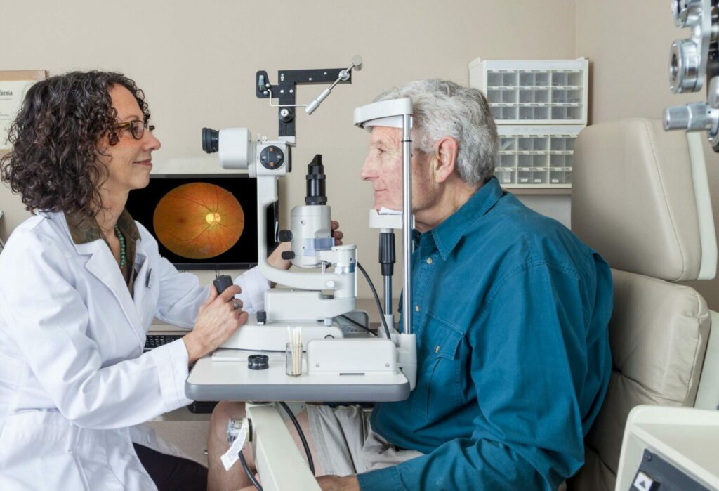
Dilated Eye Exam
Undergoing a dilated eye exam allows an eye care professional to look inside at the back of your eyes, including retina and vitreous fluid, in order to spot conditions like cataracts, glaucoma and macular degeneration – it should be recommended annually for people at risk for these diseases.
Once the eye drops have taken effect, you are asked to remain still while your doctor uses a special camera known as a slit lamp to take images of your retina with an ultra-small microscope that provides bright illumination of inner eye tissues for examination by doctors. Images captured with this type of camera can reveal various issues including:
Your doctor will use a dye, usually fluorescein, to detect areas of damage or abnormality in the retinal cells of your eye. This could appear as discolored splotches near the center of the retina and could indicate disease such as diabetic retinopathy which damages small blood vessels in your retina.
Expect your vision to temporarily blur following an eye exam, making reading and focusing easier when looking at nearby objects than those further away. Your doctor may give you disposable sunglasses to use during your exam or you can bring your own; either way, arrange transportation back home as it is not safe to drive with dilated eyes.
After having their eyes dilated during an eye exam, your vision should typically return to its usual state within 4-6 hours; some individuals may experience dilation that lasts longer.
A dilated eye exam should not be painful; you may only feel some mild stinging from when the drops are first placed into your eye, but otherwise there should be no discomfort or adverse side effects from this examination. After receiving the drops, you may experience temporary blurriness which should clear up within hours after your exam has completed. You might also become more sensitive to light afterward and feel tightness in your eyelids which should ease over time; thus it would be wise to bring sunglasses or arrange transportation home from this appointment.
Amsler Grid
The Amsler grid is an invaluable tool used to detect distortion of straight lines – often an early indicator of wet age related macular degeneration (Wet AMD). Untreated Wet AMD can lead to rapid vision loss. Wavy lines on an Amsler grid could indicate leakage of fluid or blood into the macula and swelling retinal cells which cause straight lines on an Amsler grid to appear wavy as well as missing sections on it. If your Amsler grid displays any distortions or areas are missing contact your eye care practitioner immediately for an eye exam with dilated pupils for diagnosis.
The Amsler Grid is an easy and noninvasive test you can perform at home to detect changes to your central vision. The Macular Disease Foundation Australia recommends it for individuals at risk of wet AMD, such as those with family histories of it or individuals over 50 who wish to remain vigilant of early signs of vision loss.
At its core, using an Amsler grid is simple: sit or stand 12-14 inches from it and gaze upon its central dot with an open eye; try to focus on this dot without shifting your gaze, and check whether both eyes can see its edges. Also note any dark or missing spots on the grid and take note.
Marc Amsler, a Swiss ophthalmologist, first proposed the Amsler grid test in 1945. Consisting of printed horizontal and vertical lines that form an irregular grid pattern, the Amsler grid serves to monitor central vision – particularly macular region retina and optic nerve head health – through observation of central visual fields such as retinal macular region or optic nerve head head. Furthermore, its presence helps detect metamorphopsia or scotoma in visual fields caused by damage to macula or epiretinal membrane and abnormalities of visual pathways leading to brain.
Optical Coherence Tomography (OCT)
Optic Coherence Tomography (OCT) is a noninvasive imaging technique that offers high resolution micrometer scale cross-sectional images of tissues at micron scale. OCT’s applications in ophthalmology utilize near infrared light to view cornea, iris and lens with resolutions as small as several millimeters – it may also help detect or monitor various retinal diseases or conditions.
As soon as your pupil is dilated, an eye doctor can perform OCT scans of your retina using a handheld instrument using light waves to measure thicknesses of retinal layers and macular pigment epithelium (MPE) structures – this information helps determine if someone may have glaucoma or related conditions that may contribute to it.
OCT can also be used to detect diabetic retinopathy (damage to the retina caused by diabetes) and macular edema (accumulation of fluid within the macula). Furthermore, OCT has also been utilized in patients suffering from glaucoma to detect and track choroidal neovascularization over time.
OCT angiography allows for more complete analysis of retinal circulation and extent of vascular disease in diabetic patients, especially through measurement of ocular blood flow using OCT. A recent innovation known as swept source OCT or SS-OCT has drastically increased scanning speeds while also providing detection of deeper structures like the choroid; which are key features when monitoring vascular diseases of retina.
As part of the scanning process, a series of line scans are collected which are then combined and processed by software into a 3D image. This allows a high-resolution view of retinal architecture and can reveal subtle pathologie, including drusen, macular holes and other abnormalities that might otherwise be difficult to identify on standard line scans. Spaide et al reported enhanced depth imaging, or EDI, as another feature of SS-OCT back in 2008. This feature brings the zero delay line closer to the choroid for more accurate thickness measurement and tracking progression of choroidal neovascularization over time. EDI can currently be found on Zeiss Cirrus 5000 OCTA devices and will soon become standard feature of the next generation of SS-OCT.
Fluorescein Angiography
Fluorescein angiography is a special photographic test that allows doctors to observe blood flow between the retina and choroid layers at the back of your eye, and shows blood vessel growth from these two regions. Fluorescein angiography can help diagnose various eye diseases as well as evaluate treatment progress – it is often employed in diabetic retinopathy diagnosis and age related macular degeneration detection as well as to detect problems like capillary microaneurysms, choroidal neovascularization or retinal vein blockage.
NaFluorescein, a fluorescent dye injected directly into your arm or hand, reflects blue light while absorbing green-yellow light as it travels through the body, reflecting back out to reflect blue light back out as it absorbs green-yellow light from an attached camera-like device that takes pictures as the dye moves through your blood vessels and retina, so the doctor can view patterns of flow and leakage of dye in its travels through these blood vessels and retina.
As part of your test, you may experience a brief hot flush in either arm/hand during which time a few seconds-long hot flush may develop before it quickly fades away. Once injected, fluorescein dye will spread to retinal arteries first before moving on to capillaries, filling retinal veins and finally seeping into vitreous cavities between retina and outer layer of eye (vitreous cavity) where leakage indicates there is some type of abnormality present, typically such as broken tight vascular junctions or diabetic retinopathy has caused new blood vessel growth (choriocapillaris).
Healthy eyes typically feature retinal blood vessels of average size and no new leakages or blockages of blood flow within them. Photographs taken to assess an issue will allow your doctor to diagnose its source and devise the most appropriate course of action; if current treatment methods appear effective and safe to continue; otherwise abnormalities in retinal vessels could be used as maps for laser or other treatment strategies to target abnormal areas within them.


