

Eye Exams and Macular Degeneration Treatment
Regular eye exams are the key to early macular degeneration detection and slowing its progress. Signs include blurry, hazy or straight-ahead vision with blind spots appearing later.
Glaucoma causes irreversible vision loss, with treatment designed to lower eye pressure and prevent further optic nerve damage and vision loss.
Open-angle glaucoma
Open-angle glaucoma is the most prevalent form of glaucoma, affecting approximately 3 million Americans and accounting for 90% of glaucoma cases. It occurs when fluid within the eye cannot drain fast enough, leading to increased pressure within. Over time, this buildup of pressure damages the optic nerve and may eventually lead to blindness; due to this slow progression it often goes undetected leading to vision loss. Regular eye exams are crucial in early detection and treatment to protect vision loss from Open-angle Glaucoma development; early detection and treatment could prevent vision loss from developing further and damage the optic nerve over time.
Glaucoma can be effectively managed with medications that reduce eye pressure. These may be taken orally in pill form or topically as eye drops; either one works by decreasing production of fluid within the eye and improving drainage through drainage meshwork, providing relief. For maximum effectiveness, medications must be taken consistently over an extended period of time.
Laser surgery may also help lower eye pressure. This procedure, known as endoscopic cyclophotocoagulation (ECP), works by creating a hole in the drainage canal or decreasing fluid production thereby decreasing intraocular pressure and is the go-to treatment option for glaucoma patients. It has become one of the most widely-recommended glaucoma therapies.
Glaucoma can cause irreparable vision loss if left untreated, and may require artificial lenses or laser therapy treatments in order to control. Your eye doctor may suggest an artificial lens or laser therapy as treatment options for your condition.
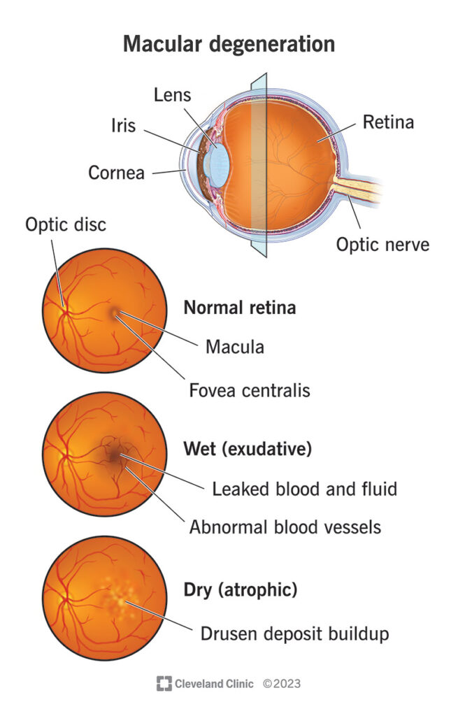
Macular degeneration is a condition in which the macula becomes thinner or fades away, leading to blurry vision. There are two primary forms of macular degeneration – dry and wet macular degeneration; dry is more prevalent while wet is less prevalent but more severe as abnormal blood vessels form and leak into its area of the macula resulting in rapid vision loss without treatment.
Retinal cameras can help to detect changes to the macula. Your eye doctor may also inject harmless orange-red dye into a vein in your arm, with special cameras taking photos as it travels through blood vessels to reach your retina.
Macular degeneration cannot be reversed, but there are steps you can take to reduce further vision loss. Your eye doctor can suggest nutritional supplements like vitamin C, E, lutein and zeaxanthin as well as encouraging regular physical exercise, healthy diet and eye exams.
Normal-tension glaucoma
Primary open-angle glaucoma is the most prevalent form of glaucoma. This occurs when drainage canals become blocked, leading to an increase in pressure within the eye that builds up over time and damages optic nerves, ultimately leading to vision loss. Most people don’t initially experience symptoms; if left untreated it could eventually cause tunnel vision and eventually blindness if left untreated; primary treatments include medication and regular eye exams.
Normal-tension, angle-closure and pigmentary glaucomas are among the various types of glaucoma. Pigmentary glaucoma occurs when an obstruction to eye drainage becomes caused by pigment which prevents the flow of aqueous fluid. Angle-closure glaucoma is especially risky since it can suddenly arise and rapidly cause vision loss – its symptoms include sudden increases in eye pressure, blurred vision, nausea and headaches.
Normal-tension glaucoma (NTG) does not involve elevated intraocular pressure; however, its symptoms resemble those of primary open-angle glaucoma in that they include progressive optic disc cupping and visual field loss – yet without an increase in IOP that characterizes POAG patients. Some experts have speculated that NTG represents either a subvariant of POAG or even a separate disease entity altogether while others suggest it’s simply an indicator of increased risk for developing this form of eye disease.
People diagnosed with normal-tension glaucoma often display nonvisible optic disc drusen (ODD), which cannot be seen with dilaated pupils and only can be detected during an eye exam. Researchers have also linked NTG with abnormalities related to arterial hypotension and reduced blood flow to the optic nerve head.
Glaucoma treatment aims at lowering eye pressure in order to protect the optic nerve and can be accomplished via medications, laser surgery or conventional surgery. Medication treatments typically consist of pills or eye drops designed to lower pressure by either decreasing fluid production or increasing drainage rates of excess fluid from your system. When choosing treatment it’s essential to inform your optometrist of any additional drugs you take as these could interfere with existing glaucoma treatments.
Secondary glaucoma
Healthy eyes contain a fluid called aqueous humor which regulates its internal pressure, keeping the pressure constant and protecting the optic nerve from damage. When this fluid doesn’t drain correctly, eye pressure increases rapidly causing irreparable optic nerve damage that could eventually lead to blindness if left untreated.
Glaucoma comes in many forms, all leading to reduced clarity and quality of vision. Open-angle glaucoma is the most prevalent type of this condition; drainage or circulation of aqueous fluid within the eye may become restricted and increased pressure causes optic nerve damage as well as vision loss.
Normal-tension glaucoma, in which no apparent problems exist with aqueous fluid flow but nonetheless damage is done to the optic nerve, is the second most prevalent form of glaucoma and more likely found among African American and Hispanic patients.
Wet macular degeneration is a type of glaucoma in which abnormal blood vessels form underneath the retina and leak, disrupting central vision. If you have wet macular degeneration, medication will likely be prescribed to slow its progression by stopping abnormal vessel growth and preventing leaks; additionally, you could take supplements containing lutein, zeaxanthin, zinc or copper to lower your risk.
Glaucoma can typically be treated successfully using either medication or surgery. Medication may help control eye pressure by restricting production of aqueous humor or increasing its outflow from the eye; surgery typically creates an outlet for this fluid by cutting or damaging tissues surrounding either your iris or cornea, depending on which form of glaucoma is diagnosed.
Laser therapy may also be effective. This procedure uses laser light to damage cells within the ciliary body that produce aqueous humor, speeding and improving its flow through your eye, which in turn lowers eye pressure.
Congenital glaucoma
Genetic mutation of CYP1B1 causes blockage of the trabecular meshwork and increased intraocular pressure, often only present in one eye but sometimes both eyes can be affected. In most cases, only peripheral vision loss and generalized haze that blurs sharp edges and contrast in visual field occur as symptoms; an enlarged optic disc with cupping; ocular enlargement; corneal edema; Haab striae, and axial length anomalies may also occur as further symptoms.
Congenital glaucoma differs from open-angle glaucoma in that there are no initial symptoms, yet is still progressive disorder with gradual loss of side vision unless IOP levels are controlled; untreated, it will ultimately lead to total blindness. Treatment options include medications but the drug of choice for managing IOP is typically steroids which lower pressure while not protecting or protecting further damage to optic nerve.
Glaucoma should be detected early as it can be difficult to diagnose in young children whose eyes are still growing. Children often cannot communicate their symptoms to parents and doctors and therefore no concerns are identified by either party. Parents should ensure their child visits a pediatrician regularly as well as consulting a specialist if glaucoma is suspected in children.
Research into eye pressure reduction also includes work that protects and preserves the optic nerve. Medication to restore lost functionality to it are being developed; regular dilated eye exams can also help slow or stop progression of this condition.
To best prevent glaucoma, it’s wise to visit your family doctor regularly for eye exams that include dilated fundus examinations and regular check-ups with eye drops. This is particularly important if family history includes people who have suffered from it. Aside from glaucoma, other eye diseases which can be detected early through dilation exams include:


