

Protecting Your Vision with Cutting-Edge AI
The condition of our eyes is a critical factor that is frequently disregarded in the field of managing diabetes. If untreated, diabetic retinopathy, a disorder that affects the eyes and progresses over time due to persistently high blood sugar levels, can cause vision loss and perhaps blindness. Fortunately, early detection and treatments of diabetic retinopathy are now more effective than ever thanks to tremendous advancements in medical technology. We go into the subject of diabetic retinopathy screening in this extensive guide, looking at its importance, the screening procedure, eligibility requirements, treatment options, and more.
What is diabetic retinopathy?
People with diabetes are susceptible to the dangerous eye disorder known as diabetic retinopathy. It happens when persistently high blood sugar levels damage the blood vessels in the retina, the light-sensitive tissue in the back of the eye. As the situation worsens, these damaged blood vessels may leak, causing the retina to enlarge and aberrant blood vessels to form. This might eventually cause vision loss.
Risk Factors, Causes, and Symptoms
Prolonged hyperglycemia (high blood sugar) is the main cause of diabetic retinopathy. High cholesterol and blood pressure are other factors that can make the problem worse. Regular screenings are important since early signs of diabetic retinopathy may not be seen. People may get impaired vision, floaters, difficulties seeing at night, and even total vision loss as the condition worsens.
Diabetic retinopathy develops and worsens as a result of numerous risk factors. These include smoking, prolonged diabetes, ineffective blood sugar management, high blood pressure, high cholesterol, and gestational diabetes. Knowing these risk factors can empower people to control their diabetes and lower their risk of developing diabetic retinopathy.
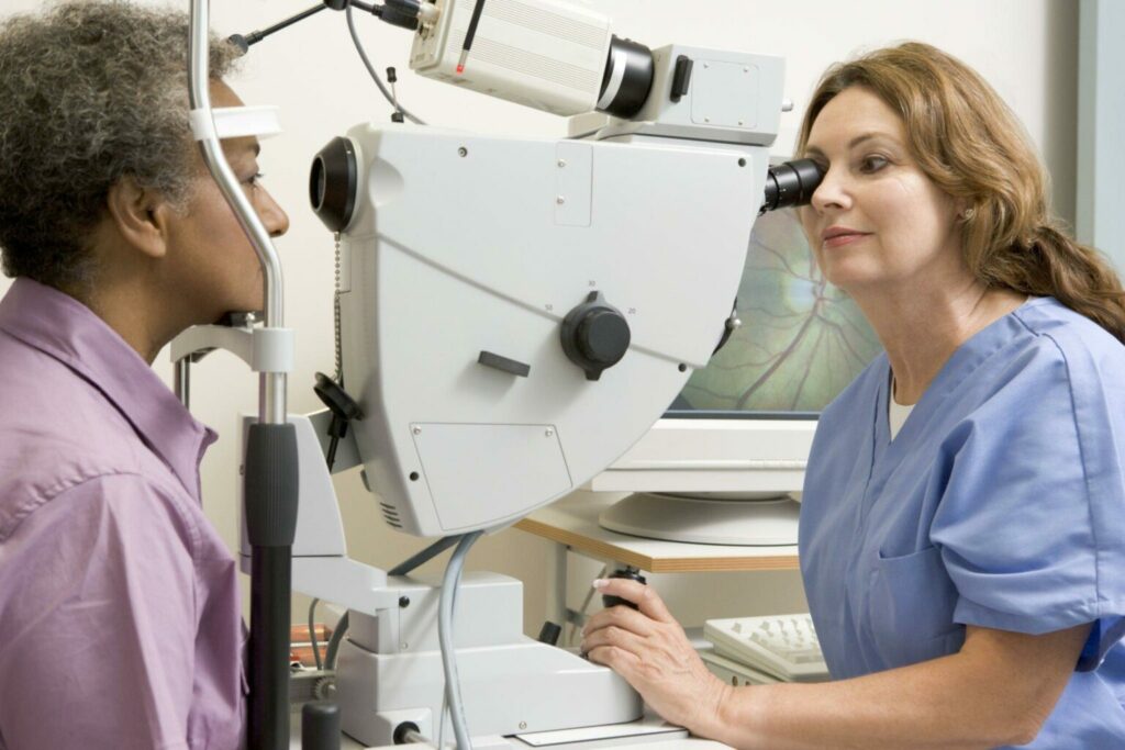
The screening Process
Screenings for diabetic retinopathy are important for the early identification and mitigation of vision loss. A thorough eye exam is often part of the screening procedure, during which an eye care specialist dilates the pupils to look for damage to the retina. Any abnormal blood vessels, retinal edema, or other diabetic retinopathy symptoms can be found during this examination.
Additional tests for screening for Diabetic Retinopathy
Several more tests and procedures may be used in addition to the dilated eye exam to evaluate the effects of diabetic retinopathy on your eyes. These tests can offer a more thorough assessment of the problem and assist in choosing the best course of action. The following are some additional examinations and techniques that could be carried out:
Fluorescein Angiography
For this test, a specific dye called fluorescein is injected into an arm vein. Your retina’s blood vessels are visited by the dye, and as it passes through them, a number of pictures are captured. This test aids in locating regions where blood vessels are dilated or otherwise aberrant, offering crucial information for treatment selection.
OCT
Optical coherence tomography is a non-invasive imaging procedure that uses light waves to produce detailed cross-sectional images of the retina. It enables eye care specialists to see the retina’s layers and spot any swelling or abnormalities. Macular edema, a frequent consequence of diabetic retinopathy, can be found using optical coherence tomography (OCT).
Color fundus photography
Using this imaging method, the retina is captured in fine detail in color. By offering distinct pictures of the retina’s blood vessels, hemorrhages, and other changes over time, it aids in documenting the development of diabetic retinopathy.
Visual Acuity
This test evaluates your ability to see clearly at different distances. It entails using an eye chart to read letters or numbers. Testing your visual acuity is crucial for determining how well your eyes are working and whether diabetic retinopathy has impaired your vision.
Tonometry
This examination measures intraocular pressure, or the pressure inside your eyes. Another eye problem that diabetics are more likely to acquire is glaucoma, which can be identified by elevated intraocular pressure.
ERG
An electroretinogram (ERG) is a test that gauges how electrically active the retina is in reaction to light. This test offers important information regarding the severity of retinal injury by evaluating the health and functionality of the retinal cells.
Ultrasound
The retina and its surroundings may occasionally be viewed via ultrasound imaging. This is especially helpful when bleeding or other abnormalities obscure the retina’s view.
Widefield Retinal Imaging
This cutting-edge imaging method offers a broad view of the retina, enabling the detection of peripheral alterations and abnormalities that might not be detectable with conventional imaging techniques.
It’s important to remember that not every person with diabetic retinopathy may require all of these tests. Various elements, including the severity of the condition, the existence of particular symptoms, and the advice of your eye care specialist, will influence the selection of tests and operations. You may assist in ensuring that your eyes are adequately examined and controlled by maintaining regular communication with your medical team and adhering to advised screening schedules.
Diabetic Retinopathy Screening is Being Revolutionized by AI
The landscape of diabetic retinopathy screening has changed recently as a result of technological developments, particularly in the area of artificial intelligence (AI). Artificial intelligence (AI)-enabled algorithms can examine retinal scans and find minute variations that might point to the presence of diabetic retinopathy. With the use of this technology, screening procedures are now much more accurate and efficient, allowing for early diagnosis and intervention.
What part does AI play in any of these tests?
AI (Artificial Intelligence) is becoming more and more important in some of the tests and methods used in the diagnosis and screening of diabetic retinopathy. Here are some examples of how AI is being used in these tests:
OCT Analysis
The imaging method known as optical coherence tomography (OCT) produces cross-sectional images of the retina. These images can be examined by AI algorithms to spot minute variations in the thickness of the retina, spot areas of fluid buildup (edema), and determine the degree of retinal damage. The accuracy and effectiveness of diagnosing diabetic macular edema, a frequent consequence of diabetic retinopathy, are improved by AI-powered OCT analysis.
Automated Grading Systems
In certain screening systems, retinal pictures obtained during routine screenings are automatically graded using AI algorithms. Based on the presence of distinctive features such as microaneurysms, hemorrhages, and exudates, these algorithms can distinguish between different phases of diabetic retinopathy. This automated grading procedure speeds up the evaluation process and makes sure that early detection of possible diabetic retinopathy patients is achieved.
Fluorescein Angiography Analysis
AI may be able to help with fluorescein angiography image analysis. Artificial intelligence (AI) systems can support ophthalmologists in identifying regions of concern and directing treatment choices by detecting aberrant blood vessel development, leakage, and other alterations.
AI can be used to improve the quality of retinal scans, making tiny abnormalities more visible and facilitating correct diagnosis. Artificial intelligence-powered image enhancement methods can change the brightness, contrast, and sharpness to make details visible that might not otherwise be visible.
Visual field test data can be analyzed using AI algorithms to find patterns of vision loss that are suggestive of diabetic retinopathy. This can make it easier to track how the problem is developing and how well therapies are working.
Telemedicine Screening
Using image-based analysis, telemedicine platforms with AI can help find possible instances of diabetic retinopathy. Patients can use smartphones or portable fundus cameras to capture retinal images, and AI algorithms evaluate these images to determine whether additional evaluation by an eye care professional is necessary.
AI algorithms may assess a patient’s medical background, lab findings, and other data to forecast the likelihood of developing diabetic retinopathy and the course of the condition. Individualized screening plans and actions can be influenced by this information.
One should keep in mind that these algorithms are intended to support healthcare professionals rather than replace them, even though AI has shown promising outcomes in helping with the analysis and interpretation of various tests. Making accurate diagnoses, evaluating results in the context of the patient’s overall health, and selecting the best course of therapy still require human knowledge.
AI has a lot of potential to help with early detection, diagnostic accuracy, and ultimately vision preservation in diabetic retinopathy screening. We may anticipate future developments in the application of AI across various facets of eye care as technology continues to grow, which will be advantageous to both patients and healthcare professionals.
Availability and Eligibility
The kind of diabetes, how long it has been present, and the patient’s general eye health all have a role in how frequently diabetic retinopathy screenings should be performed. People with Type 1 diabetes should typically have their initial screening within five years of diagnosis, however, people with Type 2 diabetes should get screened right away. Annual screenings after that should take place.
The presence of diabetes is the main factor in determining eligibility for screening for diabetic retinopathy. Anyone with diabetes is at risk of getting diabetic retinopathy, so it’s important to get frequent eye exams to keep track of their eye health. Those who have gestational diabetes while pregnant might also need to be screened.
Gold Standard Tests and Screening Authorities
Ophthalmologists or optometrists, specialized eye care providers trained to recognize and manage a variety of eye problems, frequently conduct diabetic retinopathy tests. To achieve precise and thorough assessments of the retina, these experts use cutting-edge instruments and technology.
The dilated eye examination is the gold standard technique for detecting and tracking diabetic retinopathy. Eye drops are used during this operation to make the pupils bigger so that the retina’s blood vessels and other features can be seen in great detail. The most reliable way for identifying early indicators of diabetic retinopathy is still a dilated eye exam.
Options for Treatment and Prevention
The window of opportunity for a variety of therapeutic choices that can delay or even prevent vision loss is opened by the early identification of diabetic retinopathy. These alternatives include surgery in more severe cases, medicines to reduce swelling, and laser therapy to seal leaking blood vessels. The best course of action is still prevention, though.
The risk of developing diabetic retinopathy can be considerably reduced by managing diabetes through lifestyle changes, frequent exercise, eating a balanced diet, monitoring blood sugar levels, and taking prescribed medications as directed. Additionally, maintaining healthy blood pressure and cholesterol levels can help to protect one’s vision.
Conclusion
Diabetic retinopathy screening is a lifeline for preserving your priceless gift of sight, not only a preventive measure. We have the means to fight this potentially blinding disorder through routine tests, early identification, and technological developments like AI-powered analysis. People with diabetes may take charge of their eye health and ensure a clearer, brighter future by appreciating the value of screenings, being aware of the prerequisites, and staying up to date on the most recent developments.
FAQs
What is the diabetic retinopathy screening test?
A thorough eye exam, which involves pupil dilation so that eye care specialists can inspect the retina for signs of damage or abnormalities, serves as the main screening test for diabetic retinopathy.
How frequently are diabetic retinopathies screened?
Factors including the type and severity of diabetes will affect how frequently testing are conducted. For those with Type 1 diabetes, the initial screening should take place within five years after diagnosis, and for those with Type 2 diabetes, the first screening should take place at the time of diagnosis and be followed by yearly screenings.
Who is eligible for a screening for diabetic eye disease?
Diabetic retinopathy screening is open to anybody with a diagnosis of diabetes, Type 1 or Type 2. Those who have gestational diabetes while pregnant might also need to be screened.
Who performs diabetic retinopathy screenings?
Eye care experts who specialize in recognizing and treating various eye disorders, such as ophthalmologists and optometrists, frequently conduct diabetic retinopathy screenings.
What is the best way to diagnose diabetic retinopathy?
A dilated eye exam is the best way to diagnose diabetic retinopathy. This includes using eye drops to make the pupils bigger so that the retina’s blood vessels and other features may be thoroughly inspected.
Can diabetic retinopathy be avoided?
While diabetic retinopathy cannot always be totally avoided, it can be greatly decreased by treating diabetes with a healthy lifestyle, regular exercise, and adequate medication adherence. Controlling cholesterol and blood pressure is also essential for prevention.
How has AI affected the screening for diabetic retinopathy?
AI technology has improved the efficiency and accuracy of diabetic retinopathy screening. Artificial intelligence (AI)-powered algorithms can examine retinal images for minute changes enabling early detection and treatment, ultimately protecting vision and improving patient outcomes.


