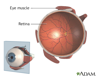


Macular degeneration occurs when the central part of your retina (known as the macula) deteriorates and results in blurry central vision, often with no symptoms initially appearing. Diagnosing macular degeneration may be difficult; as its symptoms may not manifest immediately.
NYU Langone ophthalmologists utilize both visual and imaging tests to detect early signs of macular degeneration. Furthermore, these exams can determine whether you have wet form of macular degeneration which is more serious than dry type macular degeneration.
Drusen
Drusen are yellowish deposits found under the retina, the layer that processes light. Anyone with drusen may be at increased risk for wet macular degeneration, which can lead to permanent vision loss. Wet AMD occurs when drusen block the flow of nutrients that feed blood vessels that leak or burst. When diagnosed, treatment can significantly slow its progress.
Most adults over the age of 50 typically have small drusen scattered throughout the macula that don’t lead to noticeable changes in vision. When multiple large drusen appear clustered together and visible on an Amsler grid, however, it could be an indicator of dry macular degeneration.
An ophthalmologist can inspect someone’s eyes for drusen by performing a dilated eye exam, which involves placing drops into their eye to dilate their pupil and allow for better visibility into their eye. Once inside, they can search for yellowish-white spots known as drusen. In addition, an Amsler grid-style checkerboard test may be conducted in order to assess central vision; any lines appearing wavy or curved could indicate wet AMD.
If a person with early-stage dry AMD does not exhibit symptoms from their drusen, an ophthalmologist can monitor his or her progress by regularly taking SD-OCT images of both retina and choroid layers that show any drusen, pigment changes, or any areas of geographic atrophy. These images allow professionals to detect these areas early enough.
At its heart lies noninvasive imaging using infrared light waves to produce cross-section photographs of retina and choroid using infrared lightwaves. These photos can provide important details regarding drusen, retinal structure, new blood vessels, hemorrhage and hemorrhage that an ophthalmologist can use to recommend a course of action to a patient with large drusen in the center of macula; for instance they may recommend laser treatments in order to prevent further damage in that area of macular area.
Amsler grid
An Amsler grid is used by doctors to monitor changes to central vision caused by macular degeneration, using thin black lines arranged in a grid pattern. If grid lines appear wavy or distorted, this may indicate progression from dry to wet macular degeneration and should be reported immediately. Patients are recommended to check this grid regularly and report any significant changes to their eye care provider immediately.
This test should not replace an eye exam conducted by an ophthalmologist or retina specialist, but it can detect changes to vision earlier so they can be treated quickly. People over 50 with family histories of macular degeneration are at higher risk of macular degeneration while smoking and cardiovascular disease also increase this risk.
A doctor will begin by dilation drops and using a special instrument to examine the retina closely, using special lenses known as biomicroscopes and magnifiers to view it more closely. They’ll look out for yellow deposits beneath the retina known as drusen that indicate macular degeneration; furthermore they will test central vision with Amsler grid – a set of straight lines like checkerboard with missing or irregular lines indicating macular degeneration if these occur wavily, crookedly or completely; otherwise.
To take an Amsler grid test, ensure you’re in a well-lit room and seated comfortably. Wear glasses as usual (if applicable). Focus on the center dot of the grid to detect distortions or waviness and repeat with each eye; don’t move either your head or eyes while taking this test; note any significant changes in appearance of grid. Texas Retina Associates offer this tool for early identification of macular degeneration.
Optical coherence tomography (OCT)
Optic coherence tomography (OCT) is an imaging test which uses light waves to scan retinal tissue and produce high-resolution cross-sectional images of the eye, without sound or radiation exposure. OCT scans are noninvasive procedures used for diagnosing macular degeneration, glaucoma and diabetic retinopathy as well as helping detect any changes or detect changes within eyesight itself – it provides valuable data that ophthalmologists use when making decisions regarding ongoing treatments for their patients.
This non-invasive test uses an invisible beam of light to enter your eye, measuring its reflection back out and creating an accurate picture of its structures, such as optic nerves, maculae, choroides and retina. Our doctors can see detailed images which allow them to detect areas affected by fluid build-up or structural damage and pinpoint them quickly and precisely.
OCT can be used to detect macular holes or retinal edema. Additionally, OCT can help our doctors decide the best course of treatment for you.
Your chin will be supported on a support while you look at a green target on the screen, then a laser scans your eyes painlessly in about 15 minutes.
OCT imaging shows characteristic features of macular holes such as a narrow and vertical central pit, hyperreflective band at the base of retinal break and wide area of peripheral attenuation (darkness). Other signs can also indicate retinal holes such as thin hyporeflective band or gaps between epiretinal membrane and inner retina.
OCT imaging technology can also be used to detect eye fluid build-up, including vitreous haemorrhage and subretinal bleeding, which can lead to macular edema with blurry vision, as well as cystic detachments (characterized by convex protrusion of posterior vitreous cavity, which must be distinguished from tractional detachments which have peaked configuration). OCT can help identify these conditions before they lead to macular edema with macular edema with blurry vision; OCT also helps detect other eye problems such as cystic detachments that indicate other issues, including cystic detachments (which can lead to macular edema), macular detachments (which lead to blurred vision) as well as eye conditions such as cystic detachments which is associated with macular edema with blurry vision), which leads to macular edema which causes blurry vision; cystic detachments are usually identified as being differentiated from their counterpart tractional detachments which typically exhibit peaked configuration.
Fluorescein angiography
This test uses an orange dye and rapid sequence photography to assess blood flow, vessels, and tissues in the retina and choroid (the pigmented layer behind the retina). Your doctor will inject harmless orange-colored dye into one of your arm veins; it travels rapidly through all blood vessels including those found within your eye before being photographed using special filters on a camera. Although painless, vision may become temporarily blurry due to your pupils dilation during this procedure; you may require someone to drive you home afterwards as your vision may remain blurry until normalization returns in 24 hours time.
Fluorescein angiography helps doctors detect circulation problems, swelling, leaks or abnormalities in retinal blood vessels that could compromise circulation or lead to macular degeneration. Furthermore, this technique reveals if fluid has collected in the central macula or if an epiretinal membrane has covered and compressed retinal blood vessels.
Additionally, this test can determine if your macular degeneration is related to diabetes or another systemic disease affecting the retina. Furthermore, it can reveal any choroidal neovasculation, which could indicate leaky blood vessels from macular holes or hemorrhages that leaking blood vessels from under your retinal surface are present.
Ultra-widefield fluorescein angiography provides a more detailed look at the blood vessels behind the eye, revealing more leakages in vessels than regular fluorescein angiography and also showing other indicators of macular degeneration such as irregular vessel tortuosity and dilation of eye vessels.
Both tests can be repeated multiple times without any negative impact to the patient, though risks of an allergic reaction to dye or discomfort during testing are very minimal. Your eyes may become sensitive to light for up to 12 hours post-procedure; to stay safe and avoid bright lights during this period. It is advisable that someone drive you home.


