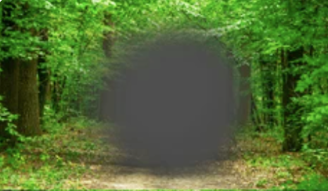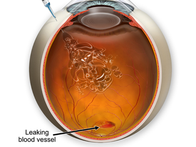
About 10% of people living with dry age-related macular degeneration progress to wet form (also known as exudative AMD). With wet AMD, abnormal blood vessels grow under the retina and macula and leak fluid into them, distorting central vision or creating blind spots.
Medical treatments may help slow the progression of wet macular degeneration and preserve your vision, including medications to block Vascular Endothelial Growth Factor, which promotes leaky blood vessels in wet AMD.
Anti-VEGF Injections
At present, one of the most effective treatments for wet age-related macular degeneration – also known as Neovascular AMD – is injections of anti-VEGF drugs. These medications block vascular endothelial growth factor protein that plays a major role in blood vessel formation both under retina and elsewhere in body. By inhibiting it, these anti-VEGF medications prevent abnormal blood vessels that lead to fluid leakage and permanent vision loss from forming and hinder their formation by blocking it effectively.
Neovascular macular degeneration was once an incurable blinding disease; with modern treatments it has become manageable, with most patients only suffering mild to moderate vision loss. Unfortunately, however, these drugs don’t treat the root cause and many will require regular injections throughout their lives in order to stop further vision loss.
Most patients using anti-VEGF drugs require monthly injections in order to maintain stable, functional vision. A typical treatment session lasts only seconds; your eye doctor will use numbing drops on your lower lid before injecting medication into your clear jelly-like substance inside of your eye (vitreous) through injection through white part of eye (sclera). Most patients find the process relatively painless; you may feel some gritty or irritated sensation that usually subsides within 24 hours.
Anti-VEGF therapy may lead to the growth of new blood vessels within the eye, known as choroidal neovascularization. While this can be desirable, its management can be challenging as new vessels may leak or hemorrhage and result in visual loss. Therefore, any patients receiving this therapy must undergo careful evaluation with fluorescein angiography and retinal thickness measurements prior to receiving therapy.
Studies suggest that long-term use of anti-VEGF therapy could contribute to geographic atrophy in some patients with neovascular AMD and dry macular degeneration, prompting drug manufacturers to create medications with altered binding affinity, extended longevity, as well as sustained delivery systems that eliminate frequent injections. This has spurred new drug innovations with enhanced binding affinity that are both long-acting and require fewer frequent injections.
Fluorescein Angiography
Under wet age-related macular degeneration, abnormal blood vessels develop and begin leaking fluid under the retina and macula, swelling this area and leading to distortion or blurring of central vision. Any change to central vision should prompt an appointment at our office for a dilated eye exam and immediate treatment, or it could quickly result in rapid and irreversible vision loss. Wet AMD is driven by a chemical called vascular endothelial growth factor (VEGF). There have been medications developed that block this chemical and help control abnormal blood vessel growth in the eye. These medications are administered via small needles with self-sealing entry sites to create anti-VEGF injections, commonly referred to as Eylea, Bevacizumab (Avastin) or Brolucizumab (Beovu) treatments; another drug called Ranibizumab (Lucentis) not only blocks VEGF but also inhibits another protein involved with blood vessel formation.
These treatments can significantly slow the rate of progression of wet macular degeneration and may even improve quality of vision, but are typically expensive and aren’t covered by Medicare or many private insurance plans. That’s why we provide financial assistance programs to make treatment more cost-effective.
Wet AMD follows the same process as dry macular degeneration, but abnormal blood vessels lead to fluid leakage and permanent vision loss in most cases. It is the leading cause of irreversible legal blindness among adults over 55 in the US. It often develops faster and worse than its dry counterpart.
Effective wet AMD treatments consist of immediately treating any signs of fluid leakage and ocular hemorrhage detected through fundus photography or an Amsler grid. An eye doctor may then administer fluorescent dye injections into the bloodstream before taking pictures with sequenced images that highlight any movement from within retinal blood vessels and allow him to visualize abnormalities more clearly.
Optical Coherence Tomography
Optic Coherence Tomography (OCT) is a noninvasive retinal imaging test that offers high-resolution images of the macula and optic nerve head. Our Retina specialists use OCT to visualize changes associated with wet age-related macular degeneration; specifically when small blood vessels form within retinal pigment epithelium (RPE), which supports light-sensitive photoreceptor cells which translate light into sight for interpretation by brain; over time these photoreceptor cells begin breaking down and degenerating, leading to reduced vision deterioration or difficulty performing activities such as driving or reading.
OCTA scans reveal the microscopic structure of the retina and detect abnormalities such as fluid accumulation. This is an early sign of neovascularization that could eventually lead to macular edema or distortion, providing valuable data about wet AMD treatment. Used together with FA, this imaging technique offers valuable tools for monitoring and managing wet AMD.
This technique employs near-infrared light to image the retina. It offers high-resolution three-dimensional images of both retinal structures and optic nerve heads with no pain associated with using this noninvasive and painless technology – an invaluable asset when monitoring injection treatments post procedure.
Early models of OCTA utilized a continuous scanning laser to create images of the retina, which proved less precise as its accuracy depended on patient cooperation; for this reason, it’s vital that patients keep their gaze steady. Newer OCTA machines now employ swept-source laser technology which enables faster scan times while eliminating movement artifacts found with earlier systems.
Studies have demonstrated that OCTA can detect choroidal neovascularization and fibrovascular capillary loops. Furthermore, OCTA can determine macular edema’s extent and identify microaneurysms in neovascular AMD cases. Furthermore, this technique can be used to evaluate anti-VEGF treatments by measuring how much fluid remains within an eyeball.
Retina Specialists of Houston are highly experienced ophthalmologists in wet age-related macular degeneration and offer cutting edge treatment options. Contact us for more information or a consultation; early detection of macular degeneration is the key to protecting vision loss and protecting vision from further loss.
Radiofrequency Ablation
Wet age-related macular degeneration, accounting for 10% of cases of AMD, can result in severe loss of central vision. Vision rehabilitation services can assist you with this difficult challenge and help adjust to living with limited sight.
Wet age-related macular degeneration (AMD) is caused by new blood vessels that form under the retina and macula and leak fluid or bleed, known as choroidal neovascularization (CNV). If this condition develops in your central vision it could result in distortion, blurriness or blind spots which require prompt medical treatment before further complications develop. If wet AMD occurs you must seek prompt attention as its progression can progress quickly.
Your eye doctor will perform a comprehensive eye exam and ask about your symptoms. They may also request a list of your medications; aspirin, clopidogrel, and any other blood thinners must be stopped prior to ablation in order to decrease risks of bleeding and other side effects from this process.
People who smoke have an increased risk of wet AMD than nonsmokers and tend to experience more central vision loss from it. To help combat wet macular degeneration, you should quit smoking, maintain a balanced diet and participate in regular physical activity; use an Amsler grid to monitor your vision regularly before discussing results with an eye care provider.
Eye care specialists will administer an injection of yellow dye into a vein in your arm, which will highlight your retina’s delicate vascular network. A certified ophthalmic angiographer will then take time-dependent photographs of your retina showing where leaky blood vessels exist – this allows our doctors to target these areas for treatment.
Wet macular degeneration can be devastating, yet you may be able to slow its progress with anti-VEGF medicines such as Eylea (Aflibercept), Bevacizumab (Avastin) and Ranibizumab (Lucentis). Talk with your eye doctor about whether these may help your vision and ask if any are suitable.