
Medically reviewed by Shaun Larsen, OD, Doctor of Optometry, Southern College of Optometry.
Last clinically reviewed: July 2026 | Next scheduled review: July 2027
In brief: the optic cup is the pale hollow at the centre of the optic nerve head, and everyone has one. Its size depends heavily on how large your optic disc is, so a big cup is often entirely normal anatomy rather than disease. What distinguishes glaucomatous cupping from physiologic cupping is not size but shape, rim pattern, symmetry between the eyes, and whether it changes over time. Cupping caused by glaucoma reflects permanent loss of nerve fibres and the visual field lost with it does not return, although the cup itself can partially reshape after a large pressure reduction, most markedly in children.
One of our most valuable senses, vision, allows us to see the environment around us. The transmission of visual data from the eye to the brain by the optic nerve is crucial to this process. However, cupping of the optic nerve can cause a number of visual problems. We will go further into the world of optic nerve cupping in this complete guide, learning about its causes, symptoms, diagnosis, available treatments, and connection to glaucoma. We will also clarify how low vision services can significantly improve eyesight for folks who are impacted by optic nerve cupping.
Understanding Optic Nerve Cupping
What the cup actually is
Roughly 1.2 million retinal ganglion cell axons converge at the optic nerve head and turn backwards out of the eye through an opening in the sclera called the scleral canal, passing through a sieve-like collagen plate called the lamina cribrosa.
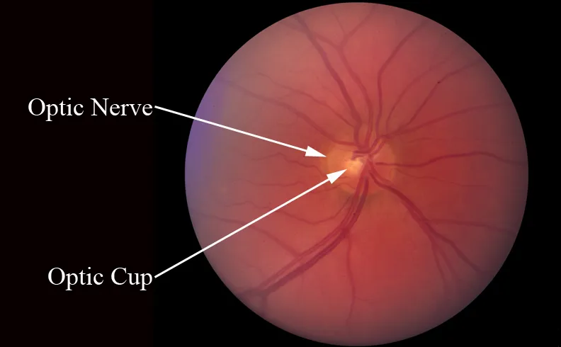
Looking at the disc through an ophthalmoscope, two zones are visible. The neuroretinal rim is the ring of orange-pink tissue at the edge, made of the nerve fibres themselves. The cup is the paler, slightly hollowed centre where fewer fibres lie and the underlying lamina is closer to the surface. The cup-to-disc ratio simply expresses the diameter of the cup as a fraction of the diameter of the whole disc, usually quoted vertically.
Everyone has a cup. It is a normal anatomical feature, not a finding in itself. What matters is whether it is proportionate to the disc, correctly shaped, symmetric with the other eye, and stable over time.
Causes of Optic Nerve Cupping
A structural alteration to the optic nerve head is referred to as optic nerve cupping, also known as cupping of the optic nerve. The optic nerve becomes cupped or indented as a result of this alteration, which may impair vision. Elevated intraocular pressure, which over time may harm the optic nerve, is the major cause of optic nerve cupping. This is a defining characteristic of the dangerous eye ailment glaucoma.
Two qualifications matter here. First, raised pressure is the principal modifiable risk factor rather than a requirement: normal-tension glaucoma produces identical cupping at pressures that never leave the normal range. Second, the largest single cause of a large cup is not disease at all, but simply having a large optic disc, which is discussed in the next section.
How glaucomatous cupping develops. Retinal ganglion cell axons are lost progressively, so the neuroretinal rim thins and the cup enlarges to occupy the space. Alongside this, the lamina cribrosa bows backwards and remodels under mechanical stress. The two processes together produce the excavated appearance. The important consequence is that the cup enlarges because tissue has been lost, not because tissue has been compressed, which is why the visual field does not recover.
Is Optic Nerve Cupping Normal?
Optic nerve cupping is not always an indication of glaucoma, despite the fact that this condition might sometimes be indicated by it. Without getting glaucoma, some people may have a bigger cupping of the optic nerve. This is referred to as “physiologic cupping,” and it is regarded as a typical variation in the structure of the optic nerve. But it’s important to distinguish between physiologic cupping and glaucoma-related cupping.
The key relationship: cup size scales with disc size
This is the concept that makes physiologic cupping comprehensible, and its absence is the biggest gap in the current page.
The amount of nerve tissue crossing the disc is broadly similar between healthy eyes, because axon counts are similar. The size of the opening those axons pass through is not: optic disc area in normal eyes ranges from roughly 0.8 mm² to about 6.0 mm², a severalfold spread. When a similar quantity of nerve fibre passes through a wider canal, it does not fill it, and the space left over is the cup.
Large disc leads to large physiologic cup leads to a high cup-to-disc ratio that is entirely normal. Ratios in normal populations extend from 0.0 to approximately 0.9. A vertical ratio of 0.7 in a genuinely large disc can be completely healthy.
Small disc leads to small cup, which can conceal real glaucomatous damage. A ratio of 0.4 in a small crowded disc may already represent significant rim loss.
The documented consequence is an asymmetry in diagnostic error: clinicians tend to over-diagnose glaucoma in large discs and under-diagnose it in small ones. Disc size also varies by ancestry, being on average largest in people of African ancestry and smallest in those of European ancestry, and larger in high myopia.
The practical question to ask at your appointment is therefore not "what is my cup-to-disc ratio" but "is my disc large, average or small, and is my nerve fibre layer normal for that disc size?" The ratio means very little without the first answer.
Optic Nerve Cupping Without Glaucoma
It’s significant to remember that glaucoma isn’t always a factor in situations of optic nerve cupping. Optic nerve cupping may also be influenced by additional factors, such as myopia (nearsightedness) or inherited factors. Even though these conditions might not eventually develop into glaucoma, they nevertheless need to be monitored by an eye care specialist.
Non-glaucomatous causes worth knowing about, because some require completely different investigation:
Physiologic cupping in a large disc. By far the most common explanation, and not a disease.
Compressive lesions of the anterior visual pathway, including pituitary adenoma, meningioma and aneurysm. These can mimic glaucomatous cupping and are the reason the red flags below matter.
Hereditary optic neuropathies, such as Leber hereditary optic neuropathy and dominant optic atrophy.
Prior ischaemic or inflammatory injury, including arteritic anterior ischaemic optic neuropathy and past optic neuritis.
Toxic and nutritional optic neuropathy, and methanol poisoning.
Congenital anomalies including optic disc pit, coloboma, morning glory anomaly and megalopapilla.
Features that should prompt neuro-imaging rather than continued glaucoma monitoring: optic disc pallor out of proportion to the degree of cupping (in glaucoma the remaining rim keeps its normal colour); visual field loss respecting the vertical midline, or bitemporal defects, since glaucoma characteristically respects the horizontal midline; early loss of colour vision or central acuity, both of which glaucoma preserves until late; young age; rapid progression, or progression despite well-controlled pressure; and accompanying headache, other neurological symptoms or endocrine disturbance.
The Spectrum of Optic Nerve Cupping
Mild to severe optic nerve cupping is a spectrum condition. Clinical tests and imaging modalities are frequently used to gauge the degree of cupping. For selecting the best course of action and therapy, determining the degree of cupping is essential.
Normal range: the actual numbers
Because this is the question most readers arrive with, here are the figures rather than a description of the fact that figures exist.
Optic disc area: approximately 0.8 to 6.0 mm² in normal eyes, mean around 2.1 to 2.8 mm². Average vertical diameter about 1.9 mm, horizontal about 1.75 mm (the disc is normally a slightly vertical oval).
Disc size categories: small below about 1.5 mm vertical diameter, large above about 2.2 mm.
Vertical cup-to-disc ratio: mean around 0.4 in normal populations, with the normal range extending from 0.0 to approximately 0.9 depending on disc size. Roughly 2.5% of the healthy population has a ratio above 0.7.
Neuroretinal rim area: approximately 1.97 mm² on average, and it scales with disc area, so larger discs have larger absolute rim area.
Inter-eye asymmetry: a difference of 0.2 or more in vertical cup-to-disc ratio between the two eyes is a recognised warning sign even when both individual values sit in the normal range, unless the two discs genuinely differ in size.
The ISNT rule is the standard rim contour check: in a normal disc the neuroretinal rim is thickest Inferiorly, then Superiorly, then Nasally, then Temporally. Violation of that order is a red flag. It holds less reliably in very large discs and tilted myopic discs, so it supports the assessment rather than deciding it.
What distinguishes glaucomatous from physiologic cupping, stated plainly: physiologic cupping is round or horizontally oval, concentric, symmetric between eyes, has a rim of even healthy colour, and does not change over years. Glaucomatous cupping enlarges preferentially in the vertical meridian, notches the rim focally at the inferotemporal and superotemporal poles, produces a rim that is pale as well as thin, may show baring or bayoneting of vessels and acquired peripapillary atrophy, and above all progresses.
A disc hemorrhage at the rim margin (Drance hemorrhage) is one of the strongest single predictors of progression and should never be dismissed as incidental.
Recognizing the Signs and Symptoms
The most important thing to say about symptoms is that there usually are none. Glaucomatous cupping develops silently. The early field loss is mid-peripheral, where the fellow eye and the brain’s perceptual filling-in conceal it completely, so patients typically notice nothing until a large proportion of nerve fibres has already been lost. Waiting for symptoms means presenting at the stage where damage is irreversible, and that is why the condition is found by examination rather than by how the eyes feel.
Common Symptoms
Depending on the severity and underlying cause of optic nerve cupping, the symptoms and indicators can change. Typical signs include:
Patients may notice a gradual shrinking of their visual field due to a loss of peripheral vision. This occurs late. Earlier defects are typically arcuate scotomas or a nasal step, which are invisible to the person who has them.
Reduced capacity to perceive in low light circumstances due to difficulty with night vision.
Acute symptoms in angle-closure, which is the exception to the silent rule: severe eye pain, a red eye, haloes around lights, headache, nausea and vomiting, with rapidly blurring vision. This is an emergency requiring same-day care, and permanent damage can occur within hours.
Late-stage functional difficulties once field loss is substantial: bumping into objects and door frames, difficulty judging kerbs and steps, missing items in peripheral view, and problems with night driving and glare recovery.
Diagnostic Methods and Tools
A thorough eye exam is often required to diagnose optic nerve cupping. Eye care specialists employ a variety of instruments and methods, such as:
Ophthalmoscopy: A specialist instrument is used to look at the optic nerve head. In practice, stereoscopic examination at the slit lamp with a 78 D or 90 D condensing lens, which gives the depth perception that direct ophthalmoscopy cannot. Disc photographs provide the baseline against which future examinations are compared, and are one of the most valuable things that can be done at a first visit.
Visual field testing: This determines how much peripheral vision has been lost. Standard automated perimetry. Two baseline tests are usually performed because the first is affected by a learning effect. Key indices are mean deviation (average sensitivity loss), pattern standard deviation (how uneven the loss is, which distinguishes localised glaucomatous damage from diffuse cataract effect) and visual field index.
OCT: A high-resolution imaging technique that offers thorough information on the optic nerve. Specifically it measures peripapillary retinal nerve fibre layer thickness, which is largely independent of disc size and therefore resolves the ambiguity that cup-to-disc ratio creates; macular ganglion cell-inner plexiform layer thickness, which assesses cell bodies and often detects damage earliest; and Bruch’s membrane opening minimum rim width, a parameter developed specifically to account for disc size. Serial scans compute rate of loss per year, which is the number that actually determines management.
Tonometry with pachymetry. Pressure interpreted alongside central corneal thickness, since thin corneas under-read the true pressure and independently raise risk.
Gonioscopy, to determine whether the drainage angle is open or occludable. This drives the entire treatment pathway and cannot be inferred from pressure or disc appearance.
Early identification of optic nerve cupping is crucial because it enables prompt management and intervention.
One caution about OCT reports. The color coding (green, yellow, red) indicates where your measurement falls against a normative database. These are statistical flags, not diagnoses. False positives are common in high myopia, large or tilted discs, and eyes with coexisting macular disease, because such eyes are under-represented in the reference populations. A red flag is a prompt to look at the underlying scan and correlate with the disc and field, not a verdict.
Treatment and Reversal of Optic Nerve Cupping
Can Cupping of the Optic Nerve Be Reversed?
This deserves a careful answer, because the honest position is more nuanced than either "yes" or "no", and the current page attempts both within one paragraph.
The nerve fibres do not come back. Retinal ganglion cells that have died are not replaced, and visual field lost to glaucoma does not return. No current treatment restores it. Any claim otherwise should be treated with scepticism.
But the cup itself can partially reshape. Structural reversal of cupping after substantial pressure reduction is a genuine, documented phenomenon. It is most striking in infants and young children with congenital glaucoma, where the elastic immature eye allows the disc to remodel markedly after successful surgery, and younger age at surgery predicts greater reversal. It has also been documented in adults, typically after large reductions (in the order of 25 to 30% or more) achieved by trabeculectomy, and confirmed with OCT.
What that reversal actually represents, and this is the crucial qualification: it reflects biomechanical and glial remodelling, principally forward movement of the lamina cribrosa and shrinkage of the scleral canal, rather than regrowth of axons. Studies have repeatedly found that cupping reversal is not accompanied by improvement in visual field, and in paediatric cases some eyes showed continued nerve fibre layer thinning despite a visibly reversed cup. It is best understood as a marker that pressure has been reduced effectively, not as recovery.
The practical takeaway: treatment prevents future loss. That is a genuinely valuable outcome, since glaucoma treated early rarely causes disabling vision loss within a lifetime, but it is a different promise from restoration. The title of this article uses the word "reversal", and the article should be explicit that the term refers to disc appearance rather than to recovered sight.
Glaucoma-Related Optic Nerve Cupping
Treatment for glaucoma cases with optic nerve cupping generally focuses on controlling intraocular pressure to stop additional damage. In glaucoma patients, medications, laser therapy, and surgery are frequently used to manage intraocular pressure and delay the advancement of optic nerve cupping.
The options, in the order they are usually considered:
Topical medication. Prostaglandin analogues (increase uveoscleral outflow, once nightly, first-line for most patients), beta-blockers, alpha-2 agonists, carbonic anhydrase inhibitors and Rho-kinase inhibitors.
Selective laser trabeculoplasty (SLT). The LiGHT randomised trial found SLT as initial therapy achieved comparable disease control to drops with a lower rate of subsequent glaucoma surgery, moving it to a reasonable first-line option. It is repeatable, and it removes the adherence problem that undermines drop therapy.
Laser peripheral iridotomy, where the angle is occludable. This addresses a different mechanism and is not interchangeable with the treatments above.
Minimally invasive glaucoma surgery (MIGS). Trabecular micro-bypass stents, canaloplasty, goniotomy and subconjunctival micro-shunts. Lower risk, more modest pressure reduction, often combined with cataract surgery.
Filtration surgery. Trabeculectomy or a glaucoma drainage device, for eyes progressing despite maximal tolerated therapy or presenting with advanced damage.
Target pressure and rate of progression are what management actually turns on. The goal is to set a pressure low enough that the rate of loss will not produce disabling vision loss within the patient’s lifetime, then verify by serial testing that the rate is genuinely controlled. This is why a person aged 45 with early damage and a person aged 85 with the same findings may be treated quite differently.
Adherence is the weak link. Real-world persistence with glaucoma drops is poor, and the disease is silent, so stopping produces no feedback. If cost, side effects or difficulty instilling drops is a barrier, say so, because laser and surgical alternatives exist partly for that reason.
The Link Between Optic Nerve Cupping and Glaucoma
Examining the Relationship
A series of eye conditions known as glaucoma include optic nerve damage and are frequently accompanied by increased intraocular pressure. Glaucoma can be detected by the cupping of the optic nerve, and the two conditions are closely connected. Increased intraocular pressure in glaucoma can cause gradual optic nerve damage and cupping.
To state the relationship precisely, since it is easily misread in both directions: cupping is a sign of glaucoma, not a synonym for it. Most large cups are physiologic. Some glaucoma, particularly in small discs, presents with a cup that does not look large at all. And cupping can arise from causes entirely unrelated to glaucoma, as listed earlier. The diagnosis rests on the combination of disc appearance, nerve fibre layer measurement, visual field and, decisively, change over time.
What happens if you are told you have a large cup
"Glaucoma suspect" is an alarming label for what is frequently normal anatomy, so a realistic account of what follows is useful:
Baseline documentation. Disc photographs or OCT disc imaging, nerve fibre layer and macular ganglion cell scans, pachymetry, gonioscopy, and usually two baseline visual fields.
Risk stratification. Your clinician weighs pressure, corneal thickness, disc size and rim pattern, family history, ancestry, refraction and age. Many people are found to be low risk at this stage.
Interval monitoring, typically every 6 to 12 months, repeating imaging and fields to establish a trend. Because the purpose is to detect change, keeping appointments is the substance of the management rather than a formality.
Treatment only if indicated, meaning when calculated risk is moderate to high or progression is documented. Not simply because a cup looks large.
A stable large cup, unchanged across several years of good-quality imaging, is by definition not glaucoma. Establishing that baseline is why the first two years of monitoring matter most.
Low Vision Services and Optic Nerve Cupping
The quality of life for people with optic nerve cupping is significantly improved by low vision services. These services use specific tools and techniques to maximize a person’s remaining vision. But the type of aid that helps depends on the pattern of vision loss, and this is where generic advice goes wrong.
Glaucoma produces peripheral field loss with preserved central acuity, which is close to the opposite of macular disease. That distinction matters because magnification, the mainstay of macular rehabilitation, is often unhelpful or counterproductive here: enlarging an image spreads it across a field that is already restricted. A person with advanced glaucoma may read 20/25 comfortably and still be unable to cross a road safely.
What actually helps in peripheral field loss:
Orientation and mobility training, which is the single most valuable intervention for constricted fields, covering scanning technique, cane use where appropriate, and safe travel strategies.
Systematic scanning training, teaching deliberate eye and head movements to compensate for the missing field.
Prism systems, including peripheral prisms for hemianopic field loss, which shift awareness of objects from the blind area into the seeing field.
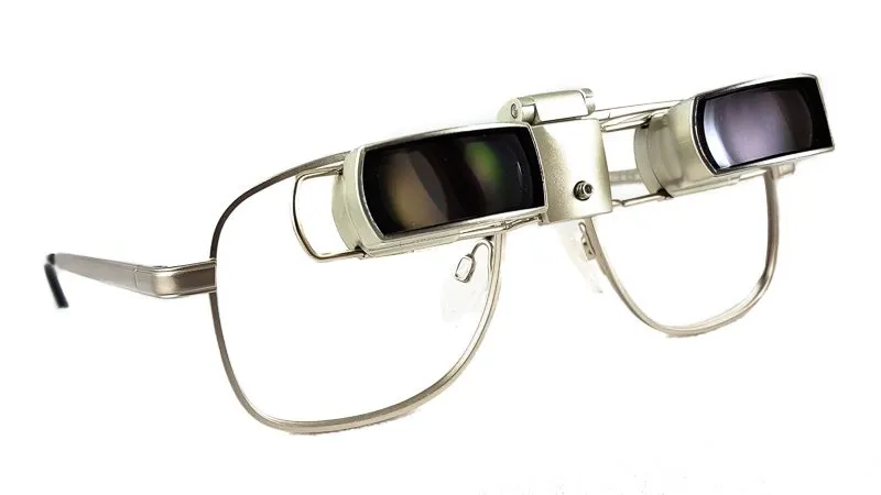
Reverse (minifying) telescopes for severely constricted tunnel vision, which compress a wider scene into the remaining field. Useful for spotting rather than continuous wear, and not suitable for everyone.
Lighting and contrast management. Glaucoma degrades contrast sensitivity and dark adaptation disproportionately, so even task lighting, glare control, and high-contrast marking of steps and edges make a substantial practical difference.
Falls prevention. Field loss and impaired contrast are established risk factors for falls, so home hazard assessment, stair marking and appropriate footwear belong in the plan.
Driving assessment. Field requirements are set independently of acuity in every state, and a driver can fail on field while passing on acuity. This is the usual situation in advanced glaucoma and needs an honest conversation rather than avoidance.
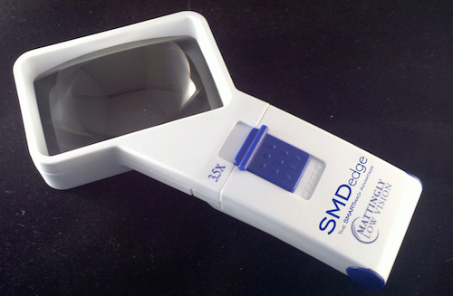
Magnification and electronic aids remain useful where central acuity has also been affected in late disease, or where another condition such as cataract or macular degeneration coexists.
Natural ways to slow optic nerve cupping
Optic nerve cupping or glaucoma cannot be totally prevented or reversed naturally, but certain dietary and lifestyle choices and routines may help limit their growth and preserve general eye health. While speaking with an eye care specialist is necessary for a thorough treatment plan, here are some natural ways to maintain eye health:
An honest framing before the list. No diet, supplement, exercise regimen or stress-reduction technique has been shown in a randomised controlled trial to prevent glaucoma or slow its progression. The only intervention with proven benefit is lowering intraocular pressure. The measures below are worth doing for general health, and a few have a plausible mechanism, but they are additions to prescribed treatment and monitoring, never substitutes. Presenting them as a way to "slow cupping" overstates what they can do.
Routine Exercise:
Regular physical activity can enhance blood flow to the optic nerve and assist control intraocular pressure. Be sure to check with your doctor before beginning a new workout regimen. Regular aerobic exercise does produce a modest measurable reduction in IOP, so this is one of the few items on the list with direct supporting evidence. Two caveats: certain inverted yoga positions transiently raise pressure substantially and are worth avoiding in established glaucoma, and in pigment dispersion syndrome vigorous exercise can trigger pressure spikes.
Don’t Smoke:
Smoking increases the chance of developing glaucoma and can harm the visual nerve. Smoking cessation can be beneficial for eye health.
Observe the prescription instructions:
Take medications as advised by your doctor if you have been diagnosed with glaucoma or another eye ailment. These drugs can aid in regulating intraocular pressure. This is the item that matters most on this list by a wide margin, and it belongs at the top rather than tenth.
Frequently Scheduled Eye Exams:
Plan routine comprehensive eye exams with a qualified eye care expert. To preserve vision, eye diseases must be promptly identified and treated. Because glaucoma is diagnosed and managed by detecting change, attending scheduled reviews is not merely good practice, it is the mechanism by which the disease is controlled.
Avoid unnecessary corticosteroid exposure
A significant minority of people are steroid responders whose pressure rises markedly on corticosteroid exposure, and this applies to tablets, inhalers, nasal sprays, skin creams and eye drops alike. Tell your eye specialist about any steroid use in any form. This is a genuinely modifiable risk factor and it is frequently overlooked.
Discuss blood pressure medication timing
Nocturnal hypotension, where blood pressure dips too low overnight, has been associated with progression in normal-tension glaucoma through reduced optic nerve perfusion. If you take antihypertensives at night, mention the timing to both your physician and your eye specialist.
It’s important to remember that these natural methods are not a replacement for medical care, even though they might improve overall eye health and even halt the advancement of eye diseases. A customized treatment strategy and competent medical attention are necessary for glaucoma and optic nerve cupping. Always seek advice from your eye care professional to choose the best course of action for your unique needs.
Conclusion
The complex eye disease known as optic nerve cupping can significantly impair one’s eyesight. Early detection through routine eye exams is crucial for prompt management and intervention. Optic nerve cupping is not itself a disease: it is a physical sign, and in most people it reflects normal anatomy. Where it is caused by glaucoma, the nerve fibre loss behind it is permanent, treatment prevents further loss rather than restoring what is gone, and the visual field does not recover even in the cases where the cup itself partially reshapes. It is essential to comprehend the origins, signs, diagnosis, and available treatments for optic nerve cupping in order to maintain eye health and preserve eyesight.
Frequently Asked Questions
What does the term “ocular nerve cupping” mean?
The term “cupping of the optic nerve” refers to a structural alteration that creates an indentation or cup in the optic nerve head. It might indicate glaucoma or another eye issue. Note that everyone has some cup: the term describes a normal anatomical feature, and only becomes a clinical concern when the cup is disproportionate to the disc, wrongly shaped, asymmetric between the eyes, or enlarging over time.
Can cupping of the optic nerve be normal?
Yes, some people may suffer from physiological cupping or their cups are naturally larger from birth. Physiologic cupping is common rather than exceptional, and it is largely explained by disc size: a large optic disc has a large cup because a similar quantity of nerve fibre passes through a wider opening. A large disc with a concentric cup, an intact ISNT rim pattern, normal OCT nerve fibre layer and a normal visual field is normal, and typically needs periodic monitoring rather than treatment.
What is a normal cup-to-disc ratio?
Mean vertical cup-to-disc ratio in normal populations is around 0.4, but the normal range extends from 0.0 to approximately 0.9 depending on disc size, and roughly 2.5% of healthy people have a ratio above 0.7. The ratio cannot be interpreted without knowing your disc size: 0.7 in a large disc may be entirely normal, while 0.4 in a small disc could already represent significant loss. Ask whether your disc is large, average or small before drawing any conclusion from the number.
Does optic nerve cupping always mean glaucoma?
No. The most common explanation for a large cup is a large optic disc, which is normal anatomy. Cupping can also arise from compressive lesions, hereditary optic neuropathies, previous ischaemic or inflammatory injury, and congenital disc anomalies. Features suggesting a non-glaucomatous cause include disc pallor out of proportion to the cupping, early colour vision loss, field defects respecting the vertical midline, young age, and progression despite well-controlled pressure.
Can optic nerve cupping get worse?
Physiologic cupping does not progress: that stability is precisely what distinguishes it from disease. Glaucomatous cupping does progress if the pressure is inadequately controlled, which is why serial imaging and visual fields matter more than any single examination. The rate of change, not the current ratio, is what determines how aggressively it needs treating.
Is optic nerve cupping hereditary?
Disc size and physiologic cup size are largely genetically determined, so a large cup often runs in families without any disease being present. Separately, glaucoma risk is substantially higher with an affected first-degree relative. If you have been told you have a large cup or are a glaucoma suspect, telling your siblings and adult children to get screened is one of the most useful things you can do.
References
1. Jonas JB, Gusek GC, Naumann GO. Optic disc, cup and neuroretinal rim size, configuration and correlations in normal eyes. *Invest Ophthalmol Vis Sci*. 1988;29(7):1151-1158.
2. Jonas JB, Budde WM, Panda-Jonas S. Ophthalmoscopic evaluation of the optic nerve head. *Surv Ophthalmol*. 1999;43(4):293-320.
3. Varma R, Tielsch JM, Quigley HA, et al. Race-, age-, gender-, and refractive error-related differences in the normal optic disc. *Arch Ophthalmol*. 1994;112(8):1068-1076.
4. Harizman N, Oliveira C, Chiang A, et al. The ISNT rule and differentiation of normal from glaucomatous eyes. *Arch Ophthalmol*. 2006;124(11):1579-1583.
5. Greenfield DS, Siatkowski RM, Glaser JS, et al. The cupped disc: who needs neuroimaging? *Ophthalmology*. 1998;105(10):1866-1874.
6. Structural reversibility of optic-disc cupping in glaucoma: pathophysiology, imaging assessment, and clinical implications. Review, 2025.
7. Shirakashi M, Nanba K, Iwata K. Changes in reversal of cupping in experimental glaucoma: longitudinal study. *Ophthalmology*. 1991;98(8):1201-1206.
8. Reversal of optic disc cupping after trabeculotomy in primary congenital glaucoma. *Can J Ophthalmol*. 2002;37(6):319-325.
9. Chauhan BC, Burgoyne CF. From clinical examination of the optic disc to clinical assessment of the optic nerve head: a paradigm change. *Am J Ophthalmol*. 2013;156(2):218-227.
10. Gazzard G, Konstantakopoulou E, Garway-Heath D, et al. Selective laser trabeculoplasty versus eye drops for first-line treatment of ocular hypertension and glaucoma (LiGHT). *Lancet*. 2019;393(10180):1505-1516.
11. Gordon MO, Beiser JA, Brandt JD, et al. The Ocular Hypertension Treatment Study: baseline factors that predict the onset of primary open-angle glaucoma. *Arch Ophthalmol*. 2002;120(6):714-720.
12. American Academy of Ophthalmology. Primary Open-Angle Glaucoma Suspect Preferred Practice Pattern. aao.org.






 Shop Best Low-Vision Aids with FREE Doctor Consultation.