

Learn about the distinctions between wet and dry macular degeneration, as well as how the symptoms, causes, and treatments can differ. Over 10 million people in the United States suffer from age-related macular degeneration (AMD). This is the most common cause of visual loss in older people.
The macula is the central portion of the retina. This area is in charge of center vision, which is required for fine detail vision. Wet and Dry AMD causes cells beneath the macula to deteriorate and die, impairing the eye’s ability to see in the center. People with AMD can still see objects in their peripheral vision, but they have difficulty seeing objects directly in front of them.
Dry (atrophic) and wet AMD are the two kinds of AMD (neovascular or exudative). The dry kind of AMD is the most common, although it can advance to the wet type in 10-20% of people. Although age-related macular degeneration is frequently bilateral (i.e., it affects both eyes), it does not always advance at the same rate. As a result, you can have the wet form in one eye and the dry form in the other.
Dry macular degeneration
When the cells in the macula start to thin and break down, this is known as dry macular degeneration. It’s marked by a drusen buildup and usually gets worse over time.
Dry AMD is less severe in general, although it can still cause significant vision loss if it progresses.
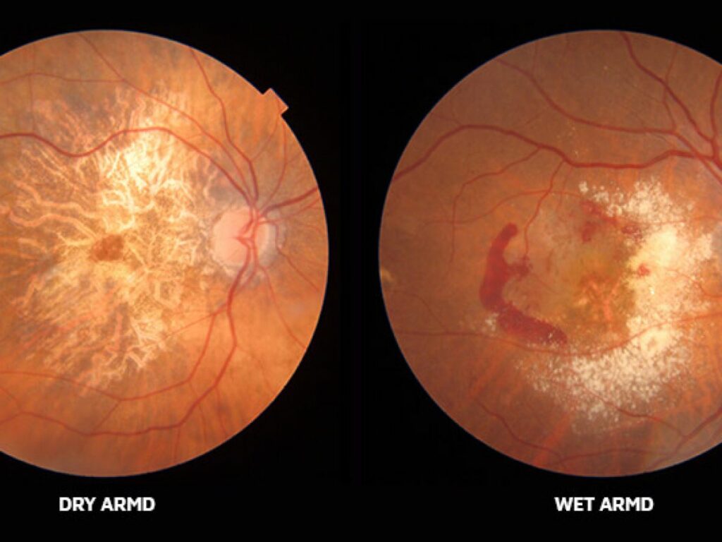
About 90% of people with Age-related Macular Degeneration (AMD) have “dry” AMD, which is a condition in which the macula’s layers including photoreceptors and the (RPE) retinal pigment epithelium, thin out over time and function less and less. This is referred to as atrophy. The pigmentation or color of the macula changes in the early stages of dry AMD. On the retina, tiny drusen appear: these are microscopic mounds of waste products from the eye’s cells. The drusen itself may cause retinal degeneration and atrophy. Pigment darkening and the development of drusen are common symptoms of dry AMD. Almost everyone over the age of 50 has at least one tiny drusen.
Drusen are a symptom of dry AMD. Waste materials have accumulated behind the retina in the form of tiny yellow deposits. Cholesterol, protein, and lipids make up their composition. Typically, when drusen initially appear, they don’t cause any vision loss. They are, nevertheless, a risk factor for developing visual loss. There are significantly more of these little yellow deposits and they are larger when a person has advanced dry macular degeneration.
Because it does not entail the exudation – or leakage – of fluids from blood vessels, dry AMD is also known as non-neovascular AMD and non-exudative AMD. Geographic atrophy (GA) is a term used to describe advanced forms of AMD, often known as “late dry” AMD, in which extensive regions of the retina that are well defined (geographies) stop functioning.
How is Dry Macular degeneration diagnosed?
To effectively diagnose Dry AMD, doctors must inspect the inside of the eye. A doctor can tell if the AMD is wet or dry after a thorough examination.
The cell layer beneath the retina will change in those with dry AMD. They will almost certainly have drusen deposits in their eyes, as well as retinal damage.
Fluid buildup surrounding the retina and waste deposits near the macula are common in people with wet AMD. Bleeding below the macula may cause a gray or green discoloration in some of these people. Finally, bleeding within or near the macula may be visible.
An Amsler grid test will very certainly be used to diagnose wet or dry AMD. A patient looks at a grid of straight lines during this test. If any of these lines appear bent or distorted, the patient may have AMD.
What are the symptoms of dry macular degeneration?
Dry AMD patients may encounter vision issues such as a dark or hazy patch in the center of their vision. However, the symptoms that people with dry AMD experience are usually milder than those that people with wet AMD experience.
People with dry AMD may have no symptoms at all in the early stages. Some signs of dry AMD that progress to the intermediate stage include slight blurriness in the center vision or difficulty seeing in dim light.
Advanced dry AMD symptoms are comparable to wet AMD symptoms. they both may have a blurry and wavy vision or even scotomas which are like a blank spots.
Treatment of dry AMD
Researchers are continuously working on a therapy for dry AMD that is successful. Your doctor may suggest an AREDS vitamin regimen, which is a combination of vitamins designed to lower the risk of severe dry AMD.
There is currently no FDA-approved therapy for dry AMD. However, some promising therapeutic trials have provided clinicians with guidelines for treating people with the disease. Certain nutrients may decrease the growth of dry AMD, according to the Age-Related Eye Disease Study (AREDS). Vitamins such as Beta carotene, vitamins C and E, lutein, and omega 3 fatty acids are among the nutrients highlighted in the study. Diet and exercise, like so many other aspects of health, are critical to our long-term well-being.
It’s also crucial to follow lifestyle choices that are thought to slow the growth of dry age-related macular, such as eating a diet rich in leafy greens, exercising regularly, quitting smoking, and maintaining a healthy weight and blood pressure.
Other dry AMD therapies are being researched. Two of these medications target a portion of the body’s immune system that attacks retinal cells.
Some researchers are also looking at using stem cells to restore the cells in the retina that have died as a result of dry AMD.
Monitoring changes in macular degeneration at home
Dry AMD can progress from a dry to a wet type, which is substantially more severe. Individuals with dry AMD should use an Amsler grid to monitor their vision on a regular basis and report any abnormalities to their eye specialist. Other home-monitoring options have emerged; ForeseeHome is the first FDA-approved system for home-based monitoring of patients who are at risk of vision loss due to wet age-related macular degeneration, as well as the first biotelemetry device in the field of ophthalmology.
Wet macular degeneration
Wet AMD is a disease in which abnormal blood vessels form behind the retina in the choroid layer. Choroidal neovascularization, or CNV, is the name for this condition. The new blood vessels are fragile, leaking fluid, lipids (a component of cell structure), and blood. The leakage seeps into the retina’s layers, including the macula’s layers, causing scar tissue to develop and retinal cells to quit working.
Approximately 10% of all cases of dry Age-related Macular Degeneration (AMD) become “Wet” AMD (typically a patient has dry AMD first and progresses toward wet).
The wet/neovascular form affects about 10-15% of people with age-related macular degeneration, but it accounts for almost 90% of all cases of severe vision loss caused by the illness. Wet AMD progresses much more quickly and causes severe central vision loss. The retina produces a protein called VEGF when the macula degenerates in this way. The retina produces this to defend itself and to develop new blood vessels. This attempt, however, is unsuccessful since the new blood vessels are irregular. These irregular blood vessels rupture, bleed, and leak fluid in wet AMD, causing damage to the macula. After a while, a scar forms over the entire macula, producing severe central vision loss.
How is wet macular degeneration diagnosed?
Fundus fluorescein angiography is another important diagnostic test (FFA). FFA is performed by injecting a dye into a blood vessel which then goes to the eye. There is no ionizing radiation used in this test.
This dye can aid doctors in determining whether there is any blood vessel leaking in the eye. If this is the case, you have wet AMD.
What are the symptoms of wet macular degeneration?
Symptoms of wet macular degeneration include blurred, wavy vision. The vision usually declines quite quickly when it converts from the dry form to the wet form so a fairly sudden change for the worse in your vision may be a symptom. Also due to the fluid that leaks under the retina, your vision may seem wavy, like a straight door frame may appear curved, or lines on the page may be distorted.
Wet Macular Degeneration treatments
Wet AMD patients can choose from a variety of therapy methods. Wet AMD can be treated with Anti-Vascular Endothelial Growth Factor (VEGF) medications since it is caused by irregular blood vessel leaking. These drugs are injected into the eye to halt bleeding or leakage from dysfunctional blood vessels. Lucentis, Avastin, and Eylea are some of the brand names for the medications. These injections are fantastic news for patients suffering from wet macular degeneration because they can cause serious visual loss if left untreated.
Newer medications are always being researched in order to increase these beneficial outcomes even further. Brolucizumab and abicipar pegol are two new anti-VEGF medications that showed promise in phase 2 studies and are currently being evaluated in phase 3 trials.
This can help prevent aberrant blood vessels from developing and obstructing eyesight. Scientists are also looking into gene therapy to assist people with wet AMD regain normal vision.
Risk factors for wet and dry macular degeneration
People who have a family history of wet or dry Age-Related Macular Degeneration are more prone to get the disease. People who smoke or who are exposed to too much UV light are also at a higher risk, according to research.
The major risk factor for wet or dry AMD is your age. Wet and dry AMD is more common in people over the age of 55, and the risk grows with age.
According to a review of AMD studies published in 2020, Caucasians have a higher risk of developing both wet and dry AMD. They are likely to develop this ailment due to a hereditary predisposition.
Light skin and blue eyes are also risk factors.
Finally, according to certain research, wet and dry AMD is more common in women. The study on sex and AMD, on the other hand, is scarce and inconclusive. More research is required to back up this finding and determine the reasons for it.
How to Prevent Macular Degeneration
The most important thing you can do to avoid visual loss from the disease is to catch it early. Everyone should see their ophthalmologist or optometrist for an annual eye exam at least once a year.
Because smoking is one of the leading causes of macular disease, quitting or refraining from smoking can help avoid the disease.
Regular exercise can also assist to minimize the risk.. Maintaining a healthy lifestyle helps individuals lower their blood pressure and cholesterol, lowering their risk of AMD even further.
People with wet and dry AMD can improve their quality of life for years to come by living a healthy lifestyle and having their eyes checked by a doctor on a regular basis.
FAQ’s
Which is worse: dry or wet macular degeneration?
Although the wet form is usually the worst, the dry form can be just as bad for a visual outcome because if caught early enough the wet form can be treated whereas the dry form can’t.
How can you tell if you have wet or dry macular degeneration?
The easiest way is to schedule an appointment with your eye doctor.
Which is treatable wet or dry AMD?
The wet form is treatable but that doesn’t mean that the prognosis is always better.
What causes macular degeneration to go from dry to wet?
Due to the atrophy of the retinal cells, they don’t function as well. Often leaving the remaining tissue starved for oxygen. Then abnormal blood vessels grow to provide more oxygen. The leaky vessels cause the conversion to the wet type.
Can you have both dry and wet macular degeneration forms at the same time?
Yes, one eye could have one and the other eye the other type. Within the same eye, it’s considered one or the other.
What foods should I avoid with macular degeneration?
Fatty foods that are high in cholesterol.


