


Initial symptoms of macular degeneration are blurry central vision, making it hard to read, drive or recognize faces. Straight lines may appear wavy or curved.
Age-related macular degeneration is most prevalent among those over 60 and may worsen over time, yet typically does not cause total blindness as it only affects central vision.
Dilated Eye Exam
An eye exam with dilation drops is one of the best ways to detect macular degeneration. Your eyes will be dilates with eyedrops so the doctor can examine your retina and other structures within your eye, providing him with information such as whether you have wet or dry forms and their stages, floating spots, visual light flashes or blurry vision. Additionally, this examination may reveal other symptoms you are unaware of such as floating spots, visual flashes or blurry vision which you might not otherwise be aware of.
Your doctor will first check for signs of dry AMD such as drusen – small deposits of metabolic waste under the retina that accumulate over time – which is one of its telltale characteristics, along with dark clumps or patches of missing pigment that have emerged under it. They may also conduct vision tests using charts with straight lines and ask you whether you see any wavy, distorted, or blank spots.
If your doctor suspects wet type macular degeneration, they will conduct a fluorescent dye test which detects any leakage or bleeding of blood vessels within your retina. This test is more effective when conducted over an extended period.
Optometric coherence tomography (OCT) is a noninvasive technique that utilizes infrared light to produce cross-section images of the retina and choroid layer, enabling doctors to view different layers of retinal layers as well as determine its thickness. OCT may also detect areas in which retinal thickness has decreased due to advanced macular degeneration or geographic atrophy.
At this test, the doctor places a probe over your eye and shines a light onto it, which illuminates your retina and causes light-sensitive cells to fire – this allows them to assess their health as a means to understand how quickly macular degeneration progresses and whether any treatment options should be pursued.
Optical Coherence Tomography
Optical coherence tomography (OCT) is the go-to high-resolution imaging test for macular degeneration and other eye conditions that result in vision loss, such as diabetic retinopathy or glaucoma. With near cellular resolution images delivered quickly in under a minute and no discomfort or radiation exposure involved, OCT has become a cornerstone of multimodal ophthalmic testing for these eye diseases and more – especially since its invention by OHSU Casey Eye Institute physician-scientist David Huang MD OCT has revolutionized how people living with macular degeneration or other eye diseases receive care and treatment.
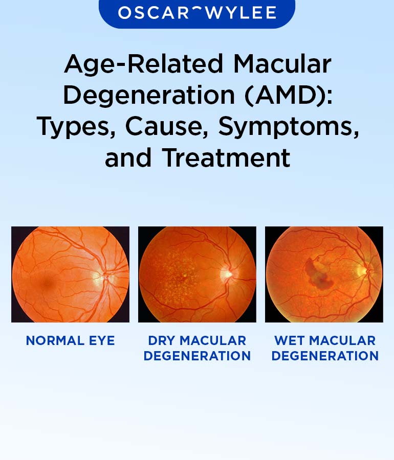
At the early stages of dry macular degeneration, protein deposits called drusen begin to build up under the retina, distorting light signals sent from retina to brain and blurring central vision. Although symptoms aren’t immediately evident in early stages, it’s still important to visit your physician regularly for monitoring purposes.
As macular degeneration progresses into its intermediate stage, the drusen grow larger and clump together, impairing central vision gradually and leading to straight lines appearing wavy or crooked. At this point, your doctor may recommend daily vision checks using an Amsler Grid as well as various tests designed to assess your macular degeneration progress.
Age-related macular degeneration (AMD), also known as wet AMD (neovascular or exudative AMD), occurs when abnormal blood vessels form beneath the retina in the choroidal plexus and leak fluid into the macula, potentially leading to permanent damage and blindness. With wet AMD, symptoms typically show up more quickly than with dry AMD; one eye often compensates for another in order to hide symptoms until treatment arrives.
Fluorescein angiograms involve injecting fluorescent dye into your arm and taking pictures using a special camera of how that fluorescent dye reflects light differently in your retina, making it easier for doctors to identify any leaky blood vessels and assess possible macular degeneration treatments.
Fluorescein Angiogram
At this test, your doctor injects fluorescent dye into your arm before taking pictures of your eye as the dye passes through its blood vessels. This allows an ophthalmologist to identify any leaky blood vessels associated with wet macular degeneration as well as assess its severity. While the procedure is typically safe and noninvasive, some individuals may experience mild to severe adverse reactions that include nausea or anaphylaxis symptoms.
The macula is the central portion of your retina, the light-sensitive layer of tissue responsible for providing clear vision in front of you. Macular degeneration deteriorates this central portion, making fine detail more difficult to discern or reading more difficult, along with dark spots, blank spots in vision and distortion of straight lines or loss of central vision. There’s no cure for macular degeneration; however certain vitamins and nutrients may slow its progression.
Early on in macular degeneration, your doctor may observe a loss of central vision without any obvious symptoms. They can identify drusen – tiny protein-lipid deposits under your retina – which indicates dry macular degeneration or wet macular degeneration depending on which form it takes.
Wet macular degeneration occurs when the macula becomes thinner and small blood vessels sprout beneath the retina, leaking fluid and leading to sudden changes in central vision. It can be detected with regular eye exams or by noting sudden losses of central vision.
if you experience signs or symptoms of macular degeneration. NYU Langone retina specialists can diagnose the condition and provide treatment advice, performing various tests such as dilated eye exams, optical coherence tomography scans and fluorescein angiograms to test and observe for changes to eye color or pupil shape during your examinations. During their exam they may ask about any symptoms as well as detect changes to color of eyes or changes in pupil shape during an exam visit.
Amsler Grid
The Amsler Grid is an effective test to detect early and intermediate macular degeneration symptoms. After administering numbing eye drops, a physician places a grid over each patient’s eyes before asking them to look at it; any straight lines appearing wavy or missing could indicate progression of disease progression and therefore progress monitoring should begin immediately. In some instances, copies can even be given out so patients can self-monitor at home.
Step one in treating age-related macular degeneration involves taking vitamin and mineral supplements, known to slow its progression. This is especially essential in cases involving “dry” macular degeneration characterized by yellowish deposits known as drusen under the retina; patients usually experience vision loss gradually over time.
VRMNY doctors specialize in treating wet macular degeneration, which occurs when new abnormal blood vessels proliferate and leak fluid and blood into the macula, by administering ranibizumab (VEGF inhibitor). This drug works by targeting and blocking abnormal blood vessel formation. When combined with photodynamic therapy – where an arm injection binds low-density lipoprotein proteins within the eye – photodynamic therapy may eliminate leakage of fluid into macula and improve vision in many patients.
Another effective treatment for wet macular degeneration is injection of anti-vascular endothelial growth factor drugs into the eye. Lucentis and Eylea, both FDA-approved anti-VEGF therapies, have proven successful at improving vision in wet macular degeneration patients. At VRMNY, our doctors will discuss all available treatment options to find the optimal solution for your wet macular degeneration condition. This may involve regular appointments or at-home monitoring using an Amsler grid ensuring you receive effective care for wet macular degeneration. VRMNY also actively participates in research to develop new and exciting treatments which have the potential of significantly improving visual outcomes in many patients with this disease.


