

Macular Degeneration Optomap and the early detection of macular degeneration
The biggest cause of blindness in the United States is age-related macular degeneration (AMD), which most frequently affects persons over 60. Your risk of developing AMD increases as you become older, are overweight or have a family history of the condition.
As we get older, we have an increased chance of developing certain eye disorders including dry eye and impaired vision as well as age-related macular degeneration, cataracts, diabetic retinopathy, and glaucoma. By 2050, there will be more than 88 million Americans aged 65 or older, up from more than 40 million now. Not surprisingly, this increase will coincide with a projected doubling of the number of Americans suffering from age-related eye illnesses. To preserve vision, early identification and treatment are essential.
Some people with AMD progress so slowly that visual loss takes a very long time to develop. Others experience a quicker progression of the illness, which might result in the loss of vision in one or perhaps both eyes. Simple daily tasks, such as the ability to drive, read, write, or perform close detailed work, such as cooking or home maintenance, might be hampered by AMD’s loss of central vision.
Dry AMD to Wet AMD
From Dry AMD to Wet AMD as the disease advances through the asymptomatic stage. The light-sensitive cells in the macula that transmit visual information to the brain and the supporting tissue below the macula gradually deteriorate in geographic atrophy (dry AMD). Neovascular AMD (wet AMD) is marked by the abnormal growth of blood vessels beneath the retina. These veins may leak fluid and blood, which might cause the macula to expand and get damaged. It’s critical to evaluate the likelihood of the illness moving from its dry to wet forms.
Signs of disease Before Drusen
Numerous cutting-edge technologies are at the disposal of optometrists, such as optical coherence tomography (OCT), which is incredibly useful in imaging a variety of retinal illnesses, including AMD. OCT, meanwhile, focuses solely on the structure. In fact, because of OCT’s high level of accuracy in detecting structural change, eye care professionals are now more likely to focus on gathering this type of diagnostic information at the expense of keeping track of their patient’s functional vision. An OCT scan of a patient with a few minor drusen may be deceptive and may understate the severity of the disease, depending on how it is interpreted. When OCT shows photoreceptor ellipsoid layer thinning, the retinal function is probably already severely compromised.
Research Indicates AMD Is Underreported
A recent study shows how often dilated eye tests fail to detect AMD cases. Doctors overlook AMD roughly 25% of the time, according to cross-sectional research that comprised 1,288 eyes from 644 adult patients who participated in the Alabama Study on Early Age-Related Macular Degeneration (ALSTAR).
The authors set out to find out how often ODs fail to recognize AMD when it is truly present. Adults 60 years of age or older medical records were examined for the research. The patient’s medical record from the most recent thorough dilated examination must not contain a diagnosis of AMD in either eye nor may the remarks in the medical record mention AMD symptoms.
Digital color fundus pictures of each patient in the ALSTAR trial were collected, and the presence or absence of AMD findings using the Clinical Age-Related Maculopathy Staging system was assessed by qualified graders wearing masks. Also observed were the different kinds of lesions connected to AMD.
The findings showed that despite possessing macular features suggestive of AMD in the fundus pictures, one in four eyes investigated did not get an AMD diagnosis during the dilated fundus examination. Seventy-eight percent of the 320 undiagnosed eyes had 10 or more small, intermediate, or both drusen, and eighty-six percent of the undiagnosed eyes (30.0%) had big drusen.
This study shows that even primary eye care physicians with the best training might overlook AMD. Missing this diagnosis might have detrimental effects on the patient, including significant eyesight loss.
Additionally, there was no difference in the study’s undiagnosed AMD prevalence between ophthalmologists and optometrists.
Since many of these individuals would have been candidates for therapeutic intervention, the researchers of this study point out that improved AMD detection procedures may be required in primary eye care clinics, even if they acknowledge that the causes for the missed diagnosis are still unknown.
In summary, existing techniques such as just using a dilated eye exam may encourage complacency and may not always allow us to identify AMD at an early enough stage. When dark adaptation testing is available, this may be overcome. Also using new technologies such as an Optomap may also aid in earlier diagnosis. Next, we will discuss how Optos and macular degeneration Optomap screening can help.
Optos macular degeneration Screening
Even while there have been several advancements in our knowledge of AMD’s origins, including linkages to genetics, there is still a great deal we don’t know about this complex, degenerative condition. The retinal periphery has been investigated in AMD more readily thanks to the development of multi-modality ultra-widefield imaging (which is what OPTOS is) to ascertain the utility in the identification and/or monitoring of the illness. The structure and function of the Retinal Pigment Epithelium (RPE), which is where AMD occurs within the eye, are both captured by color optomap® imaging. According to recent research, 97% of AMD patients display signs of illness outside of the central pole. This result showed that the majority of eyes had drusen and reticular alterations, clearly suggesting that AMD is a disorder that affects the whole retina rather than just the macular area. A follow-up investigation is being conducted to ascertain if these peripheral alterations are connected to the development of the illness.
What is an Optomap?
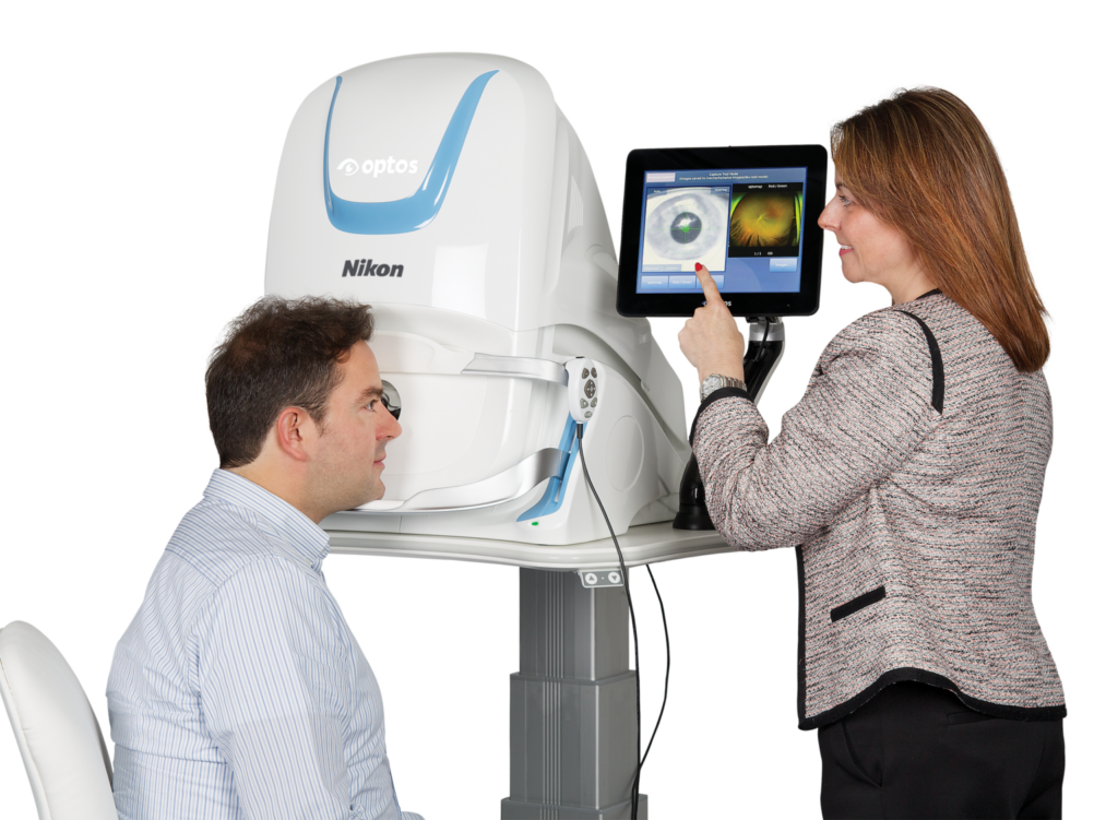
An Optomap picture may be obtained quickly, painlessly, and comfortably. Nothing ever touches your eye. It is appropriate for the entire family. A flash of light will signal that the gadget has successfully captured an image of your retina after you just glance into it with one eye at a time (similar to peering through a keyhole).
Normal conditions could exclude the need for dilating drops, but your eye doctor will determine if this is essential for you based on the condition of your eyes. It just takes a fraction of a second for a picture to be captured, and then it is instantaneously available for viewing your own retina. Even in a 3D animation, you see precisely what your eye care professional sees.
Early disease symptoms may be evident in the retina’s periphery and go unnoticed for a very long period when diagnosed using conventional techniques. While conventional imaging techniques normally only display 15% of your retina at a time, the Optomap ultra-widefield retinal picture is a novel technology that captures more than 80% of your retina in a single image.
The advantages of having an macular degeneration Optomap image
Having an Optomap ultra-widefield retinal picture taken has the following advantages:
- Optomap makes it easier to prevent vision loss or blindness before it happens.
- Early diagnosis of conditions including cancer, stroke, and cardiovascular disease that are fatal
Your eye care professional can identify early indications of retinal diseases more quickly and accurately than with conventional eye exams because of the unique Optomap ultra-widefield image.
Early identification lowers the risk to your sight and health and allows for the administration of effective remedies.
According to research that was published in Eye and Brain, integrating an optomap into a conventional BIO test might increase sensitivity by up to 30%. According to the findings of this cross-sectional investigation, image-assisted and conventional fundus examinations had a fair level of agreement. Over 70% of the time when there was a disagreement, the adjudicator sided with the image-assisted technique. This shows that the capacity of the examiner to detect or rule out lesions can be increased by up to 30% by including nonmydriatic imaging, in this instance Optomap UWF SLO, during the clinical examination.
Comparison of Traditional Fundus Examination vs Image-Assisted Fundus Examination
Eye and brain | 2014 Brown, Sewell, Trempe, Peto, Travison
When compared to a typical fundus examination alone, image-assisted fundus examination may increase the identification of retinal abnormalities by 30%.
The image-assisted and conventional fundus examinations showed high agreement in this cross-sectional investigation.
- In this study, the image-assisted technique had a greater identification rate of posterior pole lesions (90.1%).
- 92.2% of all abnormalities at the vitreoretinal interface were found using the image-assisted approach, compared to 54.7% using the conventional examination.
- 90.6% of the posterior pole’s drusen were found using an image-assisted technique, vs 43.8% using a standard fundus examination alone.
- The image-assisted technique was right in 75% of the cases where the methods differed for any type of lesion.
According to the type of lesion, there was excellent agreement between image-assisted and traditional fundus examinations for staphyloma (kappa 0.76), fair agreement for suspected cupping (kappa 0.66), drusen in the posterior pole/macula & mid-to-peripheral retina (0.45,0.41), (RPE) retinal pigment epithelial changes in the posterior pole/macula (0.54), peripheral retinal degeneration (0.50), cobblestone ( (0.53).
The results showed that when the methods disagreed, the image-assisted examination had a statistically significant advantage in identifying suspicious cupping (P = 0.04), drusen in the posterior pole/macula and mid-to-peripheral retina (P = 0.004, P,0.001), retinal pigment epithelial changes in the posterior pole/macula (P = 0.04), nevi in the posterior pole/macula and mid-to-peripheral (P, 0.001).
According to earlier research, the sensitivity of dilated ophthalmoscopy ranges from 32% to 82%.
Summary of using Optomap for macular degeneration diagnosis
As the studies listed above have shown, AMD along with other retinal diseases can easily be missed with just a dilated fundus exam by the practitioner. Having a broad field view of the retina to examine in detail instead of relying on small views that the doctors have through their ophthalmoscopes is very helpful to not miss the retinal disease. In addition, being able to see peripheral drusen and degeneration also has clinical significance and is believed to be early signs for some that they may be at risk for macular degeneration. Having an Optomap macular degeneration screening on a yearly basis for those that have a familiar risk or are over the age of 60 is probably a good idea.
FAQs for macular degeneration optomap
Why is a retinal examination so crucial?
Your retina may show some of the early indications of conditions including stroke, diabetes, and even some types of cancer before you notice other symptoms. They are easier to see using an Optomap.
Can kids use an Optomap without harm?
Yes. In fact, many vision issues start in childhood, making it crucial for kids to get regular, high-quality eye care.
What is the Optomap?
The Optomap is a retinal digital picture created by the scanning laser technology of Optos. It is the only technology that can simultaneously record 82% of your retina.
Is it painful?
No. The scan only takes a split second and is absolutely painless.
How can Optomap help me?
Your eye doctor may be able to identify issues more quickly and easily using the ultra-widefield Optomap. The Optomap picture may be retained for subsequent comparisons, unlike conventional retinal examinations.
How frequently should an Optomap be done?
Your doctor should decide whether to do this. However, it is often advised that you have an Optomap every time you get your eyes checked.
Are there any negative effects?
Low-intensity scanning lasers that are non-intrusive produce Optomap pictures. In more than 150 million sessions, there have been no recorded negative health impacts.


