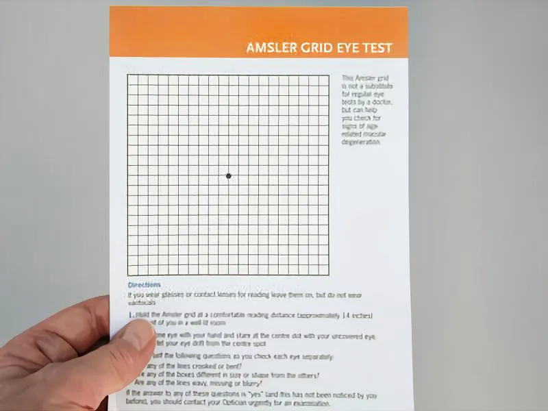

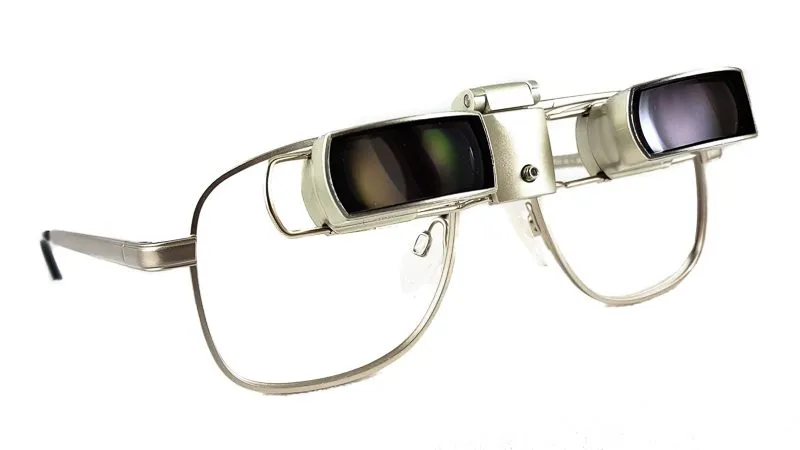
Medically reviewed by Shaun Larsen, OD – Doctor of Optometry. Low vision rehabilitation specialist.
Last clinically reviewed: August 2026 | Next scheduled review: August 2027 | Reading time: 18 minutes
Editorial standard: every clinical claim in this article is sourced to a peer-reviewed journal, a national eye institute, or a professional society guideline. Sources appear as bracketed numbers [1] matching the reference list at the end.
If you have been diagnosed with age-related macular degeneration (AMD), your eye doctor has probably handed you a small square of graph paper and told you to check it regularly. That square – the Amsler grid – has been the standard home test for macular disease since 1947 [1]. Used correctly, it costs nothing and takes under a minute a day. But you should also know what the evidence shows: the grid misses roughly one in three conversions to wet AMD [9], and validated home-monitoring technology now exists that performs substantially better [10]. This guide covers how to use the grid properly, what changes on the grid mean inside your retina, what the research says about its accuracy, and when to move to a monitored program.
What is the macular degeneration grid called?
The Amsler Grid <https://lowvisionaids.org/amsler-grid-online-low-vision-test/> is the grid test people refer to when monitoring their macular degeneration at home. It is named for Marc Amsler (1891-1968), the Swiss ophthalmologist who formalized the chart in a 1947 paper in Ophthalmologica [1] and later described its use for catching the earliest symptoms of macular disease [2].
What is the Amsler Grid?
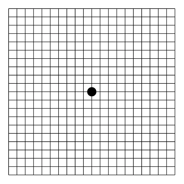
The Amsler grid is an easy-to-use home monitoring test for macular degeneration <https://lowvisionaids.org/blog/macular-degeneration-testing/> . Download your free Amsler grid here and use the Amsler grid for home monitoring.
The Amsler grid is a test used by optometrists and ophthalmologists to diagnose visual impairments caused by macula damage (the central part of the retina). The standard chart is a 10 x 10 cm square divided into 5 mm boxes with a fixation dot at the center. Held at about 30 cm (12 inches), each small square covers 1 degree of visual angle, so the full grid maps the central 20 degrees of your visual field – 10 degrees to each side of fixation [3]. That is precisely the territory served by the macula, the retinal region responsible for reading, face recognition, and all fine-detail vision.
After a diagnosis of a retinal condition, the Amsler grid may be used to identify early indicators of the disease and track changes in vision. Amsler actually published seven chart variations: the familiar grid (chart 1), a version with diagonal lines to help patients who cannot see the central dot hold fixation (chart 2), a red-on-black grid that probes the long-wavelength cones in conditions causing red desaturation (chart 3), a dots-only chart used to tell a true blind spot from distortion (chart 4), and three others for specialized use [3].
If you have dry macular degeneration (AMD) <https://lowvisionaids.org/macular-degeneration-amd/> , an Amsler grid may help you keep track of your vision. It can flag progression from dry to wet AMD <https://lowvisionaids.org/blog/wet-amd/> – but it is not a safety net. In pooled studies the grid detects only about two-thirds of wet AMD cases [8,9], so it supplements, and never replaces, scheduled dilated examinations and, for higher-risk eyes, the monitored home devices described below.
Why lines bend: what grid changes mean inside your retina
Everything the grid shows has a physical explanation in the retina. The macula's photoreceptors sit in a precise mosaic, and the brain assumes that mosaic is flat and evenly spaced. Anything that lifts, stretches, or compresses it makes straight lines appear bent – a symptom called metamorphopsia (visual distortion) [5].
Wavy or bent lines (metamorphopsia). Drusen – deposits of lipid and protein under the retinal pigment epithelium – or leaked fluid physically displace photoreceptors. When receptors are pushed apart or dragged together, the image is mapped onto a distorted receptor grid and straight edges warp [5].
Squares that look smaller (micropsia) or larger (macropsia). If photoreceptors are spread apart by swelling, an object stimulates fewer receptors than expected and looks smaller; if receptors are crowded together, it looks larger [5].
Gray, black, or blurred patches (scotoma). A blind spot means photoreceptors in that area are not signaling – separated from their blood supply by fluid, blood, or atrophy. A new scotoma is the most urgent finding on the grid [3].
Missing corners while staring at the dot. This suggests a defect beyond the very center of the field and also warrants examination.
How to use the macular degeneration grid (Amsler Grid)
Using an Amsler grid to test your eyes is simple and takes just a few minutes. Here’s how you do it:
Do the test with good lighting you would normally read under. Use the same lamp and the same spot in your home every time – changing conditions changes the test.
Wear your regular reading glasses (even if you just have store-bought reading glasses).
Hold the Amsler grid about 12 to 14 inches (30-35 cm) from your face. The chart is calibrated so that each square equals 1 degree of visual field at 30 cm (12 in); holding it farther away shrinks the area of retina you are actually checking [3].
Each eye should be tested separately: While testing one eye, cup your hand over the other.
Answer the following questions while keeping your gaze fixed on the dot in the grid’s center:
Is there anything wavy, blurry, or distorted about any of the lines in the grid?
Do all of the squares in the grid seem to be the same size and shape?
Is the grid free of “holes” (missing sections) or dark spots?
Can you perceive all of the grid’s corners and edges (while maintaining your gaze on the center dot)?
Cover the other eye and repeat on the other side.
Test at about the same time of day, and mark any abnormal area directly on a paper copy with the date.
Hold the grid at a distance that allows you to get most of the lines in focus while wearing reading glasses. If you use it with both eyes open, the good eye will compensate for the wet AMD eye, and you won’t notice any abnormalities. This is why one-eye-at-a-time testing is non-negotiable: many patients who lose vision to wet AMD in one eye noticed nothing for months because the fellow eye filled in the picture.
All lines will seem straight to an eye without wet AMD. Wet AMD, on the other hand, causes parts of the lines to seem bent or even blacked out by a gray, white, or black zone. This is caused by fluid accumulating inside or behind the retina, which may form a blister, giving the appearance of curved straight lines. The fluid may sometimes create a gray, white, or black “blind spot” in or around the center of your visual field by interfering with retinal function.
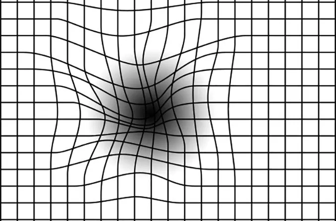
When using the Amsler Grid to test your vision, if any holes or blurry patches emerge, or if the lines seem wavy, fuzzy, crooked, warped, or broken, you should contact your eye care practitioner right away; these are all possible indicators of AMD (Age-related Macular Degeneration) <https://lowvisionaids.org/macular-degeneration-amd/> . Specifically: call the office, say you have a new change on your Amsler grid, and ask to be seen within 24 to 48 hours. New distortion in an eye with dry AMD is presumed wet AMD until an OCT scan proves otherwise. The Amsler grid is not a replacement for routine examinations: the American Academy of Ophthalmology's Preferred Practice Pattern recommends dilated examination every 6 to 18 months for intermediate AMD (sooner at the higher-risk end) and prompt examination whenever new symptoms appear [18].
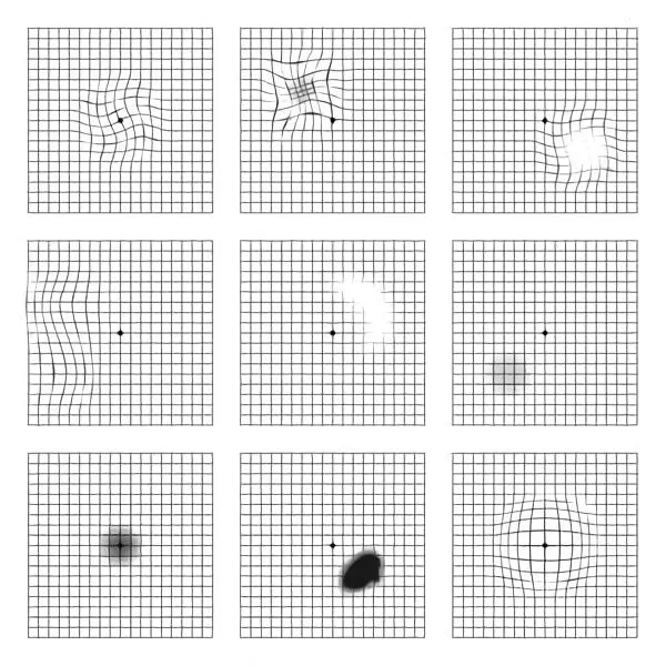
If an eye with wet AMD is left untreated for longer than a few months, retinal scarring may occur. Because scarring may cause irreparable vision loss in sections of the visual field, it’s ideal to check your vision with the grid at least once a week at home and notify your retina doctor if anything changes. What exactly is a change? If your optometrist informs you that you don’t have wet AMD, glance at the grid right thereafter. That’s your starting point. Due to large “drusen,” semi-solid deposits that accumulate behind the retina in AMD, the lines may be somewhat wavy in a few areas. If you then check again and believe the waviness has worsened, a new region has emerged, or you have a new “blind spot,” it’s time to see a retina doctor. Most retina specialists now advise daily rather than weekly testing – the check takes under a minute, and the benefit of home monitoring in trials came from frequent, consistent use [10]. Time matters because injection outcomes are consistently better when treatment starts while the abnormal vessel complex is still small: in the CATT trials, larger baseline lesion size independently predicted worse vision at five years [19], and patients treated within about 48 hours of diagnosis retain more vision long-term than those treated after weeks of delay [20].
How accurate is the Amsler grid? What the evidence actually shows
The honest answer: less accurate than most patients – and many clinicians – assume. Three findings matter.
1. Sensitivity is modest. A 2023 systematic review and meta-analysis in JAMA Ophthalmology (10 studies; 425 wet-AMD eyes, 1,262 dry-AMD eyes, 203 healthy control eyes) pooled the grid's diagnostic performance: sensitivity 67% and specificity 99% against healthy controls, but sensitivity 71% and specificity only 63% against the comparison that actually matters clinically – eyes with dry AMD [9]. An earlier meta-analysis reached similar figures, at levels its authors judged typically not recommended for monitoring purposes [8]. Put plainly, roughly one wet-AMD eye in three passes the grid test. And these were supervised, clinic-based studies; unsupervised home performance is likely worse [6].
2. Your brain fills in the gaps. The visual cortex is built to complete regular patterns across small defects – a phenomenon called perceptual completion, or filling-in. Achard and colleagues showed that patients with proven central scotomas often report a fully intact grid because the brain paints the missing squares back in; defects smaller than about 6 degrees are frequently invisible on the chart [7,3]. The grid's regular, predictable pattern is exactly the stimulus the brain completes best – a design weakness identified decades after Amsler introduced the test.
3. Absence of evidence is not evidence of absence. That was the title of Crossland and Rubin's 2007 review in the British Journal of Ophthalmology, which collected reported sensitivities below 50% in some series and concluded the chart cannot be recommended as the sole method for detecting progression [6]. A normal grid is reassuring, but it is never proof that nothing has changed.
None of this makes the grid worthless. It is free, fast, and better than no monitoring at all – and because a definite new change is rarely a false alarm in a healthy eye, an abnormal grid is a strong signal to act. It simply cannot carry the whole weight of surveillance in an eye at real risk of conversion.
Beyond the grid: validated home-monitoring technology
Because of the grid's limitations, the past decade has produced FDA-reviewed home devices with randomized-trial and real-world evidence behind them. If you have intermediate dry AMD, ask your eye doctor whether you qualify.
ForeseeHome (preferential hyperacuity perimetry)
ForeseeHome (Notal Vision) is a tabletop device that tests hyperacuity – the visual system's extreme sensitivity to misalignment of dots – across roughly 500 points in the central 14 degrees of field, in about 3 minutes per eye [10]. It was validated in the AREDS2-HOME randomized trial of 1,520 patients with intermediate AMD, which was stopped early for benefit: among eyes that converted to wet AMD, 94% of patients using the device as directed still had 20/40 or better vision at detection, versus 62% with standard care that included Amsler grids; median loss was 4 letters versus 9 [10]. The real-world ALOFT analysis (2,123 patients, 3,334 eyes, up to 10 years of monitoring, average 5.2 tests per week) found 84% of converting eyes maintained 20/40 or better at conversion, versus roughly 34% in national IRIS Registry standard-care data [11]. The program is prescribed through your eye doctor and run by the Notal Vision Monitoring Center, which alerts the practice when the algorithm flags a change. Medicare has covered the monitoring since 2016 for eligible patients – generally intermediate dry AMD with best-corrected acuity of 20/60 or better [12].
Home OCT (SCANLY)
Optical coherence tomography (OCT) – the in-office scan your retina specialist uses to see fluid inside the retina – is now available as a self-operated home device. In May 2024 the FDA granted De Novo marketing authorization to the Notal Vision SCANLY Home OCT, the first patient-operated OCT, previously designated a Breakthrough Device, for monitoring eyes already diagnosed with wet AMD between office visits [13]. Patients self-scan in about a minute per eye; images upload automatically to a monitoring center, where an AI algorithm estimates retinal fluid and alerts the prescribing specialist. Pivotal data published in 2025 showed 97% of wet-AMD patients could successfully self-image, averaging 5.9 scans per week, with home scans diagnostically equivalent to in-office OCT for fluid detection [14]. Dedicated CPT codes for remote OCT exist (0604T-0606T); broad Medicare payment was still being established as of this writing, so availability runs through participating retina practices [13,14].
Smartphone apps
Several app-based hyperacuity tests are FDA-cleared or CE-marked, including Alleye (a dot-alignment task with an area under the curve of about 0.85 for detecting wet AMD in validation studies) and Home Vision Monitor (formerly myVisionTrack) [15,16]. These are reasonable adjuncts for motivated patients when prescribed and reviewed through an eye-care provider. Be cautious with the many free 'Amsler grid' apps: screen size, viewing distance, and brightness are uncontrolled, almost none have published validation, and a digital grid inherits every limitation of the paper one. An unvalidated app is not a monitoring strategy [15].
Who should be monitoring – and how closely
Roughly 20 million US adults live with some form of AMD, about 1.5 million of them with vision-threatening late disease [4] – but the risk of converting from dry to wet AMD is far from uniform. The AREDS Simplified Severity Scale scores one point per eye for large drusen and one for pigment changes; the 5-year risk of advanced AMD climbs from about 0.5% with zero factors to 12% with two, 25% with three, and 50% with four [17]. Patients with high-risk intermediate AMD – the group this article matters most for – convert at several percent per year, and once one eye has wet AMD the fellow eye needs the closest watching [10,18].
Early AMD, low risk: dilated examination every 1-2 years; grid or app testing provides useful baseline awareness [18].
Intermediate AMD: dilated examination every 6-18 months, daily home monitoring (grid at minimum; ForeseeHome if eligible), and AREDS2 supplementation, which reduced 5-year progression to advanced AMD by about 25% in this group [17,22,18].
Wet AMD in one eye: device-based monitoring of the fellow eye rather than the grid alone; discuss home OCT for the treated eye [10,13,18].
Who should not rely on the grid: people with significant cataract or uncorrected near vision (blur invalidates the test), poor central fixation from an existing scotoma, or memory or consistency problems that make reliable self-testing unrealistic – and eyes already under regular anti-VEGF treatment with scheduled OCT, where the office scans, not the grid, drive decisions. If you cannot test reliably, tell your doctor; a monitored program removes the burden of self-judgment.
FAQs
Is an Amsler grid necessary for early-stage macular degeneration?
Early detection is the key to preserving vision, and daily self-monitoring should begin as soon as intermediate AMD is diagnosed. If you are consistent with using the test you will know early on when your vision is changing and can make an appointment with your eye doctor or low vision specialist <https://lowvisionaids.org/low-vision-specialist-optometrist-near-me> to treat it as soon as it is warranted. Just remember the limits: a normal grid does not rule out progression [6,9], so keep every scheduled dilated exam regardless of what the grid shows.
Is the Amsler grid for wet macular degeneration?
Yes, although once the disease has converted to the wet form and you’re being treated for it, the test isn’t as beneficial. It will show you progression or hopefully regression during treatment. For eyes already diagnosed with wet AMD, home OCT (SCANLY) was designed for exactly this situation – it measures the fluid itself rather than your perception of it [13,14].
What causes dry Macular degeneration to change to the wet form?
In dry AMD, drusen and a failing retinal pigment epithelium impair oxygen and nutrient exchange between the choroid (the blood-vessel layer beneath the retina) and the photoreceptors. The resulting stress – relative ischemia plus chronic inflammation – drives retinal cells to secrete vascular endothelial growth factor (VEGF), which stimulates neovascularization: fragile new vessels sprouting from the choroid into and under the retina. These new leaky blood vessels leak fluid into areas of the retina that can cause irreversible vision loss. These areas if left untreated can develop retinal scarring and further vision loss. This VEGF pathway is exactly what modern treatment blocks: intravitreal anti-VEGF agents – ranibizumab, aflibercept 2 mg and the high-dose 8 mg formulation, off-label bevacizumab, and the bispecific antibody faricimab – can hold many treated eyes stable on injections spaced every 8 to 16 weeks [21,23].
What does it mean when you see wavy lines?
Obviously, the lines on the grid are supposed to be straight horizontal or vertical lines. If the lines are wavy it could mean you have drusen as in the case of dry AMD or it could be the early signs of the wet form of the disease. In either case, it's because there is something in or under the retina distorting it. If it is a change from your baseline, it should be checked within 24 to 48 hours [18,20].
What does the grid look like with intermediate macular degeneration?
Intermediate AMD is a defined stage – large drusen (over 125 microns), or pigment changes together with at least medium drusen – that sits between early and advanced disease; it is not the end stage of dry AMD. The late form of dry AMD is geographic atrophy, in which patches of the retina and its pigment layer die off [17,18]. In this stage, you may notice some waviness to the lines on the grid but probably not any gray or missing areas.
Why do lines on the macular degeneration grid appear double?
If the lines look double you may have some large drusen distorting the retina. If this is a change from what the lines looked like on the Amsler grid test before, see your eye doctor. Doubling that persists with the other eye covered can also come from corneal or lens problems, and doubling only with both eyes open suggests an alignment issue – both are reasons for an exam rather than watchful waiting.
How long do you look at the Amsler grid?
You only have to do the test long enough to determine if there is a change from your baseline. If just starting to monitor your vision you will do it long enough to determine if any of the lines are wavy or missing, if there are any grey or black spots and if you can see all four corners. Do each eye separately and at the same distance consistently. In practice that is 10 to 30 seconds per eye.
How do they diagnose macular degeneration?
Macular degeneration is usually diagnosed when your eye doctor performs a dilated eye exam and looks at the macula with a special magnifying lens. The first signs of it are yellow spots called drusen. The wet form is diagnosed when fluid is seen under the retina either by this same type of exam or by a test called an OCT. OCT (optical coherence tomography) is a non-contact scan that images the retina's layers in cross-section and detects fluid far earlier than symptoms appear; OCT angiography and, less commonly now, dye-based fluorescein angiography map the abnormal vessels themselves [18].
Is the macular degeneration grid test used to test for other forms of vision loss?
The Amsler test <https://lowvisionaids.org/amsler-grid-online-low-vision-test/> can also be used for screening for progression of macular swelling, including cystoid macular edema <https://lowvisionaids.org/blog/cystoid-macular-degeneration/> and diabetic macular edema from diabetic retinopathy. It can be helpful in screening for disorders that cause red desaturation like pituitary tumors, toxic maculopathy from medications (notably hydroxychloroquine), and toxic optic neuropathy. For those conditions, Amsler's chart 3 – red lines on a black background – is used because it specifically stimulates the long-wavelength cones. Chart 4, which replaces the lines with scattered dots, distinguishes a true blind spot from distortion [3]. Central serous chorioretinopathy, epiretinal membrane, and macular holes also produce grid changes – another reason new findings need an examination rather than self-diagnosis [3].
Should I document the results for my doctor?
Having a copy of the grid is a great idea, if you notice changes in your vision and defects on the test, you can draw on the copy what you see or don’t see. This will aid your doctor in knowing what may be occurring. Date each marked copy and bring the series to your appointment; a dated sequence showing progression is genuinely useful triage information for the retina team.
If I notice defects on the test should I go to my doctor immediately or wait to see if it resolves?
Do not wait. A new defect on the grid in an eye with AMD is presumed conversion to wet AMD until proven otherwise, and it will not resolve on its own. Call your eye doctor or retina specialist the same day and ask to be seen within 24 to 48 hours. The treatment window is real: anti-VEGF outcomes are consistently better when injections begin while the lesion is small [19], patients treated within about 48 hours of diagnosis do better than those treated after longer delays [20], and in trial data, eyes whose conversion was caught early kept driving-level vision at nearly twice the rate of standard care [10].
Will new glasses help to fix the problems detected with the Amsler grid test?
No, getting new glasses won’t help with whatever condition is causing the defects on the test. It is probably a good idea to wait until your doctor tells you your condition is stable before spending the money on new glasses. Distortion from retinal disease is a photoreceptor-position problem, not a focusing problem – no lens can straighten lines the retina itself is bending.
I have AMD in the family, should I do this test to monitor it for myself?
Early detection is always the goal, however, your eye doctor will probably detect the disease before you will doing the test. With a family history, get a baseline comprehensive dilated exam by age 40 and follow your doctor's recommended interval – typically every 1 to 2 years from age 65 [18]. Quit smoking (the largest modifiable risk factor, roughly doubling AMD risk), and eat a diet rich in leafy greens and fish [24]. One correction on supplements: AREDS2 vitamins are proven only to slow progression in eyes that already have intermediate or advanced AMD; they do not prevent AMD in people who merely have a family history, so do not start them without a diagnosis and your doctor's advice [22].
Living with macular degeneration: low vision rehabilitation
Monitoring exists to protect the vision you still have – but if AMD has already cost you central vision, function does not end there. Low vision rehabilitation works around a central scotoma using magnification, contrast, lighting, and trained eccentric viewing (learning to use a healthy patch of retina just off-center) [24]. Depending on your acuity and goals, that can mean high-add reading glasses, handheld and stand magnifiers, video magnifiers (CCTVs) that enlarge and boost contrast at the same time, bioptic telescopes for distance tasks and – in many states – driving, and head-worn electronic devices. A low vision specialist <https://lowvisionaids.org/low-vision-specialist-optometrist-near-me> can match the tool to the task; the evaluation is worth scheduling as soon as everyday tasks like reading mail or recognizing faces become work.
This article is for educational purposes and does not replace an examination by a qualified eye care professional. If you notice new distortion, blurring, or blind spots on an Amsler grid or in daily life, contact your eye doctor promptly rather than acting on anything you read here.
References
1. Amsler M. L'examen qualitatif de la fonction maculaire. Ophthalmologica. 1947;114(4-5):248-261. https://karger.com/oph/article-abstract/114/4-5/248/248477
2. Amsler M. Earliest symptoms of diseases of the macula. Br J Ophthalmol. 1953;37(9):521-537. doi:10.1136/bjo.37.9.521
3. Tripathy K, Salini B. Amsler Grid. In: StatPearls Internet. Treasure Island (FL): StatPearls Publishing; updated August 25, 2023. https://www.ncbi.nlm.nih.gov/books/NBK538141/
4. Rein DB, Wittenborn JS, Burke-Conte Z, et al. Prevalence of age-related macular degeneration in the US in 2019. JAMA Ophthalmol. 2022;140(12):1202-1208. doi:10.1001/jamaophthalmol.2022.4401
5. Midena E, Vujosevic S. Metamorphopsia: an overlooked visual symptom. Ophthalmic Res. 2015;55(1):26-36. doi:10.1159/000441033
6. Crossland M, Rubin G. The Amsler chart: absence of evidence is not evidence of absence. Br J Ophthalmol. 2007;91(3):391-393. doi:10.1136/bjo.2006.095315
7. Achard OA, Safran AB, Duret FC, Ragama E. Role of the completion phenomenon in the evaluation of Amsler grid results. Am J Ophthalmol. 1995;120(3):322-329. doi:10.1016/s0002-9394(14)72162-2
8. Faes L, Bodmer NS, Bachmann LM, Thiel MA, Schmid MK. Diagnostic accuracy of the Amsler grid and the preferential hyperacuity perimetry in the screening of patients with age-related macular degeneration: systematic review and meta-analysis. Eye (Lond). 2014;28(7):788-796. doi:10.1038/eye.2014.104
9. Bjerager J, Schneider M, Potapenko I, et al. Diagnostic accuracy of the Amsler grid test for detecting neovascular age-related macular degeneration: a systematic review and meta-analysis. JAMA Ophthalmol. 2023;141(4):315-323. doi:10.1001/jamaophthalmol.2022.6396
10. AREDS2-HOME Study Research Group; Chew EY, Clemons TE, Bressler SB, et al. Randomized trial of a home monitoring system for early detection of choroidal neovascularization: Home Monitoring of the Eye (HOME) study. Ophthalmology. 2014;121(2):535-544. doi:10.1016/j.ophtha.2013.10.027
11. Mathai M, Reddy S, Elman MJ, et al. Analysis of the long-term visual outcomes of ForeseeHome remote telemonitoring: the ALOFT study. Ophthalmol Retina. 2022;6(10):922-929. doi:10.1016/j.oret.2022.04.016
12. Notal Vision. ForeseeHome AMD Monitoring Program: coverage information and patient eligibility. Accessed August 2026. https://foreseehome.com/doctor/rs/how-it-works/coverage-information
13. Notal Vision. FDA grants AI-powered Notal Vision Home OCT 'SCANLY' De Novo marketing authorization. Press release, May 16, 2024. https://notalvision.com/assets/press-releases/May-16-2024-FDA-Grants-AI-Powered-Notal-Vision-Home-OCT-22SCANLY22-De-Novo-Marketing-Authorization.pdf
14. Notal Vision. Pivotal data validates diagnostic equivalence of SCANLY Home OCT and in-office OCT in wet AMD management. July 2025. https://glance.eyesoneyecare.com/press-releases/pivotal-data-validates-diagnostic-equivalence-of-scanly-home-oct-and-in-office-oct-in-wet-amd-management/
15. Balaskas K, Drawnel F, Khanani AM, Knox PC, Mavromaras G, Wang YZ. Home vision monitoring in patients with maculopathy: current and future options for digital technologies. Eye (Lond). 2023;37(15):3108-3120. doi:10.1038/s41433-023-02479-y
16. Mobile apps and visual function assessment: a comprehensive review of the latest advancements. Ophthalmol Ther. 2025. doi:10.1007/s40123-024-01071-1
17. Ferris FL, Davis MD, Clemons TE, et al; Age-Related Eye Disease Study (AREDS) Research Group. A simplified severity scale for age-related macular degeneration: AREDS report No. 18. Arch Ophthalmol. 2005;123(11):1570-1574. doi:10.1001/archopht.123.11.1570
18. American Academy of Ophthalmology. Age-Related Macular Degeneration Preferred Practice Pattern. 2024 (updated). https://www.aao.org/education/preferred-practice-pattern/age-related-macular-degeneration-ppp
19. Ying GS, Maguire MG, Pan W, et al; CATT Research Group. Baseline predictors for five-year visual acuity outcomes in the Comparison of AMD Treatment Trials. Ophthalmol Retina. 2018;2(6):525-530. https://pubmed.ncbi.nlm.nih.gov/29938247/
20. Treatment of neovascular age-related macular degeneration within 48 h from diagnosis improves long-term functional outcome. Biomed Pharmacother. 2023. https://www.sciencedirect.com/science/article/pii/S0753332223001567
21. Heier JS, Khanani AM, Quezada Ruiz C, et al. Efficacy, durability, and safety of intravitreal faricimab up to every 16 weeks for neovascular age-related macular degeneration (TENAYA and LUCERNE): two randomised, double-masked, phase 3 trials. Lancet. 2022;399(10326):729-740. doi:10.1016/S0140-6736(22)00010-1
22. Age-Related Eye Disease Study 2 (AREDS2) Research Group. Lutein + zeaxanthin and omega-3 fatty acids for age-related macular degeneration: the AREDS2 randomized clinical trial. JAMA. 2013;309(19):2005-2015. doi:10.1001/jama.2013.4997
23. Regeneron Pharmaceuticals. Eylea HD (aflibercept 8 mg) approved by FDA with dosing intervals up to 5 months for wet AMD and DME. August 2023. https://www.drugs.com/newdrugs/eylea-hd-aflibercept-approved-fda-first-only-injectable-anti-vegf-dosing-intervals-up-5-months-wet-6763.html
24. National Eye Institute. Age-Related Macular Degeneration (AMD). Accessed August 2026. https://www.nei.nih.gov/learn-about-eye-health/eye-conditions-and-diseases/age-related-macular-degeneration








 Shop Best Low-Vision Aids with FREE Doctor Consultation.