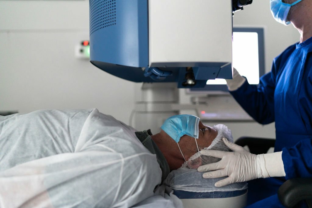
What is Goniotomy?
A goniotomy is a surgical procedure used to create a better outflow of a glaucoma patient’s inner eye fluid and lower the interocular pressure. The glaucoma procedure can be used either alone or in conjunction with medicine or eye drops. A goniotomy opens up the trabecular meshwork, the first layer of the eye’s natural drainage system. This releases pressure by facilitating the fluid’s easier passage away from the eye. Goniotomy is a possibility if eye drops or laser treatment is not sufficient to reduce intraocular pressure. Controlling ocular pressure by a goniotomy may be possible with or without the use of glaucoma drugs. Goniotomy offers a bleb-forming glaucoma surgery alternative for eligible patients, replacing tube shunts or trabeculectomy (Xen).
With a goniotomy, who is a candidate?
When undergoing cataract surgery, glaucoma patients are treated with goniotomies. It may also be applied to patients in various situations who have:
- Aniridia ( No Iris can be seen)
- Juvenile rheumatoid arthritis and uveitic glaucoma
- Open-angle glaucoma in children (JOAG)
- Rubella syndrome in mothers
For many patients with open-angle glaucoma seeking a long-term solution, it is also an excellent therapy option. It is the best option for children with congenital glaucoma.
The outcome of a successful goniotomy can help prevent permanent damage to your optic nerve, relieve the intraocular pressure caused by glaucoma, and lessen your need on medication.
Ophthalmologists had a lot to consider after the Ocular Hypertension Treatment Study (OHTS), even though it offered a foundational idea for patient management.
The study found that using ocular hypotensive medicine to lower intraocular pressure (IOP) lowers the chance of developing primary open-angle glaucoma.
It also showed that about 90% of people with high intraocular pressure may not develop optic nerve injury or a reduction in their visual field.
For whom, therefore, do the advantages of treatment surpass the drawbacks, expenses, and risks? Finding the optimal solution necessitates weighing the effectiveness of the various treatment choices against life expectancy and quality of life in addition to taking IOP and test findings into account.
How Do They Perform Goniotomies?
Anesthesia is used during goniotomy surgery, and before the procedure, numbing medicine is administered to the eyes. Incisions are produced in the front of the eye after devices are used to make sure the eye stays open. After that, specialized lenses are applied to the eye to look at the fluid’s natural outflow. Open channels are made to let fluid drain from the eye after a piece of the obstruction in the drain has been removed. You won’t feel any discomfort, but you will see a lot of brilliant lights during the process. Usually, the procedure takes five minutes or less.
What Are the Surgical Steps?
Under anesthesia, the procedure is carried out in the operating room. After cleaning the eye, numbing medicine is administered. The eyelids are then opened using an apparatus. To see the eye’s natural outflow, tiny punctures are made on the front of the eye, and a special gonio lens is applied. The section of the wall obstructing the drain is taken down. This opens up a pathway for fluid to exit the eye. Goniotomy procedures typically take 20 minutes, although they will take longer if cataract surgery is also being done at the same time.
What to expect following Goniotomy?
During the procedure, you might see bright lights, but you shouldn’t experience any pain. A transparent plastic shield covering the operated eye will be removed upon your release. You will be given sedatives, so you will need an adult to drive you home.
Post Goniotomy Mechanical Results
The natural drain of the eye creates an open route through which fluid exits the eye. As a result, the ocular pressure is reduced, and following surgery, one or more glaucoma drugs may be discontinued.
Follow up care
The day following surgery, approximately one week later, and several weeks afterward are when you will see your doctor. You may need more or fewer visits, depending on how your eye heals.
Post Op Medications
For the majority of patients, steroid and antibiotic eye drops will be prescribed. The level of inflammation in the eyes determines how often the steroid eye drops should be used.
Will Glaucoma Meds be needed afterward?
Depending on how your eye heals, your doctor will advise you on which drops to use going forward and how often at each visit. Patients may be able to cut back on how many eye drops they take at times. If your pressure is lower after the surgery, the procedure is successful, even if you continue to use the same glaucoma medications. The duration of eye drops required following this treatment varies widely and is dependent upon the kind of glaucoma you have and how quickly it is progressing.
Is goniotomy going to cure my glaucoma?
The short response is no. There is no cure for glaucoma, it is a chronic condition, glaucoma necessitates ongoing care and monitoring. Your eye pressure will decrease as a result of the goniotomy treatment. That being said, it won’t stop eyesight loss that has already started.
What do I do if my IOP is still High after surgery?
The amount of scarring in your eye, the kind of glaucoma you have, what is deemed a “safe” pressure for your eye, and other factors will determine whether or not you require medication or a second treatment following GATT.
Additional Treatment Options and Their Dangers
There are side effects to even the most effective treatments we have. Topical drugs may cause hyperemia or ocular surface illness, which might have consequences beyond just appearance. The expense and stress of topical drug therapy may be too much for many people.
Even though they are uncommon, the possible side effects of alternative treatment options must be taken into account. The results of selective laser trabeculoplasty (SLT) may include corneal thinning, haze, and edema.
Patients undergoing procedures requiring a filtering bleb are exposed to a wide range of possible issues, such as increased risk of infection and trauma.
The collection of choices that cause blebs reminds us of race cars whose brakes may not always perform to our satisfaction. They successfully reduce IOP, but they may have major unintended consequences that are difficult to control.
The possible dangers of utilizing a device that stays in the eye to promote aqueous outflow and reduce IOP are also now known to us. There have been reports of endophthalmitis caused by the Allergan XEN Gel Stent eroding through the conjunctiva.
Complications can also arise with the more current microinvasive glaucoma surgery (MIGS) technologies. It was discovered that the extremely promising CyPass Micro-Stent (Alcon) significantly reduced the number of corneal endothelial cells. As a result, it was taken off the market.
It’s important to remember that a patient’s definition of success could be different from a doctor’s as we strike a balance between treatment efficacy and safety and the patient’s quality of life.
Goniotomy with Cataract Surgery
Because glaucoma and visually significant cataracts have similar epidemiological traits and risk factors, they frequently coexist. In both affluent and developing nations, these conditions are the primary causes of blindness. Because cataracts impair optic nerve visualization, the vision field is compromised, and optical coherence tomography (OCT) testing is not as reliable, they can delay the timely and accurate diagnosis of glaucoma. Different types of cataracts can also cause concomitant glaucoma, such as narrow angles with angle-closure glaucoma (ACG) and lens-induced glaucomas, to occur or worsen. It is safe and successful to treat cataracts and glaucoma together surgically using lens extraction together with either conventional filtration techniques or microincisional glaucoma surgery (MIGS) procedures. It has also been demonstrated that combining surgical procedures lowers total healthcare costs.
A typical procedure for treating cataracts is clear cornea phacoemulsification; however, many patients in developing nations are unable to pay for the procedure and go untreated, which could result in blindness. An alternative method of extracting lenses at a cheaper expense is manual small incision cataract surgery (MSICS). When comparing MSICS with phacoemulsification, published literature has shown equivalent postoperative outcomes in terms of best-corrected visual acuity (BCVA) and comorbidities at a significantly lower cost. When compared to other phacoemulsification machines, MSICS is safer, more economical, and easier to perform in environments with limited resources because it doesn’t require one.
As a stand-alone treatment or in conjunction with phacoemulsification, excisional goniotomy with KDB is safe and successful in lowering intraocular pressure (IOP) and glaucoma drugs.
Goniotomy vs Trabeculotomy
Going by the results of past untreated instances, trabeculotomy and goniotomy are particularly effective in treating primary infantile glaucoma that manifests postnatally but before the age of one year. While trabeculotomy may be more technically challenging to perform flawlessly than goniotomy, it is preferable in situations where the cornea is so hazy that direct visibility is not possible to view the angle well enough for goniotomy. Both methods appear to be safe and effective overall.
What are the outcomes in the long run?
Effect of Short-Lived IOP Lowering
According to early accounts, IOP significantly decreased following trabeculotomy or goniotomy. However, because of the somewhat transient nature of the IOP-reducing effect, these methods lost appeal among adults while showing promising early results. The authors of a 1977 publication write that “Trabeculectomy is a poor choice of operation for the control of adult-onset glaucoma, in contrast to the results of congenital glaucoma.” It is believed that the ripped trabecular leaflets probably scarred over time, possibly making the outflow problem worse than it was before the trabeculotomy.
Unimpressive Outcomes
Dr. Baerveldt received a patent for the Trabectome in 2005. This device is made to remove the trabecular meshwork rather than rip through it, removing any ripped edges that can cause scarring. However, because the Trabectome was an expensive piece of technology that only a small number of surgery facilities could afford, it never became widely accepted. The outcomes were also disappointing: following surgery, the ultimate IOP tended to remain in the upper teens.
The Kahook Dual Blade (KDB), which debuted recently, has seriously challenged the Trabectome. Similar to the Trabectome, the KDB eliminates trabecular meshwork, which lessens the possibility that lingering leaflets will eventually impede outflow. But unlike the Trabectome, the Kahook Dual Blade is a one-time-use tool that doesn’t cost a lot of money upfront. The surgery has gained rapid acceptance among eye surgeons due to its ease of execution.
Is this the superior choice?
The trabecular meshwork is destroyed in GATT, Trabectome, and KDB, regardless of one’s concern about residual trabecular leaflets. If the trabecular meshwork was only a clogged drainage grate, then this might not be an issue. Nonetheless, there is mounting evidence that the trabecular meshwork serves a much more important purpose than as a drainage grate and needs to be conserved. Murray Johnstone has produced a significant body of work that demonstrates dynamic pulsatile flow. Put differently, the trabecular meshwork functions as a pump. The pump is broken when the trabecular meshwork is removed. Furthermore, it is now recognized that there is a sophisticated mechanosensory regulating mechanism in place, where tension applied to the trabecular meshwork controls outflow. Lastly, since the trabecular meshwork is the site of action for several novel pharmaceutical drugs including RHOPRESSA® (netarsudil ophthalmic solution 0.02%), it is doubtful that these agents will be helpful if the trabecular meshwork has been destroyed. No Rhopressa benefit, no trabecular meshwork. If there are no other viable possibilities, why delete a whole class (or future classes) of TM-dependent therapy options?
FAQs
What kinds of goniotomies are there?
kinds. There are two methods for doing goniotomy: the goniotomy surgery. Goniotomy with laser (gonio photoablation)
What distinguishes a canaloplasty from a goniotomy?
A procedure called canaloplasty makes the eye’s natural drainage structure larger, improving fluid evacuation. A goniotomy, also known as a Trabeculotomy ab interno, is a surgical technique in which a tiny hole is made in the drainage system to improve the flow of fluid out of the eye.
What distinguishes a trabeculectomy from a trabeculotomy?
The episcleral venous system is omitted during a trabeculectomy. Similar to this, in congenital glaucoma, CTT may theoretically offer the following two methods for lowering IOP: trabeculectomy increases aqueous filtration while reducing trabecular meshwork resistance.
References
Goniotomy | Wills Eye Hospital. (2023, October 10). Wills Eye Hospital. https://www.willseye.org/goniotomy-2/
Alvarez-Ascencio, D., & Kahook, M. Y. (2022, October 18). Outcomes after combined excisional goniotomy and manual small incision cataract surgery. International Journal of Ophthalmology; Press of International Journal of Ophthalmology (IJO PRESS). https://doi.org/10.18240/ijo.2022.10.21
Article headline. (n.d.). Ophthalmology Breaking News. https://ophthalmologybreakingnews.com/cataract-surgery–goniotomy


