


AMD makes central vision loss incapacitating for driving, reading, cooking and even recognising faces; most individuals living with AMD retain peripheral vision.
Your eye doctor can detect early AMD with a comprehensive eye exam and Amsler grid testing. Nutritional supplements, eating healthy diet, not smoking and exercising may all help slow its progress.
Drugs
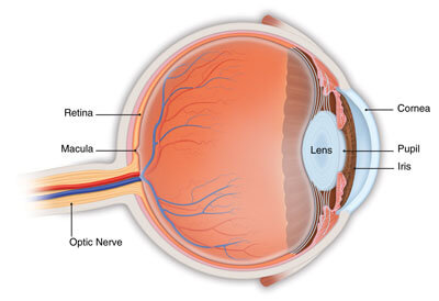
The macula is a small area on the retina at the back of each eye that provides central vision for reading, driving and seeing fine details. As AMD progresses, however, its function deteriorates and a blind spot forms in your field of vision; this condition is one of the leading causes of irreversible legal blindness in those over 60. However, there are treatment options for AMD.
“Wet” AMD occurs when abnormal blood vessels form beneath the retina and leak fluid, blurring central vision. These new vessels are caused by an chemical called vascular endothelial growth factor (VEGF), which doctors can block with eye injections of medication such as Aflibercept (Eylea), Bevacizumab (Avastin) and Ranibizumab (Lucentis), as well as faricimab-svoa (Vabysmo). Doctors administer injections in office; patients often keep their vision even after multiple sessions of injections.
Damage to light-sensing cells in the macula results in gradual vision loss, usually first seen as blurry vision in dim lighting conditions. Over time, however, blind spots develop within your field of vision, making reading or driving increasingly difficult.
Early stage dry AMD typically does not present with symptoms; if vision loss begins occurring, however, patients should consult an eye care professional immediately. Your physician should ask whether there has been any wavy or blank spots in your central vision, or objects are appearing misshaped (metamorphopsia).
Nutritional supplements containing lutein and zeaxanthin may slow progression to intermediate stage AMD. A clinical trial called AREDS (Advanced Research Evaluating Eyecare and Diabetic Retinopathy Study) demonstrated this. These pills significantly slowed development of advanced AMD. Recently, however, beta-carotene was removed due to potential link with lung cancer among smokers in its formula for use within this trial.
Studies are being done on new drugs designed to prevent or delay wet AMD, such as extended-release versions of VEGF inhibitors. Another possible therapy targets complement regulation – an area disrupted by wet AMD; one potential therapy agent being studied is POT-4 which acts on complement component C3.
Lasers
Laser therapy for AMD aims to halt the progression of dry macular degeneration and reduce the size and number of drusen, small white-yellow deposits under the retina characteristic of early AMD. Drusen size and number directly correlate with severity of dry macular degeneration as well as risk for progression to wet AMD (which could result in blindness).
Recent RCT’s have shown that traditional thermal lasers can clear drusen, though this does not prevent progression to wet AMD and its effects are short lived. Researchers are currently developing subthreshold laser energy delivered in short bursts with longer intervals between pulses (known as duty cycles) to minimise thermal diffusion into nearby tissues and achieve similar results without damage to overlying retinal pigment epithelium (RPE).
Before applying a short, low-energy laser beam to the retina, an injection of yellow or green dye into the eye is used to stimulate retinal pigment epithelium to absorb its energy and break down old RPE cells, leading to reduced drusen size and new blood vessel formation that helps slow progression to wet AMD.
Panretinal photocoagulation is an extensive laser treatment for proliferative diabetic retinopathy that uses wide areas of retina. The purpose is to restrict abnormal blood vessel growth and stop its progress into advanced forms.
Photodynamic therapy (PDT), used as an early stage treatment of wet AMD, may offer significant benefit. PDT combines laser and anti-VEGF injections in order to slow progression to advanced wet AMD while simultaneously helping preserve central vision for patients who already have early stage wet AMD. However, no treatments can prevent macular degeneration from progressing further and cause permanent vision loss; PDT cannot stop macular degeneration from taking place and help preserve central vision in early stage wet AMD patients.
Injections
When the retina, or light-sensitive layer in the back of your eye, degenerates, it can lead to fuzzy or “swiss cheese-like” vision and reduce your ability to read, drive, or recognize faces. You can delay its progression by eating healthily, taking supplements or vitamins as directed, not smoking, using proper lighting with UVA/UVB protection in place, exercising regularly and engaging in other healthy habits – as well as by consulting your physician who may perform tests to measure progress of AMD.
AMD typically presents in two forms, dry and wet. Dry AMD typically progresses slowly while wet AMD can lead to faster loss of central vision. Wet AMD occurs when abnormal blood vessels grow beneath the retina and leak fluid or blood, causing vision loss. Furthermore, new blood vessels may bleed into an eye causing even greater vision loss.
Treatment for wet AMD includes injections of anti-angiogenic drugs into the eye. These anti-angiogenic medications work to inhibit new blood vessel formation in the eye and have been shown to significantly decrease vision loss among wet AMD patients. Anti-angiogenic injections should be given every 1-2 months and usually prove well tolerated; side effects are typically mild.
Complement inhibitors may also be utilized as part of a comprehensive wet AMD treatment strategy, inhibiting proteins involved in abnormal blood vessel growth in wet AMD patients. Multiple clinical trials have demonstrated their efficacy by improving visual acuity by 7-12 percent in wet AMD patients. Available compounds have already been discovered, while more are currently in various stages of development.
Researchers from the University of Illinois Chicago have developed a compound that could serve as an effective and safe treatment option for wet age-related macular degeneration (WAMD). It targets endothelial cells lining blood vessels – known as endothelial barrier 3 or EB3 for short. When inhibited, leakage from these blood vessels stops completely, thus eliminating wet AMD. If further studies prove safe and successful for this compound, eyedrop delivery would be an incredible advance over the need for injections to treat WAMD!
Surgery
Macular degeneration is a disease of the retina, the light-sensitive portion at the back of your eye that converts light and images into nerve signals that your brain understands. Most common among those over 60, this disease deteriorates central vision making reading, driving or seeing fine details difficult. Drusen deposits under the retina cause progressive loss of central vision while it may also result in permanent peripheral (side) vision loss causing difficulty walking driving using computers recognising faces or using phones – those suffering wet macular degeneration have higher risks of going blind than those suffering dry macular degeneration due to retinal disease due to retinal nerve signals being sent back out by neurons compared with dry macular degeneration which makes reading hard as central vision fades quickly compared with dry forms.
Wet AMD occurs when abnormal blood vessels form underneath the retina and leak fluid into the macula, a process known as pathological neovascularization that quickly advances the disease.
There are medications available that can slow or stop the formation of new blood vessels, including injecting Lucentis and Avastin directly into your eye and blocking chemical substances that promote their formation. They work by stopping these abnormal blood vessel from growing and can even help stop further formation in their tracks. Injections should take place at your doctor’s office without pain – these injections should not hurt when given by qualified health providers.
Other means of diagnosing wet AMD include conducting a comprehensive eye exam and an Amsler grid test. With an eye exam, the person observes a grid with vertical and horizontal lines; any time these appear broken or faded is indicative of wet AMD.
People with a family history of AMD are at an increased risk for AMD, with smoking being another contributing factor. To reduce your chances of wet macular degeneration it is best to maintain a healthy diet, exercise regularly, and avoid smoking; regular eye exams beginning at age 50 is recommended for best protection.


