


Macular degeneration is the progressive breakdown of the macula, the part of your retina responsible for color perception, fine detail perception and straight ahead vision. Over time it can lead to blind spots or the complete loss of central vision.
Macular degeneration most commonly presents itself in its dry form; however, some individuals may develop wet AMD. This form is marked by abnormal blood vessels growing beneath the retina that leak blood and fluid into it causing rapid central vision loss.
Risk Factors
Macular degeneration is a progressive eye disease that gradually diminishes central vision. This condition results from damage to the macula – the area at the center of your retina that detects light and allows you to read, drive, recognize faces and drive safely – which detects light. While not causing peripheral (side) vision loss or total blindness, macular degeneration does make it more difficult to see straight lines and objects clearly; it is the leading cause of blindness among people over 50 and can develop over several years or quickly over weeks or months (“wet macular degeneration”). Although nobody knows exactly why this disease develops, genetics lifestyle and diet are all major risk factors for macular degeneration.
Age is the primary risk factor and unfortunately something you cannot change, however, you can decrease it by not smoking, limiting exposure to sunlight and other sources of UV radiation, eating healthy diet rich in fruits and vegetables and having regular comprehensive eye exams.
Some individuals who initially suffer from dry AMD may ultimately progress to wet macular degeneration; however, it is less prevalent and harder to treat than its dry counterpart. Wet macular degeneration occurs when new abnormal blood vessels form beneath the retina that leak blood or fluid into the macula; such abnormal vessels may lead to rapid and severe vision loss.
People suffering from wet macular degeneration will often notice straight lines wavying or completely disappearing; sudden loss of central vision; or in extreme cases even complete blindness.
Studies indicate that eating a nutritional diet and supplementing with antioxidants may slow macular degeneration progression, although further study is still needed. Diets rich in lutein and zeaxanthin may provide particular benefit; such foods could include spinach, kale, collard greens, broccoli squash oranges kiwi fruit yellow corn sweet potatoes mangos peaches (your doctor can suggest specific nutritional supplements depending on your personal needs).
Symptoms
Early stage macular degeneration typically does not exhibit noticeable symptoms; as the disease advances, however, people may notice that straight lines appear wavy or colors look faded or less vivid; central vision gradually worsens until eventually people lose the ability to read or drive and can become legally blind. There are two forms of macular degeneration: dry and wet; about 85% to 90% are dry varieties while wet forms occur when abnormal blood vessels grow into retina and leak fluid or blood into it which then scars macula leading to blurring or loss of central vision resulting in legal blindness compared with dry macular degeneration which makes wet varieties much more severe than their counterparts.
Macular degeneration’s exact cause remains unknown, although both genetics and environmental factors play a part. It tends to run in families, increasing with age and smoking status or outdoor time spent. Studies have also suggested a diet lacking certain essential vitamins may increase risk, but more research needs to be conducted in this regard.
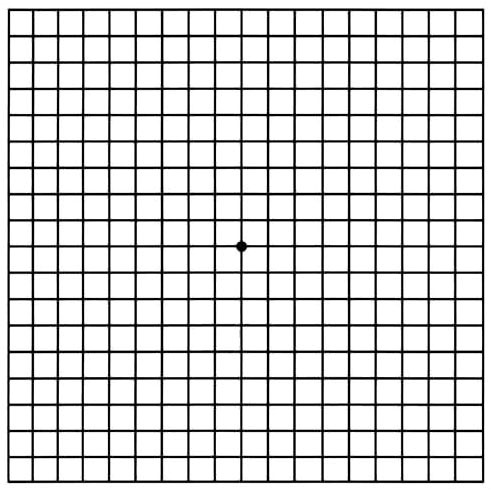
Doctors at NYU Langone use several types of eye exams to detect early signs of macular degeneration. At its earliest stage, doctors look for yellow deposits called drusen in the macula; during an exam they ask you to gaze upon a dot on a grid of horizontal and vertical lines; any time these appear wavy or faded this could indicate macular degeneration.
Ophthalmologists often employ fundus autofluorescence imaging as part of their examination of macular degeneration to take pictures of your macula. This technique takes images of retinal pigments to show doctors how the macular pigmentation changes with time.
Your ophthalmologist can also give you an Amsler grid to use at home for monitoring vision. This chart features both vertical and horizontal lines, and your doctor may ask you to look at it from approximately the same distance that you would view newspaper or book pages. If any lines appear wavy or blurry on an Amsler grid chart, this could indicate macular degeneration.
Diagnosis
Damage to retinal pigment epithelium results in macular degeneration. While for some individuals this disease progresses slowly without severe central vision loss, other may experience sudden central vision loss with symptoms including wavy or distorted straight lines, dark areas of field of vision (blind spots) and lack of detail in color vision. For accurate identification of macular degeneration it is essential that a comprehensive eye exam by an Eye Doctor be conducted.
As part of an eye exam, your ophthalmologist (Eye M.D.) may administer eye drops to dilate or widen your pupils in order to examine the back of your eye and retina with magnifying lenses in search of signs of macular degeneration and other problems.
Most people with dry macular degeneration develop small yellow deposits called drusen in the macula, thought to be caused by retinal pigment epithelium damage. Drusen typically don’t lead to any noticeable vision changes; however, large or multiple drusen that run together or focal pigmentation could indicate increased risk for wet macular degeneration and should be reported immediately to a healthcare provider.
Wet macular degeneration occurs when abnormal blood vessels form beneath the retina and leak blood and fluid into it, interfering with its function and eventually leading to permanent blindness if left untreated. If you suspect wet macular degeneration is occurring in you, regular testing using an Amsler grid may help detect fluid build-up underneath your retina and may suggest possible solutions such as taking antibiotics as soon as possible to manage symptoms.
Fluorescein angiogram is another test used by your ophthalmologist to assess the condition of your retina. This procedure involves injecting fluorescent dye into your arm and using a camera to take pictures as your eye absorbs the dye – your ophthalmologist uses these images to detect leaky blood vessels caused by wet macular degeneration, for instance. It is generally considered safe, though rarely it has caused side effects ranging from nausea to severe allergic reactions.
Treatment
Macular degeneration begins as wastes and nutrients are transported by a layer beneath the retina (called retinal pigment epithelium or RPE), but when this system breaks down, these wastes and nutrients build up, creating yellowish deposits called drusen that often do not cause severe vision loss or blindness. Over time however, as people age their macula ages more gradually the number and size of drusen increases gradually; for some this may interfere with central vision as straight lines appear wavy or as though parts are missing altogether- this could indicate wet macular degeneration.
Wet macular degeneration occurs when new blood vessels sprout, leaking fluid and debris into the retina, disrupting its function and leading to permanent central vision loss if left untreated quickly. Its symptoms include distortion or blurriness of straight lines, wavy or missing spots near your center of vision and dark or empty areas in your visual field.
Photodynamic therapy may be an effective solution to help combat wet macular degeneration, although no cure exists. Eye doctors administer this procedure using anesthetic eye drops before shining light on the retina to activate photosensitive medication that collects in abnormal blood vessels that leak fluid under the retina and clots them off to stop further damage to vision.
There is evidence to suggest that diets high in certain antioxidants may help lower the risk of macular degeneration. Foods rich in lutein and zeaxanthin seem particularly useful, like spinach, collard greens, kale, broccoli, papaya oranges kiwis sweet potatoes. Furthermore, omega-3 fatty acids could provide additional support. They’ve been found to block an enzyme which triggers new abnormal blood vessels under retina and thus reducing wet macular degeneration risk.


