AMD causes central vision to blur over time. Dry AMD develops more slowly due to protein clumps known as drusen forming under the retina and progressing more gradually than wet AMD.
Wet AMD occurs when abnormal new blood vessels form and start leaking blood and fluid. Treatment involves receiving injections of drugs to suppress this growth of abnormal vessels.
Anti-VEGF Drugs
Vascular Endothelial Growth Factor (VEGF) is a protein that stimulates the formation of new blood vessels, necessary for healing wounds and maintaining an effective circulatory system, but too much VEGF may lead to abnormalities such as leaky or swollen vessels in the eye causing decreased vision through age related macular degeneration or retinal vein occlusion diseases; treatment often includes injections of anti-VEGF drugs.
Bevacizumab (Avastin), ranibizumab (Lucentis) and aflibercept (Eylea) are FDA-approved anti-VEGF medications that inactivate the VEGF protein to decrease its effects on eye cells. They may be taken alone or together to treat neovascular AMD and retinal disorders using fine needle injection directly into your eye – your eye doctor will clean and anesthetize the area to minimize pain during this procedure; patients typically receive several injections over several months with complications such as increased intraocular pressure or subconjunctival hemorrrhage occurring as potential adverse events.
As anti-VEGF medications may increase IOP, it’s vitally important that IOP be carefully monitored. To do so safely and responsibly, the AOA suggests scheduling at least two dilated fundus examinations every year for all patients taking injections of bevacizumab, ranibizumab or aflibercept.
Studies have demonstrated that these drugs are more effective than steroids at restoring vision in patients suffering from macular edema due to retinal vein occlusion or central macular degeneration. Furthermore, they have also been associated with lower rates of RPE tears, cataract development/progression and conjunctival hemorrhage.
Keep in mind that while these drugs may be effective, they have not been shown to prevent permanent vision loss. Therefore, they should only be used as short-term therapy options along with other treatments like laser surgery and nutritional supplements.
Laser Surgery
Laser surgery allows eye care professionals to use light beams to destroy new blood vessels that form under the retina in wet AMD, thus slowing vision loss and helping protect sight. Although not suitable for every situation, speak with your eye care provider before deciding if laser surgery is right for you.
Wet macular degeneration occurs when abnormal blood vessels form under the retina and leak or bleed, damaging the macula, leading to distortion in central vision and blind spots in one or both eyes. A diagnosis of wet AMD increases your risk for it in other eyes; so, early treatment for wet AMD should be sought to avoid more severe vision loss.
Your eye care provider may recommend laser photocoagulation to treat wet AMD, which involves using a light beam to burn or destroy blood vessels in your eye care provider’s office and usually lasts less than an hour. As an alternative, photodynamic therapy might also be an option; with this procedure, verteporfin will be administered throughout your body before your provider shines a bright light onto your retina for about 90 seconds and activated with light, binding to newly formed blood vessels before closing off with more gradual vision loss over time.
Femtosecond laser photocoagulation was successfully employed in one study to treat wet AMD patients suffering from choroidal neovascularization. It significantly decreased vision loss among those who experienced CNV beneath the foveal area of retina (FAZ). The laser used here was similar to that employed during SMILE surgery techniques which employ lasers for incision and placement of precalculated mini lenses inside cornea. Not only can this kind of femtosecond laser be used against wet AMD, but other eye conditions including cataracts and Keratoconus; currently the ZEISS VisuMax system is the only commercially available option.
Fluorescein Angiography
Your retina (the membrane lining the back of your eye) transforms light into electrical impulses that your brain can interpret as sight. Your central vision comes from an area in the retina called the macula which contains millions of light-sensing cells to provide clear images. Dry AMD results from cell breakdown which leads to gradual loss of central vision over time; scientists do not yet fully understand why.
Wet macular degeneration is much less prevalent and occurs when new blood vessels form behind the retina and leak blood and fluid, moving the macula out of its usual position at the back of the eye and lifting it upward. It is a much more serious condition which may lead to rapid loss of central vision.
As part of this test, an orange dye called sodium Fluorescein will be injected intravenously and travel quickly to your blood vessels at the back of your eye, where a special camera records its progress through them. This test allows us to identify abnormal blood vessel leakage associated with wet macular degeneration, diabetic retinopathy and occlusive diseases such as retinal vein or artery occlusions.
Before your test begins, we will ask about any allergies and medications you are currently taking. A medication will then be given to prevent an allergic reaction from the dye used during this test; though mild reactions such as flushing or nausea may arise; usually this passes quickly with time. Your urine may turn orange as this dye exits your system within several hours.
Fluorescein angiography can help identify treatable vascular disease in wet macular degeneration and diabetic retinopathy, helping determine its severity as well as determining whether laser treatment may be a feasible option for you. The procedure itself is quick and painless but you may experience some slight discomfort as the dye enters your bloodstream.
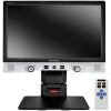
Optical Coherence Tomography
Optical Coherence Tomography (OCT) is an imaging test that uses light waves to create extremely detailed pictures of your eye, enabling your doctor to see layers within your retina and macula cross-sectionally. OCT can help detect glaucoma as well as age-related macular degeneration; furthermore it serves to monitor treatment effects as well as diagnose disease conditions such as these.
Under OCT, near-infrared light passes through your eye. A portion of it reflects off structures within it while other portions transmit back to a detector where its delay comparison determines a three dimensional image of eye tissue based on which to calculate optical coherence of your eyes.
Your retina is a paper-thin layer lining the back of your eye that transmits visual signals to your brain. The macula, located at the central portion of your retina, provides central vision that allows you to clearly see details. Age-related macular degeneration (AMD), however, slowly destroys this central vision resulting in blindness for those over 50 in the United States.
Macular degeneration comes in two varieties – wet and dry. Dry macular degeneration usually progresses more slowly, without leading to any noticeable peripheral vision loss; you may only lose straight line visibility. On the other hand, wet macular degeneration progresses more aggressively and causes central vision deterioration more quickly than dry degeneration does.
OCT allows your doctor to evaluate the thickness of your optic nerve fiber layer and ganglion cell complex as well as fluid circulation within your eye. OCT is a noninvasive procedure that does not use radiation or any harmful agents – similar to the technology cardiologists use during cardiac catheterization to inspect blockages in heart arteries.