

Glaucoma is a group of eye conditions that can lead to irreversible vision loss if left untreated. One particular type of glaucoma is secondary glaucoma, which occurs as a result of an underlying eye condition or other health issues. In this blog, we will delve into the causes, symptoms, prevention, treatment options, and prognosis of secondary glaucoma, addressing common questions related to each aspect.
Causes of Secondary Glaucoma
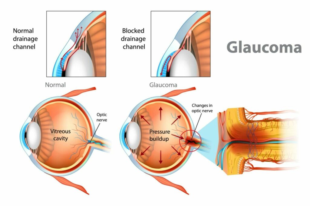
Secondary glaucoma is caused by various factors that affect the drainage of fluid (aqueous humor) from the eye. Some common causes include:
Eye trauma
Traumatic injuries to the eye can disrupt the normal flow of fluid, leading to increased intraocular pressure (IOP) The following are some of the processes by which eye damage can increase pressure:
Disruption of the drainage system
The eye has a sophisticated drainage system that keeps the fluid (aqueous humor) balance inside the eye in check. Trauma to the eye can harm components like the trabecular meshwork, which drains the fluid. IOP rises when the drainage system is compromised because the aqueous humor cannot exit the eye effectively.
Blockage of the drainage angle
Eye injuries can result in an accumulation of blood or debris in the drainage angle, which is the junction of the iris and cornea. This obstruction may prevent the aqueous humor from draining properly, increasing the pressure inside the eye.
Swelling and inflammation
Eye damage may result in an inflammatory response. IOP might rise as a result of drainage system dysfunction brought on by inflammation and edema.
Bleeding
Serious eye injuries may result in internal bleeding that builds up in the anterior chamber of the eye. The drainage routes may become blocked by blood, which can increase intraocular pressure.
It’s important to remember that eye trauma can range in severity, and the spike in IOP that follows can be modest to severe. Furthermore, the onset of secondary glaucoma may take weeks or months following the initial lesion rather than occurring right away.
Eye surgery
Certain eye surgeries, such as cataract surgery, can trigger secondary glaucoma due to changes in the drainage angle or other structures.
Eye inflammation
Conditions like uveitis, iritis, or severe conjunctivitis can cause secondary glaucoma by affecting the drainage system or producing excess fluid.
Medications
Long-term use of corticosteroids, especially in eye drops or oral forms, can increase the risk of secondary glaucoma. The exact mechanism by which corticosteroids cause an increase in IOP is not fully understood, but there are several proposed mechanisms:
Reduced outflow of aqueous humor
Corticosteroids are believed to affect the trabecular meshwork, which is responsible for draining the aqueous humor from the eye. They can alter the meshwork’s structure and function, reducing fluid outflow and leading to an accumulation of aqueous humor in the eye, thereby increasing IOP.
Increased production of aqueous humor
Corticosteroids can stimulate the production of aqueous humor by the ciliary body, which is responsible for its production. When there is excessive production of fluid and the drainage system cannot compensate, it can increase IOP.
Altered tissue response to inflammation
Corticosteroids have potent anti-inflammatory properties. While this can be beneficial in managing various eye conditions, it can also interfere with the eye’s natural response to inflammation. Inflammation can sometimes contribute to elevated IOP, and by suppressing this response, corticosteroids can indirectly lead to increased pressure.
It’s important to note that not everyone who uses corticosteroids will experience an increase in IOP. The risk is influenced by several factors, including the duration and dosage of corticosteroid use, the method of administration, and individual susceptibility.
Tumors or neovascularization
In some cases, tumors or abnormal blood vessel growth can obstruct the drainage system, leading to increased IOP. Neovascularization, also known as abnormal blood vessel growth, can occur in various parts of the body, including the eye. In the context of secondary glaucoma, neovascularization can lead to increased intraocular pressure (IOP) and contribute to the development of the condition. Some underlying factors and conditions that can cause neovascularization in the eye include:
Retinal ischemia
Retinal ischemia refers to a condition in which the blood supply to the retina, the light-sensitive tissue at the back of the eye, is reduced. When the retina does not receive adequate oxygen and nutrients, it triggers a response to promote the growth of new blood vessels. However, these new vessels are fragile and abnormal, leading to neovascularization.
Retinal ischemia can occur due to various causes, including:
Diabetic retinopathy
Diabetes can damage blood vessels in the retina, causing retinal ischemia and subsequent neovascularization.
Retinal vein occlusion
A blockage in the veins that drain blood from the retina can lead to retinal ischemia and neovascularization.
Retinal artery occlusion
A blockage in the arteries supplying blood to the retina can cause retinal ischemia and trigger abnormal blood vessel growth.
Ocular ischemic syndrome
Ocular ischemic syndrome is a condition characterized by inadequate blood flow to the eye, often due to severe carotid artery disease or other vascular disorders. The reduced blood flow can result in retinal ischemia and subsequent neovascularization.
Central retinal vein occlusion (CRVO)
CRVO occurs when there is a blockage in the central retinal vein, impeding the outflow of blood from the retina. The backup of blood can lead to retinal ischemia and the growth of abnormal blood vessels.
Rubeosis iridis
Rubeosis iridis refers to the presence of abnormal blood vessels on the iris, the colored part of the eye. It can occur due to various underlying conditions, such as:
Ischemic central retinal vein occlusion (CRVO)
The ischemia in CRVO can stimulate the growth of new vessels on the iris.
Carotid artery occlusive disease
Severe narrowing or blockage of the carotid arteries, which supply blood to the head and neck, can cause rubeosis iridis.
Chronic uveitis
Long-standing inflammation in the eye, such as chronic uveitis, can lead to neovascularization on the iris.
Common Questions:
Can secondary glaucoma develop in both eyes simultaneously?
Yes, it is possible for secondary glaucoma to affect both eyes at the same time, especially if the underlying cause affects both eyes equally.
Can secondary glaucoma be inherited?
Secondary glaucoma is typically not inherited, as it arises as a result of an underlying condition or injury. However, the predisposing factors for the underlying condition may have a genetic component.
Symptoms of Secondary Glaucoma
The symptoms of secondary glaucoma are similar to those of primary open-angle glaucoma, the most common form of glaucoma. However, symptoms may vary depending on the underlying cause. Some common symptoms include:
- Gradual or sudden loss of peripheral vision.
- Blurred vision or difficulty focusing.
- Severe eye pain or headache.
- Redness in the eyes.
- Halos around lights.
- Nausea or vomiting (in acute cases).
- Eye pressure or discomfort.
Common Questions:
Is secondary glaucoma always associated with pain?
Not always. While some individuals may experience eye pain, others may have no pain at all. The presence of pain depends on the underlying cause and the stage of the disease.
Can secondary glaucoma cause blindness?
If left untreated, secondary glaucoma can lead to permanent vision loss and blindness. Early detection and timely treatment are crucial for preserving vision.
Diagnosis of Secondary Glaucoma
Diagnosing secondary glaucoma involves a comprehensive eye examination and an evaluation of the underlying condition or cause contributing to the development of glaucoma. The diagnostic process may include the following:
Medical history and symptoms
The eye care specialist will discuss your medical history, including any previous eye conditions, surgeries, or trauma, as well as symptoms you may be experiencing, such as changes in vision, eye pain, or discomfort.
Visual acuity test
This test measures how well you can see at various distances using an eye chart. It helps determine if there are any visual impairments or changes in vision.
Tonometry
Tonometry is performed to measure the intraocular pressure (IOP). It can help identify elevated IOP, which is a key characteristic of glaucoma. Various methods can be used, including the use of a tonometer that gently touches the surface of the eye or a non-contact tonometer that measures pressure without touching the eye.
Gonioscopy
Gonioscopy is a procedure that allows the eye care specialist to examine the drainage angle of the eye. This evaluation helps determine if there are any blockages or abnormalities in the angle that may be contributing to increased IOP.
Optic nerve examination
The health of the optic nerve is crucial in diagnosing glaucoma. The eye care specialist may use various tools, such as an ophthalmoscope or a special lens, to examine the optic nerve for any signs of damage, such as cupping or thinning of the nerve fibers.
Visual field testing
Visual field testing, also known as perimetry, assesses your peripheral vision. It helps detect any loss of vision or changes in the visual field, which can indicate glaucoma.
Additional tests
Depending on the suspected underlying cause of secondary glaucoma, additional tests may be necessary. These tests can include imaging tests such as optical coherence tomography (OCT) or ultrasound biomicroscopy (UBM) to evaluate the structures within the eye, or blood tests to assess systemic conditions that may be contributing to glaucoma.
The diagnosis of secondary glaucoma relies on identifying both the signs of glaucoma (elevated IOP, optic nerve damage, visual field loss) and the presence of an underlying condition or cause that is contributing to its development. The eye care specialist will use the information gathered during the examination and tests to make an accurate diagnosis and determine the most appropriate treatment plan. Regular follow-up visits and monitoring are essential to manage and control secondary glaucoma effectively.
Prevention of Secondary Glaucoma
Although secondary glaucoma is often caused by underlying conditions that may not be preventable, certain measures can help reduce the risk or delay its onset:
Regular eye exams
Routine eye examinations can help detect early signs of glaucoma and identify any underlying conditions that may lead to secondary glaucoma.
Safety precautions
Wearing protective eyewear during sports or hazardous activities can minimize the risk of eye trauma.
Medication awareness
Discuss potential side effects with your healthcare provider when using corticosteroids or any medications known to increase the risk of glaucoma.
Common Questions:
Can lifestyle choices influence the development of secondary glaucoma?
While lifestyle choices do not directly cause secondary glaucoma, maintaining a healthy lifestyle, including regular exercise, a balanced diet, and avoiding smoking, can contribute to overall eye health.
Can secondary glaucoma be prevented if the underlying condition is treated?
Treating the underlying condition promptly and effectively can minimize the risk of developing secondary glaucoma. Regular follow-ups with your healthcare provider are important to monitor any potential changes in your eyes.
Treatment Options for Secondary Glaucoma
The treatment of secondary glaucoma focuses on managing the underlying cause and reducing intraocular pressure. Treatment options may include:
Medications
Eye drops, oral medications, or both may be prescribed to reduce eye pressure by increasing fluid drainage or decreasing fluid production.
Laser therapy
Procedures such as laser trabeculoplasty or laser iridotomy can help improve fluid drainage and reduce intraocular pressure.
Surgery
In some cases, surgical intervention may be necessary to create a new drainage pathway or remove blockages.
Common Questions:
Is surgery the only option for treating secondary glaucoma?
Surgery is not always the first-line treatment for secondary glaucoma. It is usually considered when other treatment options have failed to control the intraocular pressure or if the underlying cause necessitates surgical intervention.
Are there any alternative or complementary treatments available?
While alternative and complementary therapies may be explored, it is essential to consult with an eye care specialist before considering any such treatments. These therapies should never replace conventional medical treatment.
Prognosis of Secondary Glaucoma
The prognosis of secondary glaucoma largely depends on various factors, including the underlying cause, the severity of the disease, and the timeliness of treatment. With early diagnosis and appropriate management, vision loss can often be prevented or minimized. However, if left untreated or uncontrolled, secondary glaucoma can lead to irreversible vision impairment and even blindness.
Common Questions:
Can vision loss caused by secondary glaucoma be restored?
Unfortunately, once vision loss has occurred, it cannot be fully restored. The goal of treatment is to preserve existing vision and prevent further deterioration.
How often should individuals with secondary glaucoma undergo eye examinations?
The frequency of eye examinations will depend on the severity of the condition, the response to treatment, and the advice of the eye care specialist. Regular follow-ups are crucial to monitor the progression of the disease and make necessary adjustments to the treatment plan.
Conclusion
Secondary glaucoma is a complex condition that can arise due to various underlying causes. Early detection, timely treatment, and appropriate management of the underlying condition are vital for preserving vision. By understanding the causes, symptoms, prevention strategies, treatment options, and prognosis of secondary glaucoma, individuals can take proactive steps toward maintaining good eye health and minimizing the risk of irreversible vision loss. Remember, regular eye exams and open communication with your healthcare provider are key to managing and preventing secondary glaucoma effectively.


