An estimated 11 million Americans suffer from age-related macular degeneration. The macula in the eye, which is important for aiding our ability to effectively see minute details, degenerates as a result of this condition. People with AMD may find it challenging to continue engaging in routine activities like reading and driving. AMD can occasionally cause irreversible vision loss and possibly blindness.
Macular Degeneration is a very common eye disease that damages the central part of the retina.
The region of the retina known as the macula is where the brain receives images for your central vision. Your center vision darkens or gets fuzzy when this region is affected. It could eventually be blurred out entirely. Although peripheral vision isn’t affected by this disease, you don’t see as much detail about it.
The leading cause of blindness in American adults aged 50 and older is age-related macular degeneration (AMD). AMD is more prevalent than glaucoma and cataracts (cataracts can be fixed therefore don’t cause blindness as often). Macular degeneration is uncommon in children and young adults, but Stargardt macular degeneration is a genetic disorder that affects around 1 in every 8,000 to 10,000 people.
There are certain outside variables that can raise the likelihood of getting macular degeneration or cause the illness early in life, while genetics and age are the two factors that AMD is most strongly linked to. These include smoking cigarettes and using vasodilator blood pressure drugs (more on this later).
Macular degeneration drugs to avoid
Patients frequently inquire about the potential impact of prescription drugs on the risk of macular degeneration-related visual loss (AMD). In patients with the wet form of the condition, one class of treatments can cause more severe vision loss, but another family of medications may have a positive impact.
Warfarin effects on Wet AMD
Luckily, very few prescription medications have any negative consequences that are known to exist for persons with AMD. One exception is the anticoagulant blood thinner warfarin (Coumadin®). Doctors frequently prescribe this blood thinner to people who have specific cardiovascular diseases that might be fatal. According to studies, those who have wet AMD, which results in bleeding from irregular blood vessels in the macula, may experience more subretinal hemorrhages if they take warfarin. More severe visual loss may result from this. In people with dry AMD, warfarin does not raise the risk of visual loss.
Even if they have wet AMD, some people must take warfarin to safeguard their lives. Other blood thinners, such as aspirin, which has not been demonstrated to enhance the number of retinal hemorrhages, can be switched to by people with wet AMD. Your ophthalmologist and other medical professionals can create a treatment strategy that is ideal for you. Never modify your medicine without first consulting your doctor.
Green leafy vegetables’ ability to provide vitamin K can counteract warfarin’s anti-clotting properties, which is another problem for those taking the medication. Because leafy greens make it more difficult to control the warfarin dose, some individuals using warfarin may be advised not to eat them. However, it has been demonstrated that consuming these same leafy greens lowers the risk of AMD-related visual loss. For many people, eating a certain quantity of leafy greens each day and routinely getting a blood test for warfarin are the best solutions. The test is known as an INR (international normalized ratio), and it assesses blood clotting time to assess how effectively warfarin is functioning.
Statins Have a Positive Benefit
Statins, a family of medications that lower blood lipid levels, particularly cholesterol, may also have an impact on AMD. The research on the effects of statins is currently conflicting. The number of drusen, which are abnormal lipid/protein deposits underneath the retina in people with early dry AMD, can be reduced by taking Lipitor® (atorvastatin) at a somewhat high dose (80 mg), and in some cases, this can improve vision in those with extremely big drusen directly in the macula’s center. Which patients are most likely to benefit will be determined through a bigger (phase III) research trial.
Blood pressure medications added an increased risk for those with dry AMD
Although there is a link between high blood pressure and AMD, the data on this topic has been weak throughout the years. However, several medical studies have also linked the use of vasodilators to decrease blood pressure to an increased risk of AMD.
Any vasodilator raised the likelihood of developing early-stage AMD, according to lengthy research conducted at the University of Wisconsin School of Medicine from 1988 to 2013. Over several decades, the research looked at the eye health of 5,000 individuals. About 8.2% of those who weren’t using vasodilators such as Apresoline and Loniten showed early symptoms of age-related macular degeneration. 19.1 percent of persons who used vasodilators eventually got the condition.
Additionally, the study group discovered that oral beta blockers like Tenormin and Lopressor raised the danger of neovascular or wet AMD. About 0.5% of those who weren’t using beta blockers got wet AMD. About 1.2 percent of the individuals on beta blockers experienced this more severe type of macular degeneration.
If your doctor gives you a blood pressure medicine, you probably have a chronic illness that has to be managed in order to enhance your quality of life. In particular, if age-related macular degeneration runs in your family or you already have the disease, talk to your doctor about the potential hazards of taking this medication.
Other Medications that are Toxic to the Macula
Many medications can be toxic to the retina. Several of them may be categorized by anatomical location or kind of toxicity, but several of these drugs also have distinct effects when taken alone. We will just highlight the ones that affect the macula. Some are reversible and some aren’t. Most of them don’t actually cause macular degeneration however they can cause macular damage or add to an already strained macular health condition. For maximum macular health check out the vitamins below.

Pigmentary Retinopathy
Quinolénes
Traditional antimalarial drugs like chloroquine (Aralen) and hydroxychloroquine (Plaquenil) are now utilized to treat autoimmune disorders including (RA) rheumatoid arthritis and (SLE) systemic lupus erythematosus. Both drugs have been found to bind melanin and concentrate in the retinal pigment epithelium, ciliary body, and iris, affecting normal physiological function.
Early on, individuals may have no symptoms at all, exhibiting merely RPE granular pigmentary alterations and blunting of the foveal reflex (the fovea is the center of the macula). Symptoms of progression might include blind spots, light issues, and reduced vision.
Thioridazine
With the introduction of atypical antipsychotic medications, the usage of the piperadine antipsychotic thioridazine (Mellaril) has decreased. Since its initial description in 1980, ocular toxicity has been linked to both short- and long-term treatments.
Vision loss and visual color changes are signs of toxicity. Alterations occur in the macular pigment on clinical examination.
Deferoxamine
Hemochromatosis is one disorder that is treated with deferoxamine (Desferal), an iron-chelating medication. It can also be administered intravenously or subcutaneously to treat aluminum toxicity.
Scotomas (blind spots), dyschromatopsia (color vision issues), and vision loss are examples of ocular poisoning symptoms. The macular RPE is affected by this medication.
With the termination of the medicine, deferoxamine toxicity can be reversed, resulting in complete recovery of visual function. However, the drug is frequently prescribed for chronic illnesses that need ongoing treatment, such as thalassemia. To identify any early vision loss in these individuals, periodic ocular examinations must be performed.
Macular Edema
Latanoprost
Reversible cystoid macular edema can be brought on by latanoprost.
Epinephrine
In the United States, the sympathomimetic medication epinephrine and its precursor dipivefrin are no longer utilized as the main form of treatment for glaucoma.
Within weeks to months of starting medication, patients report impaired or reduced eyesight.
Niacin
Niacin, often known as vitamin B3, is used to treat hyperlipidemia, hypercholesterolemia, and pellagra. The most frequent systemic adverse effect is face flushing, however, patients might also experience metamorphopsia, impaired vision, and reduced eyesight.
Niacin withdrawal will progressively lessen CME and improve your vision.
Rosglitazone
An oral thiazolidinedione drug called rosiglitazone (Avandia) is used to reduce insulin resistance in type 2 diabetes. A case study from 2005 detailed how rosiglitazone caused diabetic macular edema to progress.
The DME and peripheral edema may improve quickly upon drug withdrawal, but in most cases, these conditions resolve more slowly and may necessitate supplementary therapy with focused laser photocoagulation.
Tamoxifen
A selective estrogen receptor modulator called tamoxifen is used to treat breast cancer. It is an amphiphilic substance that can build up in lysosomes and result in oxidative harm.
Patients may not initially exhibit any symptoms but later report dyschromatopsia and impaired eyesight. While therapeutic dosages are substantially lower, ocular problems have been reported most frequently at doses larger than 120 mg twice per day. The dosage for the current anti-neoplastic regimens is 20 mg per day at first, then 40 mg per day as needed.
Clinically, a fundus exam will reveal intraretinal crystalline deposits that are refractile and are largely localized in the perifoveal macula.
Annual ophthalmologic examinations are advised even though the current therapeutic dose levels are lower than those that have been found to cause crystal deposition. If crystals are noticed, it is advised to stop using tamoxifen. However, intraretinal crystalline deposits may linger over time even if visual function improves as the CME passes.
Cyanaxanthine
Previously employed as an oral tanning agent, canthaxanthine is a vitamin A derivative used in the treatment of psoriasis and eczema. After high-dose oral administration of more than 0.5 mg/kg/day, toxicity has been demonstrated. Patients are often asymptomatic, and poisoning is diagnosed based on both a clinical examination and an intake history.
Your doctor may see that the fovea is encircled by a doughnut-shaped ring of golden intraretinal deposits. Although FA, ERG, and color vision are frequently normal, OCT shows crystalline deposition in the inner retinal layers. Visual field testing, which can be useful in identifying toxicity, reveals a dose-dependent decline in retinal sensitivity.
Ophthalmologic examinations should be performed routinely to monitor patients. The retinal crystals may take years to go away once medication use is stopped, and patients frequently have no symptoms.
Digoxin
A cardiac glycoside called digoxin (Lanoxin) is used to treat congestive heart failure, atrial fibrillation, and atrial flutter. Although systemic toxicity is uncommon when plasma digoxin content is less than 0.8 g/L, it has a limited therapeutic window.
High dosages frequently cause ocular adverse effects, with signs and symptoms ranging from blurred vision to photopsias, xanthopsia, and scotomas.
Both of the visual problems gradually away once digoxin is discontinued over many weeks as the drug is metabolized.
Sildenafil
In order to treat erectile dysfunction, sildenafil (Viagra), a selective inhibitor of cGMP-specific phosphodiesterase type 5 (PDE5), relaxes smooth muscle and dilates blood vessels. While sildenafil is selective for PDE5, it also affects cGMP levels in the retina by blocking the retina-specific PDE6.
Patients may complain of hazy vision, photophobia, and cyanopsia when they first report visual difficulties. On examination, there are often no fundoscopic alterations and no decrease in visual acuity. Within four to six hours of taking the drug, symptoms go away.
Summary
Patients seldom associate visual symptoms with their medications. However, it is not unexpected that a variety of pharmaceuticals can impair vision when you consider that your eyes are actually simply an extension of your brain, an organ that is particularly susceptible to numerous chemicals.
Speak with your prescribing doctor and/or ophthalmologist if you have any questions or would like additional details about any of the medications on this list and their potential associations with macular degeneration. You might also want to talk about whether using the medicine on this list is worth the higher risk of side effects compared to using alternative therapies. Mention any prescription drugs, over-the-counter medicines, and other vitamins you are taking. What adverse effects, if any, did you encounter? Always remember that careful interactions with your healthcare providers are irreplaceable.
FAQs for Macular degeneration drugs to avoid
Which medications lead to macular degeneration?
Topical epinephrine, nicotinic acid, topical prostaglandin analogs (such as latanoprost), antimicrotubule medicines (paclitaxel, docetaxel), fingolimod, imatinib, glitazones (rosiglitazone, pioglitazone), and trastuzumab are just a few of the substances that can induce cystoid macular.
Can ibuprofen cause macular degeneration?
Results. The frequency and duration of aspirin or ibuprofen usage reported at baseline did not appear to be associated with AMD.
What Alternative Treatments Are Available for AMD?
- Beware of beta-carotene if you smoke, quit smoking.
- Increase your vegetable intake, particularly leafy greens.
- Immediately cut back on your sugar intake.
- Eat more meals high in omega-3 fatty acids, such as fish.
- Consume more fruit, particularly fiber-rich fruit.
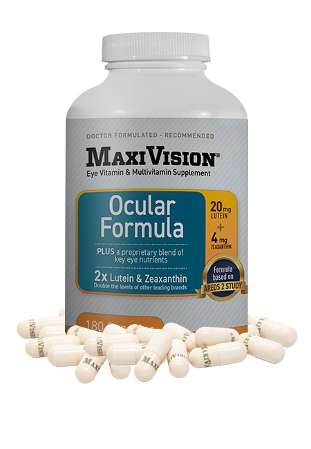
- Take a AREDS formulation ocular vitamin


