Medically reviewed by Shaun Larsen, OD – Doctor of Optometry, Southern College of Optometry.
Last clinically reviewed: July 2026 | Next scheduled review: July 2027
In brief: If you have been told you have a large optic nerve, large optic disc, or “large cup”, this most often describes normal anatomy rather than disease. The optic disc varies severalfold in size between healthy people, and because the neural tissue passing through it is broadly similar in amount, a bigger opening produces a bigger central cup. That large physiologic cup is the single most common reason people are labelled glaucoma suspects and monitored for years without ever developing glaucoma. Distinguishing a big normal nerve from a damaged one is a structural and functional question – answered by OCT, visual fields, and serial comparison – not a question that can be settled by the cup-to-disc ratio alone.
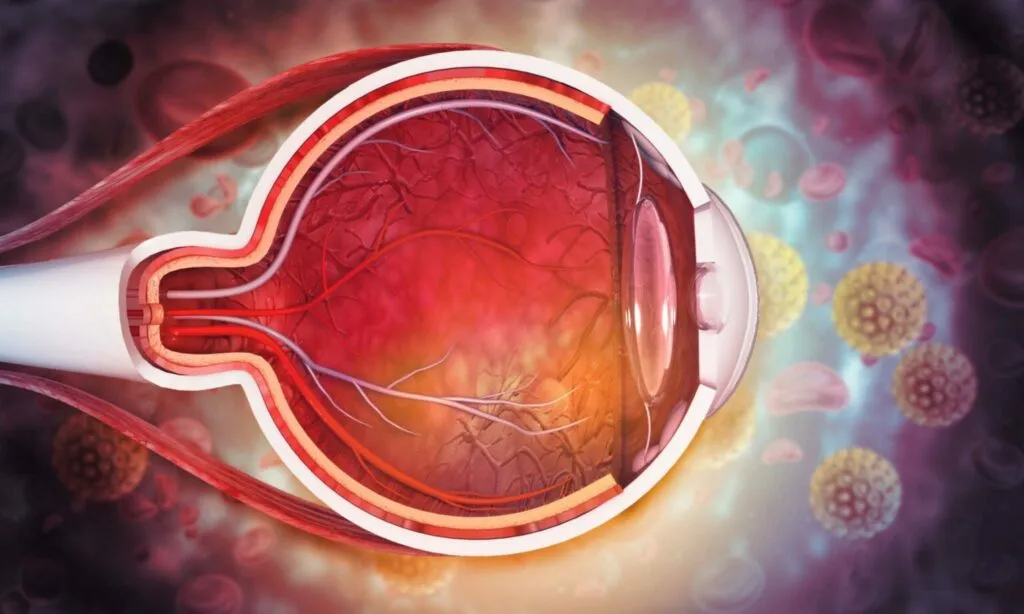
The optic nerve is one of the most important parts of the human eye, a marvel of biological ingenuity. The optic nerve, frequently referred to as the “cable” that enables eye-to-brain communication, is crucial to our capacity for visual perception. It carries roughly 1.2 million retinal ganglion cell axons from the eye to the lateral geniculate nucleus. Those axons converge at the optic nerve head and turn posteriorly through the scleral canal, passing through a sieve-like collagen structure called the lamina cribrosa. The size of that canal opening is what determines optic disc size – and it is set during embryological development, not acquired later. Some people may have a larger-than-average optic nerve, which can be a condition that raises questions about eye health. This issue is referred to as a “large optic nerve without glaucoma.” This in-depth blog post will cover every facet of the subject, from understanding the optic nerve’s architecture to examining possible causes, and implications for vision, diagnostic techniques, and the function of low vision services in improving visual outcomes.
Are you looking for “large” or “swollen”? They are different findings
A large optic disc means the disc is physically wide – a big anatomical opening. It is present from childhood, unchanging, symptomless, and in isolation requires no treatment. It matters only because it makes glaucoma harder to diagnose.
A swollen optic disc means the nerve head is elevated and edematous, with blurred margins and obscured vessels. It develops over hours to weeks and is always abnormal. Causes include raised intracranial pressure (papilledema, often bilateral), optic neuritis, non-arteritic and arteritic anterior ischaemic optic neuropathy, malignant hypertension, infiltration and compression. Bilateral disc swelling with headache, transient visual obscurations, or pulsatile tinnitus requires urgent neuro-imaging and assessment. If that is what you have been told, this article is not about your finding – seek prompt specialist evaluation.
Pseudopapilledema is a third category worth knowing: a disc that looks swollen but is not. The usual cause is optic disc drusen – calcified deposits within the nerve head, which are more common in small, crowded discs. Autofluorescence, B-scan ultrasound and enhanced-depth OCT distinguish drusen from true oedema and can spare a patient an unnecessary lumbar puncture and MRI.
A Large Optic Nerve Without Glaucoma: What Is It?
An “enlarged optic nerve head” or “optic disc,” as well as “large optic nerve,” are terms used to describe an optic nerve that seems to be larger in diameter than what is regarded as normal. When an eye care expert examines the optic nerve, the optic nerve head is the portion that is visible inside the eye.
The actual numbers. Across normal eyes, optic disc area ranges from roughly 0.80 mm² to about 6.0 mm² – the largest healthy discs are several times the area of the smallest. Mean disc area in a typical population is around 2.1–2.8 mm². In terms of diameter, the average disc is approximately 1.75 mm horizontally and 1.90 mm vertically (the disc is normally a slightly vertical oval). Conventional cut-offs place small discs below about 1.5 mm vertical diameter and large discs above about 2.2 mm, with disc areas above roughly 2.8–2.9 mm² commonly treated as the upper limit of normal in research settings. A disc that is large but otherwise structurally and functionally normal is sometimes called megalopapilla.
Disc size varies systematically by ancestry and refraction. Mean disc area is generally largest in people of African ancestry, followed by Asian and Hispanic populations, and smallest in those of European ancestry. High myopia (beyond about −8 D) is associated with larger discs; significant hyperopia (beyond about +5 D) with smaller ones. This is population-level anatomy, not pathology – but it has a direct clinical consequence: applying a single cup-to-disc threshold across all patients systematically over-refers people with naturally large discs and under-refers those with small ones.
The presence of glaucoma, a set of eye disorders defined by progressive retinal ganglion cell loss with characteristic optic nerve head and visual field change, is not always indicated by having a large optic nerve. It is critical to distinguish between a large optic nerve that is glaucoma-free and one that is glaucoma-affected, as these conditions can have quite distinct effects on vision and eye health.
The key relationship: cup size scales with disc size
This is the concept the whole topic turns on, and it is worth stating plainly.
The amount of neural tissue crossing the disc – the neuroretinal rim – is broadly comparable between healthy eyes, because axon counts are similar. The size of the opening those axons pass through is not. When a similar quantity of nerve fibre passes through a wider canal, it does not fill it, and the central space left over is the cup. So:
- Large disc → large physiologic cup → high cup-to-disc ratio that is entirely normal. Cup-to-disc ratios in normal populations extend from 0.0 to approximately 0.9. A vertical ratio of 0.7 in a genuinely large disc can be completely healthy.
- Small disc → small cup → low cup-to-disc ratio, which can conceal real glaucomatous damage. A vertical ratio of 0.4 in a small crowded disc may already represent significant rim loss.
The clinical consequence is a well-documented asymmetry in diagnostic error: clinicians tend to over-diagnose glaucoma in large discs and under-diagnose it in small discs. Neuroretinal rim area also scales with disc size – larger discs have larger absolute rim area even though the rim can look proportionally thin – which is why rim assessment must be interpreted against disc size rather than in isolation.
How disc size is actually measured. In clinic, a 90 D or 78 D condensing lens is used at the slit lamp and the vertical beam height is adjusted to the disc margins; the reading is multiplied by a lens-specific correction factor (commonly around 1.3 for a 90 D and 1.1 for a 78 D lens). With a direct ophthalmoscope, the 5-degree spot approximates an average disc – a disc contained within the spot is small, one exceeding it is large. Disc–fovea distance in disc diameters provides another chairside estimate. OCT measures Bruch’s membrane opening directly and is the most reproducible method available.
Fundamentals of Ocular Hypertension
A condition known as ocular hypertension occurs when the intraocular pressure, or IOP, is higher than it should be. Even though glaucoma is far more likely to develop in those with elevated intraocular pressure, this is not always the case. Optic disc size does not influence measured IOP. The measurement artefact that genuinely matters is central corneal thickness: Goldmann applanation tonometry assumes an average corneal thickness, so thick corneas cause pressure to be over-estimated and thin corneas cause it to be under-estimated. Pachymetry should therefore be part of any glaucoma-suspect workup.
Ocular hypertension is often identified during a normal eye exam and might occur without any obvious symptoms. To determine their risk of developing glaucoma, ocular hypertensive patients must undergo regular monitoring.
What the evidence says about risk. The Ocular Hypertension Treatment Study (OHTS) followed 1,636 participants with raised IOP and normal discs and fields. Over five years, roughly 9.5% of untreated participants developed primary open-angle glaucoma, compared with about 4.4% of those given pressure-lowering treatment. Critically, OHTS also identified which baseline factors predict conversion: older age, higher IOP, thinner central corneal thickness, larger vertical cup-to-disc ratio, and greater visual field pattern standard deviation. Combining these stratified patients into groups with five-year risk of about 1–2% at the low end and 25–35% at the high end.
That stratification is the practical takeaway: ocular hypertension is not one condition requiring one response. Many patients are safely observed; a minority warrant treatment. And note that cup-to-disc ratio appears in that risk model without correction for disc size, which is exactly why a large physiologic cup can inflate an individual’s apparent risk score and why disc size must be factored in when the model is applied to a real patient.
Physiologic Cupping vs. Optic Disc Cupping
The appearance of the optic disc, particularly the presence or absence of optic disc cupping, is one of the primary markers that eye care specialists use to distinguish between a big optic nerve without glaucoma and one with glaucoma.
The excavation or hollowing out of the optic nerve head to produce a cup-like shape is referred to as optic disc cupping. In glaucoma, this cupping intensifies as the optic nerve fibers sustain more and more damage.
On the other hand, physiologic cupping is a typical fluctuation in the optic nerve head’s appearance. Physiologic cupping that is within the normal range may be present in people with big optic nerves, showing that the optic nerve is still in good health despite its size.
What distinguishes the two is shape and rim behaviour, not size.
- Physiologic cupping is typically round or horizontally oval, concentric, symmetric between the two eyes, with a rim of even, healthy colour, and it does not change over years of follow-up.
- Glaucomatous cupping enlarges preferentially in the vertical meridian, because the superior and inferior poles – where the arcuate nerve fibre bundles enter – are damaged first. It produces focal rim notching, a rim that is pale as well as thin, baring and bayoneting of circumlinear vessels, nasal displacement of the central vessel trunk, acquired peripapillary atrophy, and – most importantly – it progresses.
The ISNT rule is the standard rim-contour heuristic: in a normal disc the neuroretinal rim is thickest Inferiorly, then Superiorly, then Nasally, then Temporally. Violation of this order is a recognised red flag, and in one long-term cohort of eyes with large cupping, ISNT violation carried among the highest hazard ratios for later conversion to glaucomatous nerve fibre layer defect. It is a useful rule with a real caveat: it holds less reliably in very large discs and in tilted myopic discs, so it supports the assessment rather than deciding it.
Inter-eye asymmetry deserves separate emphasis. Disc size and physiologic cupping are usually near-symmetric between a person’s two eyes. A vertical cup-to-disc difference of 0.2 or more between eyes is a recognised warning sign even when both individual values sit inside the normal range – unless the discs themselves differ in size, in which case the asymmetry may simply follow the anatomy. Comparing the two eyes is one of the most informative things an examiner can do, and it is free.
Compression of the anterior visual pathway and optic neuropathy
An optic nerve injury that could lead to visual loss is referred to as optic neuropathy, a broad phrase. Optic neuropathy can be caused by a variety of conditions, including glaucoma, another well-known cause, vascular problems, inflammation, or compressive lesions along the anterior visual pathway.
It is crucial to take into account the likelihood of optic neuropathy caused by causes unrelated to increased intraocular pressure while talking about the large optic nerve without glaucoma. To identify the underlying cause of optic neuropathy, a medical evaluation and diagnostic procedures like MRIs or CT scans may be required.
When should non-glaucomatous causes be actively suspected? Cupping is not exclusive to glaucoma, and compressive lesions of the anterior visual pathway can mimic it. Features that should prompt neuro-imaging rather than continued glaucoma monitoring include:
- Optic disc pallor out of proportion to the degree of cupping. In glaucoma the remaining rim generally keeps normal colour; a pale rim points elsewhere.
- Visual field loss respecting the vertical midline, or bitemporal defects – suggesting chiasmal compression rather than glaucoma, which characteristically respects the horizontal midline.
- A relative afferent pupillary defect disproportionate to the asymmetry of disc or field findings.
- Reduced colour vision or central acuity early on. Glaucoma preserves both until late.
- Young age, rapid progression, or progression despite well-controlled IOP.
- Accompanying headache, other neurological symptoms, or endocrine disturbance.
The relevant differential includes pituitary adenoma, meningioma, aneurysm, hereditary optic neuropathies such as Leber and dominant optic atrophy, toxic and nutritional optic neuropathy, and prior ischaemic or inflammatory injury. Non-arteritic anterior ischaemic optic neuropathy is associated with the opposite anatomy – small, crowded discs with little or no cup, the “disc at risk” – so a genuinely large disc is comparatively protected against it. Conversely, large discs are more prone to optic disc pit and morning glory anomaly.
The Role of Eye Disease
Retinal conditions including age-related macular degeneration and diabetic retinopathy can also have an effect on the optic nerve and cause vision issues. The health of the retina should be evaluated thoroughly in those with big optic nerves since retinal illness can develop without glaucoma.
There is also a measurement interaction worth knowing: retinal disease degrades the reliability of the OCT and visual field tests used to assess the optic nerve. Macular pathology reduces ganglion cell layer measurements independently of any optic neuropathy, epiretinal membrane and vitreous traction distort segmentation, and myopic and tilted discs frequently generate false-positive “red” nerve fibre layer sectors because the normative databases contain few such eyes. An OCT report flagged red is a prompt to examine the underlying B-scan and correlate with the disc and field – not a diagnosis in itself. This is a common source of unnecessary alarm.
Determining the Difference Between Glaucoma and the Large Optic Nerve
Using a combination of clinical observations, diagnostic testing, and the patient’s medical history, eye care experts are able to distinguish between a large optic nerve without glaucoma and a large optic nerve with glaucoma. The following are some crucial distinctions:
- Intraocular Pressure (IOP): Glaucoma is frequently accompanied by high IOP, but those without glaucoma who have a big optic nerve may have IOP that is within the normal range. Note the two-way caveat: normal-tension glaucoma occurs at IOP that never exceeds 21 mmHg, and many people with raised IOP never develop glaucoma. Pressure alone neither confirms nor excludes the diagnosis.
- Cupping: this is the point most often reversed. A large disc without glaucoma typically shows a large but stable physiologic cup – not a small one. What separates it from glaucoma is that the cup is concentric rather than vertically elongated, the rim obeys the ISNT pattern, the two eyes match, and nothing changes across serial examinations. Glaucomatous cupping enlarges vertically, notches the rim focally, and progresses.
- Visual Field Testing: Glaucoma frequently causes recognizable visual field deficiencies that are uncommon in people with a large optic nerve who do not also have glaucoma. Glaucomatous defects follow the retinal nerve fibre layer anatomy – nasal step, arcuate scotoma, paracentral defect – and respect the horizontal midline. A physiologically large cup produces no field defect at all.
- Risk factors: People who have a family history of glaucoma and several demographic characteristics, like age and ethnicity, are more likely to develop the disease. Established risk factors: age, family history in a first-degree relative, African or Hispanic ancestry, elevated IOP, thin central cornea, high myopia, low ocular perfusion pressure, and type 2 diabetes.
- Serial change is the decisive evidence. Glaucoma is defined by progression. A single examination of a large cupped disc frequently cannot resolve the question; two or three examinations spaced over one to two years, with reproducible imaging and fields, usually can. If you have been labelled a glaucoma suspect on the basis of disc appearance alone, establishing a stable baseline and returning for comparison is the management – it is not inaction.
Age, Loss of Color Vision, and Visual Acuity
Optic disc area does not increase with age. The scleral canal is established during development and disc size is essentially stable from around age 10 onwards. What does change with age is the neuroretinal rim, which thins gradually as retinal ganglion cells are lost at a normal attritional rate, producing a modest increase in cup-to-disc ratio over decades. Distinguishing that expected age-related change from early glaucomatous loss is a question of rate, and it is answered by serial measurement rather than by a single snapshot. To provide an appropriate assessment of eye health, it is crucial to take other clinical findings and the results of diagnostic tests into account in addition to age.
The evaluation of eyesight must take into account visual acuity and color perception. Even though they might not have glaucoma, people with big optic nerves may nevertheless suffer from retinal degeneration or optic neuropathy, two other underlying eye disorders, that can affect visual acuity or color vision. To enhance overall visual function, these problems should be addressed and controlled appropriately.
Why colour vision is diagnostically useful here: glaucoma characteristically preserves central acuity and colour discrimination until advanced disease. Early loss of colour vision – particularly red desaturation – alongside disc cupping points away from glaucoma and toward a compressive, inflammatory, toxic or hereditary optic neuropathy. It is a quick, informative bedside test and it changes the direction of the workup.
The Neuroretinal Rim’s Appearance
The area of the optic nerve head that encircles the cup is known as the neuroretinal rim. In people without glaucoma who have a big optic nerve, the neuroretinal rim should be structurally sound and appear to be in good condition. This is a crucial component of the evaluation to distinguish it from glaucoma, in which the rim can thin out over time due to damage to the optic nerve.
Mean neuroretinal rim area in normal eyes is approximately 1.97 ± 0.50 mm², and it correlates strongly with disc area – larger discs have larger absolute rim area even though the rim may appear proportionally narrow because it is distributed around a wider circumference. This is why “the rim looks thin” is an unreliable observation in a large disc unless it is judged against disc size and rim pattern. The rim-to-disc area ratio is a more meaningful figure than rim appearance alone.
In glaucoma, rim loss is characteristically focal before it is diffuse, and it begins at the inferotemporal and superotemporal poles. A disc haemorrhage at the rim margin (Drance haemorrhage) is one of the strongest single predictors of progression and should never be dismissed as incidental – in the long-term cohort data on large-cupped discs, a history of disc haemorrhage carried by far the highest hazard ratio of any baseline feature for later glaucomatous conversion.
The Role of OCT Scan
OCT is a useful diagnostic method for assessing the condition of the optic nerve and its supporting tissues. Eye care experts can measure the thickness of the nerve fiber layer and spot small changes indicative of glaucoma or other optic neuropathies using the extensive cross-sectional images of the retina and optic nerve head provided by OCT scans.
What OCT measures, and why it is particularly valuable in large discs:
- Peripapillary retinal nerve fibre layer (RNFL) thickness, sampled on a circle around the disc. Because it measures axons outside the disc margin, it is largely independent of disc size – which is precisely why it resolves the ambiguity that cup-to-disc ratio creates. A large disc with a large cup and a normal RNFL is normal.
- Macular ganglion cell–inner plexiform layer thickness, which assesses the cell bodies rather than the axons. Frequently detects damage earlier than RNFL in macular-involving disease, and is less confounded by disc anatomy.
- Bruch’s membrane opening minimum rim width (BMO-MRW), which measures the rim from a defined anatomical landmark rather than from a clinically judged disc margin, and accounts for disc size directly. This parameter was developed specifically for the problem this article describes and improves discrimination in large and anomalous discs.
- Serial change analysis. Modern platforms register successive scans to the same landmarks and compute rate of loss per year, separating true progression from measurement noise – the actual clinical question.
In eyes with large cupping and normal IOP, combining peripapillary RNFL and macular ganglion cell analysis has been shown to substantially improve discrimination between glaucoma and large physiologic cupping over either measure alone. A caution: OCT normative databases under-represent very large discs, high myopia and tilted discs, so false-positive “red” flags are common in exactly this population. Interpret the numbers alongside the disc, the field, and the trend – never in isolation.
Summary of Key Points
To sum up, the main ideas mentioned thus far:
- A large optic nerve head, which can be a typical anatomical difference, distinguishes a large optic nerve without glaucoma. Disc area in healthy eyes spans roughly 0.8–6.0 mm², and disc size varies with ancestry and refractive error.
- Cup size scales with disc size, so a large healthy disc has a large physiologic cup and a correspondingly high cup-to-disc ratio. Ratios up to about 0.7 can be entirely normal in a large disc.
- Elevated intraocular pressure, or ocular hypertension, is a risk factor for glaucoma. OHTS quantified five-year conversion at roughly 9.5% untreated versus 4.4% treated, and identified age, IOP, central corneal thickness, cup-to-disc ratio and field pattern standard deviation as predictors.
- What distinguishes physiologic from glaucomatous cupping is shape, rim pattern, symmetry and stability over time – not the size of the cup.
- Loss of vision can result from optic neuropathy and can happen without increased intraocular pressure. Disc pallor, early colour vision loss, or field defects respecting the vertical midline should redirect the workup toward non-glaucomatous causes and neuro-imaging.
- When assessing people with big optic nerves, retinal diseases should be taken into account.
- Evaluation of the optic nerve should be done in conjunction with age, visual acuity, and color vision. Disc area does not change with age; the neuroretinal rim thins gradually.
- Integrity of the neuroretinal rim is essential for determining the health of the optic nerve. Apply the ISNT rule, compare the two eyes, and treat any disc haemorrhage as a significant finding.
- OCT scans offer precise imaging to help in the identification and classification of disorders affecting the optic nerve. RNFL, macular ganglion cell analysis and BMO-MRW are relatively independent of disc size, which is what makes them decisive in this setting.
- Serial comparison is the diagnosis. Glaucoma is defined by progression; a stable large cup over years is, by definition, not glaucoma.
What happens if you are told you have a large optic nerve or a large cup
A realistic account of what to expect, since “glaucoma suspect” is an alarming label for what is often normal anatomy:
- Baseline documentation. Disc photographs or OCT disc imaging, peripapillary RNFL and macular ganglion cell scans, pachymetry, gonioscopy, and baseline visual fields – usually two, since the first is affected by learning effect.
- Risk stratification. Your clinician weighs IOP, corneal thickness, disc size and rim pattern, family history, ancestry, refraction and age. Many people are found to be low-risk at this stage.
- Interval monitoring. Typically every 6–12 months for a suspect, with imaging and fields repeated to establish a trend. The purpose is to detect change, so keeping appointments matters more than any single result.
- Treatment only if indicated. Pressure-lowering therapy is started when risk is judged high or when progression is documented – not because a cup looks large.
Two things worth asking directly at your appointment: “Is my disc large, average or small?” and “Is my nerve fibre layer normal for my disc size?” Those two answers usually resolve most of the anxiety this diagnosis creates.
Guidelines for Preserving Eye Health
There are a number of proactive measures you may take to maintain and improve your vision, regardless of whether you have a big optic nerve without glaucoma or deal with other eye health issues:
- Schedule routine eye checkups with an optometrist or ophthalmologist to keep track of your eye health and identify any potential problems early. For a documented glaucoma suspect this is the single most effective measure, because the diagnosis depends on serial comparison.
- Manage Systemic Health: Eye health can be impacted by diseases like diabetes and hypertension. Change your lifestyle and use your recommended medications to treat these illnesses. Nocturnal hypotension – sometimes from over-treated blood pressure taken at night – is associated with normal-tension glaucoma progression, so mention your antihypertensive timing to your eye specialist.
- When engaging in activities that increase the risk of eye injury, such as sports or home improvement chores, safeguard your eyes by wearing protective eyewear.
- Maintain a Healthful Diet: A diet high in omega-3 fatty acids, antioxidants, and vitamins A, C, and E can promote good eye health in general. Note honestly that no dietary supplement has been shown in randomised trials to prevent or slow glaucoma. Diet supports general vascular health; it is not a glaucoma treatment.
- Quit Smoking: Smoking increases your risk of developing eye conditions such as age-related macular degeneration.
- Avoid unnecessary steroid exposure. Topical, inhaled, nasal and oral corticosteroids can raise intraocular pressure in susceptible individuals – “steroid responders”. If you use steroids in any form long-term, tell your eye specialist, because this is a genuinely modifiable and frequently overlooked risk.
- Know your family history. A first-degree relative with glaucoma raises your risk substantially. Telling your siblings and children to get screened is one of the most useful things a diagnosed patient can do.
- Keep Hydrated: Maintaining the health of your eyes and the flow of tears requires adequate hydration. One caveat specific to this topic: drinking a large volume of fluid rapidly can transiently raise intraocular pressure, so spread fluid intake through the day rather than consuming large quantities at once.
Conclusion
A large optic nerve without glaucoma is a common anatomical variant, not a rare disorder – and framing it as a disease causes needless worry. What it does require is careful assessment to distinguish it from glaucoma and from other optic neuropathies, because the large physiologic cup that comes with a large disc is the most frequent reason healthy people are labelled glaucoma suspects. Working closely with eye care specialists who can offer precise diagnoses and useful management techniques is essential.
Even while a big optic nerve alone may not be a sign of a vision issue, it is crucial to treat any underlying eye diseases that can impair color or visual acuity. Utilizing cutting-edge diagnostic technologies like OCT scans can be beneficial in this situation. OCT is particularly valuable here because its key measurements – nerve fibre layer thickness, macular ganglion cell thickness, and BMO-based rim width – are largely independent of disc size, and therefore answer the question that cup-to-disc ratio cannot.
Keep in mind that preserving good eye health requires a lifetime commitment. For the best vision and eye health, regular eye checkups, a healthy diet, and protection from potential threats are necessary.
FAQs
A big optic nerve: Is this normal?
It is possible for a big optic nerve to be normal and to just be an anatomical variance. Comprehensive eye exams are necessary to distinguish between a large optic nerve without glaucoma and a large optic nerve with glaucoma. Disc area varies severalfold across healthy eyes, and large discs are common – particularly in people of African ancestry and in higher myopia. A large disc with a large but concentric cup, an intact ISNT rim pattern, normal OCT nerve fibre layer and a normal visual field is normal, and typically needs periodic monitoring rather than treatment.
Does a large optic disc mean I will get glaucoma?
Not by itself. A large disc is an anatomical variant, not a risk factor in the way raised IOP or thin corneas are. What it does do is make glaucoma harder to detect, because the naturally large cup obscures the appearance clinicians normally rely on. That is a reason for good baseline imaging and regular monitoring, not a reason to expect the disease. Note also the reverse problem: small discs make glaucoma easier to miss.
Separately – a swollen optic nerve does not indicate glaucoma either, and is a different finding entirely. Glaucoma causes the nerve head to excavate, not to swell. True disc swelling suggests raised intracranial pressure, optic neuritis, or ischaemic optic neuropathy, and needs prompt evaluation.
Why do I have a large optic disc?
Because of how the scleral canal formed during your eye’s development. Disc size is largely determined genetically, is set in early childhood, and is influenced by ancestry and by refractive error – higher myopia is associated with larger discs. It is not something you developed, not something that changes with age, and not something that can or should be altered. It is simply the size your optic nerve head is.
Can optic nerve damage occur in the absence of glaucoma?
Yes, there can be optic nerve injury without glaucoma. Optic neuropathy, also known as injury to the optic nerve, can be brought on by a number of conditions, including as vascular problems, inflammation, or compressive lesions, and it is not always linked to increased intraocular pressure (glaucoma). Features that should prompt investigation for a non-glaucomatous cause include disc pallor disproportionate to cupping, early colour vision loss, reduced central acuity, field defects respecting the vertical midline, young age, and progression despite well-controlled pressure.
Can a large optic nerve be made smaller or treated?
No, and it does not need to be. A large optic disc is a fixed anatomical dimension, like the size of any other structure in your body. There is no treatment, and none is required, because a large disc in a healthy eye causes no symptoms and no vision loss. What is monitored is whether the nerve tissue within it remains healthy over time.
The original answer here referred to steroids for optic nerve oedema – that is treatment for a swollen disc from a condition such as optic neuritis, which is an entirely different problem from a large disc, and it is not applicable to anyone reading this page about disc size.
Can stress affect the optic nerve?
Stress does not change the size of your optic disc – that is fixed anatomy. Some research has examined associations between chronic stress, autonomic and vascular regulation, and glaucoma progression, and it is plausible that ocular perfusion is involved, but the evidence is limited and no stress-reduction intervention has been shown in a randomised trial to slow glaucoma or protect the optic nerve. Managing stress is worthwhile for your general health; it is not a substitute for intraocular pressure control or regular monitoring, and it should not be presented as one.
Is a “large cup-to-disc ratio” the same as a large optic nerve?
No, and confusing the two is the most common source of unnecessary worry. Cup-to-disc ratio is a ratio, so a value of 0.7 means something quite different in a large disc than in a small one. In a large disc, 0.7 may be entirely physiologic; in a small disc, the same 0.7 would be concerning. Always ask whether your disc is large, average or small before interpreting your cup-to-disc ratio, because the number is close to meaningless without it.
Understanding the differences and complexity of the optic nerve can help you collaborate with eye care providers to achieve the greatest results for vision and eye health.
References
1. Jonas JB, Gusek GC, Naumann GO. Optic disc, cup and neuroretinal rim size, configuration and correlations in normal eyes. Invest Ophthalmol Vis Sci. 1988;29(7):1151–1158.
2. Jonas JB, Budde WM, Panda-Jonas S. Ophthalmoscopic evaluation of the optic nerve head. Surv Ophthalmol. 1999;43(4):293–320.
3. Varma R, Tielsch JM, Quigley HA, et al. Race-, age-, gender-, and refractive error-related differences in the normal optic disc. Arch Ophthalmol. 1994;112(8):1068–1076.
4. Kass MA, Heuer DK, Higginbotham EJ, et al. The Ocular Hypertension Treatment Study: a randomized trial determines that topical ocular hypotensive medication delays or prevents the onset of primary open-angle glaucoma. Arch Ophthalmol. 2002;120(6):701–713.
5. Gordon MO, Beiser JA, Brandt JD, et al. The Ocular Hypertension Treatment Study: baseline factors that predict the onset of primary open-angle glaucoma. Arch Ophthalmol. 2002;120(6):714–720.
6. Kass MA, Gordon MO, Gao F, et al. Delaying treatment of ocular hypertension: the Ocular Hypertension Treatment Study. Arch Ophthalmol. 2010;128(3):276–287.
7. Chauhan BC, Burgoyne CF. From clinical examination of the optic disc to clinical assessment of the optic nerve head: a paradigm change. Am J Ophthalmol. 2013;156(2):218–227.
8. Reis ASC, O’Leary N, Yang H, et al. Influence of clinically invisible, but optical coherence tomography detected, optic disc margin anatomy on neuroretinal rim evaluation. Invest Ophthalmol Vis Sci. 2012;53(4):1852–1860.
9. Kim NR, Lee ES, Seong GJ, et al. Comparing the ganglion cell complex and retinal nerve fibre layer measurements by Fourier domain OCT to detect glaucoma in high myopia. Br J Ophthalmol. 2011;95(8):1115–1121.
10. Eyes with large disc cupping and normal intraocular pressure: using optical coherence tomography to discriminate those with and without glaucoma. Clin Ophthalmol. 2015;9:361–368.
11. Screening fundus photography predicts and reveals risk factors for glaucoma conversion in eyes with large optic disc cupping. Sci Rep. 2023;13:33.
12. Harizman N, Oliveira C, Chiang A, et al. The ISNT rule and differentiation of normal from glaucomatous eyes. Arch Ophthalmol. 2006;124(11):1579–1583.
13. Greenfield DS, Siatkowski RM, Glaser JS, et al. The cupped disc: who needs neuroimaging? Ophthalmology. 1998;105(10):1866–1874.
14. American Academy of Ophthalmology. Primary Open-Angle Glaucoma Suspect Preferred Practice Pattern. aao.org.
Related Posts
-
Glaucoma Treatment
Current treatments for glaucoma primarily focus on lowering eye pressure through prescription eye drops or medicine, laser therapies or surgery.
-
Eye Blood Pressure Treatment For Glaucoma
At an annual eye exam, your doctor will check your intraocular pressure to detect elevated intraocular pressure that could lead to glaucoma.
-
Low Eye Pressure – What is Glaucoma?
Healthy eyes produce and circulate an eyewash known as aqueous humor that must drain properly or risk creating pressure within the eye that could result in damage to its optic…