


Dry age-related macular degeneration cannot be cured, but treatments may slow its progress. Such remedies include medications which inhibit abnormal blood vessel growth such as Avastin, Eylea and Lucentis.
These medications are administered in an office setting and should be taken regularly or every other month, in addition to low vision aids, diet changes and nutritional supplements as treatment options.
Laser photocoagulation
Laser photocoagulation is a procedure designed to seal leaky blood vessels in the retina and reduce complications, such as fluid buildup under the retina that causes macular edema (swelling of the macula). This treatment may reduce risk for vision loss while stabilizing visual acuity; it cannot restore lost vision however. Laser photocoagulation may also be used to treat retinal tears or holes or stop small detachments from worsening further.
Wet macular degeneration occurs when new types of blood vessels start growing beneath the retina, leading to fluid to leak out through tiny leaks in its walls and leading to central vision loss. Laser photocoagulation is one way to help slow its progression; by creating scar tissue with laser light that blocks new blood vessel development while slowing fluid discharge from retinal surface surfaces.
Laser treatments are performed as outpatient procedures, so no hospital stays will be required. Eye drops will be used to widen (dilate) your pupil prior to beginning laser treatments; afterward, they should remain dilated for several hours post-procedure. It is strongly advised that someone drive you home afterward.
After administering anesthetic eye drops to numb your eye, your doctor will use a special type of lens with mirrors that enable them to focus a laser beam directly onto the retina and burn small areas of your eye to seal off abnormal blood vessels – you’ll experience flashes of light with each pulse from the laser; depending on its severity.
Fluorescein angiography is used to evaluate retinal disease by checking its blood vessels using yellow dye injected into veins in your arm and taking photographs as it travels through your retina’s blood vessels, giving your eye doctor the ability to spot new blood vessel growth beneath the retina and assess neovascularization processes that might be occurring. During this test, yellow dye will be injected and pictures taken showing its progress through its blood vessels – providing your physician with useful insight as to neovascularization processes taking place or not taking place within.
Fluorescein angiography
Retinal cells at the back of our eye provide us with central vision through a network of tiny blood vessels. Unfortunately, sometimes abnormalities develop that impair how these vessels function – leading to blind spots in your field of vision. Fluorescein angiography allows your ophthalmologist to visually inspect these blood vessels using special dye and make a diagnosis or plan treatment of macular degeneration using this procedure.
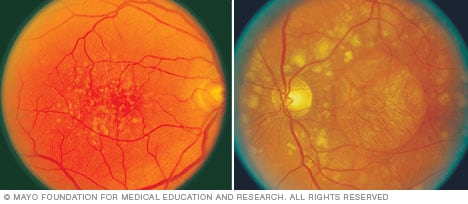
People suffering from dry macular degeneration may experience distortion or blurriness in their central vision due to an accumulation of yellow deposits called drusen beneath the retina, known as “drusen.” Drusen can cause the macula to thin and lead to vision loss known as geographic atrophy.
Typically, this stage of disease goes untreated because it does not impact peripheral vision. But an ophthalmologist can treat this condition by injecting dye into your arm that travels directly to the retina and causes it to “fluoresce”, or shine brightly, so he or she can see your blood vessels and determine whether or not they are healthy.
Wet macular degeneration occurs when new blood vessels form under the retina, leaking fluid onto retinal pigment epithelium (RPE) cells. An ophthalmologist may use thermal laser surgery to destroy these leaking vessels and stop its progression – this treatment has proven successful for approximately 10% of those diagnosed with wet macular degeneration.
Additionally to the treatments mentioned above, lifestyle modifications and nutritional supplements may also help lower the risk of macular degeneration or slow its progression. These may include not smoking, following a healthy diet and exercising regularly, as well as scheduling regular macular degeneration screenings in order to detect early and prevent further vision loss. An Amsler grid can also help identify early signs of fluid under the retina before permanent damage has taken place.
Optical coherence tomography
OCT imaging provides medical professionals with an accurate way to monitor macular degeneration progression. This noninvasive imaging technique produces high-resolution cross-sectional images of the retina that helps distinguish its layers and detect abnormalities that could indicate an underlying condition such as detached vitreous. OCT can also measure thickness of retina to assess medication efficacy – OCT may become part of care plans for people suffering from age-related macular degeneration as well as glaucoma or diabetic retinopathy.
Dry macular degeneration occurs when yellow fatty deposits known as drusen accumulate under the retina in an area called the macula, leading to gradual vision loss in central vision areas such as blurring, distortion or darkly-colored patches known as scotomas. As more retinal cells become compromised from being covered by these yellow deposits known as drusen, more retinal cells become lost leading to larger areas becoming lost altogether eventually leading to blind spots at the center of vision requiring regular check-ups with an Amsler grid in order to detect changes as early as possible in vision changes as possible.
“Wet” macular degeneration occurs when blood vessels form under the retina and leak fluid into it, damaging retinal pigment epithelium and leading to permanent vision loss. Dietary supplementation with antioxidants and zinc may reduce your risk for wet macular degeneration; laser treatment is effective against abnormal blood vessels in wet AMD but only effective in certain patients; although only effective against wet AMD in small percentage of cases; only this procedure has shown to significantly improve vision.
OCT imaging is an emerging technology capable of visualizing the choroid with unprecedented detail. Prior to OCT’s advent, however, its imagery could not adequately depict this area; now thanks to advances in OCT imaging techniques it allows better understanding of its involvement in retinal diseases.
Vitamins and minerals
Preventative measures can significantly slow down macular degeneration’s progress. Wearing sunglasses that block UV and blue light, eating fruits and vegetables regularly and taking vitamin supplements are all excellent ways to lower your risk of macular degeneration. Vitamin supplements contain organic substances essential for body functions – 13 known ones can be divided into two categories – fat-soluble vitamins such as A, D & E are stored by liver tissue until needed; water-soluble vitamins can be absorbed by cells throughout your body for transport throughout the body.
Studies have demonstrated that supplements containing vitamins C and E, lutein and zinc may reduce your chances of advanced macular degeneration. Before starting any new supplements it is wise to consult both an eye doctor and medical doctor to make sure that they will not interfere with existing health conditions or medications you take.
If you have been diagnosed with wet macular degeneration, an angiogram and optical coherence tomography test can detect abnormal blood vessel growth in the macula. Your retinal specialist can then use laser treatment to seal leaky vessels to preserve vision – this procedure takes minutes under local anesthetic at either Retina Health Center office in Fort Myers or Naples and should be painless!
No cure exists for wet age-related macular degeneration; however, early detection offers several treatment options. Anti-VEGF (vascular endothelial growth factor) drugs may help to stop leakage from abnormal blood vessels that form with wet macular degeneration; these prescription-only treatments are administered directly into anesthetized eyes in our office by needle. These anti-VEGF treatments have proven highly successful at lowering risk and even helping restore lost central vision according to studies conducted.


