

How to Prevent Macular Degeneration
Macular degeneration is an eye disease that damages central vision. This condition occurs when light-sensitive cells in your macula die off, rendering fine details unrecognizable as you see them straight ahead and at distances beyond. While macular degeneration doesn’t lead to total blindness, it does make reading, driving and recognising faces difficult tasks.
Assembled steps may help combat macular degeneration, including eating a diet rich in vegetables such as kale, raw spinach and collard greens; also taking nutritional supplements with antioxidants for additional support.
Eat a Healthy Diet
Studies have demonstrated that diets high in specific vitamins, minerals and antioxidants may help lower the risk of age-related macular degeneration while slowing its progression. People living with dry AMD may especially benefit from an intake of flavonoids and carotenoids found in fruits and vegetables containing flavonoids or carotenoids; flavonoids are especially abundant in berries, dark leafy green vegetables such as kale as well as orange or red fruits or veggies.
Macular degeneration affects an estimated 80% of patients who suffer it; most commonly the dry form, in which the macula thins and forms yellow deposits under the retina. This form typically causes gradual loss of central vision but doesn’t lead to total blindness because peripheral (side) vision remains intact.
Wet macular degeneration is less frequent but more serious, as it involves the formation of abnormal blood vessels that leak blood or fluid under the retina and cause rapid loss of central vision. It carries an increased risk of death and could even result in permanent vision impairment.
Eating well and taking supplements that support eye health are crucial steps in the fight against macular degeneration and other vision-threatening conditions, such as vitamin C, E, zinc and lutein deficiency. You can obtain these essential vitamins through foods like oranges, kale, blueberries, sweet potatoes carrots and eggs.
Apart from eating healthily and quitting smoking, another effective way of lowering your risk of macular degeneration is quitting smoking. Smokers are twice as likely to experience wet macular degeneration and therefore experience faster vision loss than non-smokers.
One of the best ways to keep both your eyes and body healthy is through regular exercise and maintaining a nutritious diet. This is particularly important for individuals at high risk for macular degeneration or cataracts; having family history increases that importance further.
Exercise Regularly
Age-related macular degeneration (AMD) is a progressive eye condition that gradually damages central vision. Affecting the retina – which lines the back of the eye – AMD is the leading cause of blindness among those over 50, as central vision is needed for reading, driving and seeing fine details clearly – yet AMD rarely results in complete blindness due to not damaging peripheral (side) vision.
Macular degeneration occurs when the macula, a small area at the center of retinal, becomes damaged. Once damaged, light-sensing cells in this part of retina become dysfunctional and it becomes difficult to see fine details. There are two forms of macular degeneration – dry and wet. Dry macular degeneration is often an early form, marked by blurry central vision accompanied by yellow deposits known as drusen underneath retina. With wet macular degeneration however abnormal blood vessels may form underneath retina which results in faster loss of central vision than its counterpart – as wet macular degeneration could potentially result in rapid vision loss and even blindness!
Exercise helps lower the risk for macular degeneration as well as other eye diseases, including high blood pressure, diabetes and glaucoma. Eating healthfully – such as including fruits and vegetables rich in omega-3 fatty acids as part of a balanced diet – may also aid in protecting against eye problems.
Exposure to UV rays increases your risk for macular degeneration and cataracts, so it is wise to wear sunglasses and a hat with a visor when venturing outdoors. Also beneficial is taking frequent breaks for your eyes using the 20-20-20 rule: every 20 minutes look at an object 20 feet away for 20 seconds for 20 seconds to keep them from becoming overworked. Finally, regular eye exams are one of the best ways to maintain eye health – make sure you inform your doctor if any family history exists so they can monitor for early signs that could mean easier treatments!
Quit Smoking
Smoking causes serious health complications for its users, from lung problems to cardiovascular diseases; but smoking also increases your risk for eye diseases like cataracts and macular degeneration. Be sure to break this bad habit now before it’s too late!
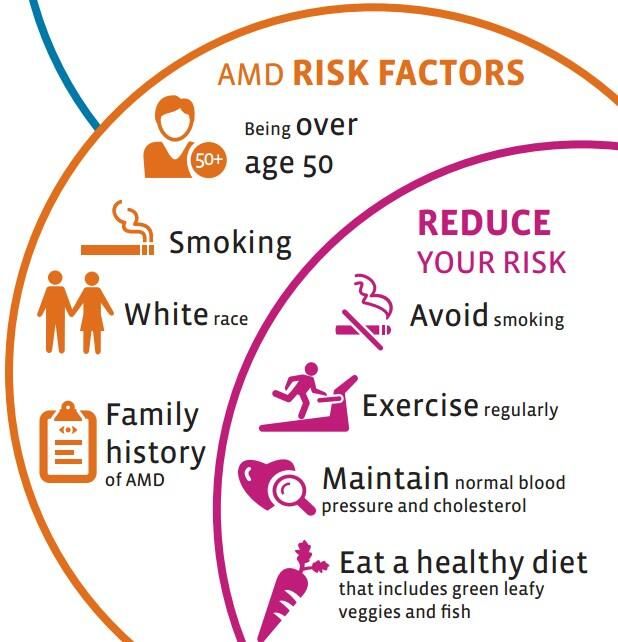
Age is the biggest risk factor for macular degeneration (AMD), yet lifestyle choices can significantly lower this chance. A diet rich in fruits, vegetables and whole grains may help lower your chances of AMD as well as other related medical problems like diabetes and cardiovascular disease.
A healthy diet can also help protect the eyes against dry eyes, which can cause blurry vision and light sensitivity. You can protect them from UV rays by wearing sunglasses and wide-brimmed hats when going outdoors.
One of the best ways to protect your vision is through regular eye exams. Your eye care professional can detect early signs of macular degeneration such as drusen and provide effective treatments.
Macular degeneration is a progressive vision loss condition which leads to central vision impairment, leaving you unable to read, drive or recognize faces; peripheral (side) vision remains normal however. Macular degeneration occurs when the macula, located at the centre of your retina, deteriorates and loses its ability to see fine details; it is the leading cause of blindness among adults over 50 and can result in significant disability.
Macular degeneration comes in two varieties, wet and dry. The former occurs more quickly and severely when abnormal blood vessels form under the retina and macula, leaking blood and fluid into the eye, while dry macular degeneration typically develops over time as deposits called drusen form under the retina, visible as yellow spots that your eye care professional can spot during a dilated exam.
Macular degeneration cannot be reversed, but eating healthily and quitting smoking are steps that may slow its progress. Modifiable risk factors such as obesity, high cholesterol, or inactivity could increase your likelihood of getting this disease.
Have Regular Eye Exams
Macular degeneration is a condition whereby the macula, an area in the retina at the back of your eye that enables you to see fine details clearly, becomes impaired. This may cause central vision to become unclear or dark while generally not impacting peripheral or side vision; left untreated it could result in serious and permanent vision loss; therefore it’s vital that regular comprehensive eye exams with an ophthalmologist become increasingly important as you age.
An eye exam should include a visual acuity test to evaluate your ability to see numbers and letters clearly, while your doctor may use an Amsler grid tool to assess central vision. If straight lines appear wavy in this grid, this could indicate macular degeneration – call us immediately if this occurs!
AMD typically takes place as a gradual loss of central vision that doesn’t cause pain, with early symptoms including distortion of straight lines when reading or looking at faces; some individuals also report experiencing blind spots in the center of their vision. Other people develop advanced AMD with wet form blood vessel growth under retina that leak fluid or raise macula and causes rapid and severe vision loss.
Macular degeneration can best be avoided with an annual eye exam conducted by your ophthalmologist. They will check for signs of diabetes, high blood pressure and brain tumors while also inspecting your retinal blood vessels to make sure they remain healthy – this step alone could prevent serious vision-related problems like glaucoma which has no early warning symptoms and could eventually lead to blindness if treated promptly.


