
Medically reviewed by Shaun Larsen, OD – Doctor of Optometry, Southern College of Optometry.
Last clinically reviewed: July 2026 | Next scheduled review: July 2027
The short answer. There is no reliable clinical evidence that homeopathic remedies slow, stop or reverse age-related macular degeneration. No homeopathic preparation has been shown in a controlled trial to preserve vision in AMD. If you have wet (neovascular) AMD, delaying anti-VEGF treatment to try an alternative therapy can cost you central vision permanently, because the damage happens over weeks and is not recoverable once scarring occurs. If you are looking for what you can do yourself – without injections – there are genuinely evidence-based answers, and they are set out in the second half of this article.
Why people search for this, and why the question is a fair one
If you have arrived here after an AMD diagnosis, the reason is usually one of a few: you have been told the dry form has no treatment beyond vitamins and you want to do something; you are facing the prospect of repeated injections into your eye and want to know whether there is an alternative; you prefer approaches you think of as natural; or you have been told nothing more can be done and you are looking anywhere for options.
All of those are reasonable. This article is not written to dismiss them. But the honest answer to the specific question of whether homeopathy treats macular degeneration is no – and because AMD can take vision that cannot be given back, you deserve that answer stated plainly rather than hedged.
What homeopathy is
Homeopathy is a system developed by Samuel Hahnemann in the late eighteenth century, built on two principles. The first is similia similibus curentur – “like cures like” – the idea that a substance producing certain symptoms in a healthy person can treat similar symptoms in a sick one. The second is potentisation: the belief that serial dilution accompanied by vigorous shaking (succussion) increases rather than decreases therapeutic power.
The dilutions used are extreme. A “30C” preparation – a common potency – has been diluted 1 part in 100, thirty times over, a dilution factor of 10⁶⁰. Avogadro’s constant is approximately 6.022 × 10²³, meaning that beyond roughly 12C, a preparation is statistically unlikely to contain even a single molecule of the original substance. At 30C the probability of any original molecule remaining is effectively zero.
This is why the debate about homeopathy is not primarily about safety. Most homeopathic preparations at typical potencies contain nothing but the diluent and are therefore inert. The issue is efficacy – and the consequences of substituting an inert preparation for a treatment that works.
One important distinction: “homeopathic” is often used loosely to mean any natural or herbal product. It should not be. Herbal medicine uses pharmacologically active plant material at doses that can have real effects, both beneficial and harmful. Homeopathy uses dilutions beyond the point where material remains. Some products marketed as homeopathic are in fact low-dilution preparations containing measurable active substance – and those can carry real risks, which is a separate concern discussed below.
What the evidence says
The Australian NHMRC review (2015) remains the most comprehensive government assessment. It examined 57 systematic reviews containing 176 individual studies across a wide range of conditions and concluded: “Based on the assessment of the evidence of effectiveness of homeopathy, NHMRC concludes that there are no health conditions for which there is reliable evidence that homeopathy is effective.” It added that homeopathy should not be used to treat conditions that are chronic, serious, or could become serious – a category into which macular degeneration clearly falls.
Specifically for macular degeneration, the evidence base is not weak – it is essentially absent. There are no adequately powered randomised controlled trials of homeopathic preparations for AMD with visual acuity, lesion growth or progression to advanced disease as endpoints. The remedies named on this page have never been tested against these outcomes in a controlled setting. This is not a case of promising early findings awaiting confirmation; it is a case of claims made without trials.
The regulatory position. The FTC’s 2016 enforcement policy statement holds homeopathic drug marketing to the same substantiation standard as other health products: efficacy claims require competent and reliable scientific evidence. In the US, homeopathic products are not evaluated by the FDA for safety or efficacy before marketing, so FDA availability is not evidence of either. In the UK, the NHS ended routine funding of homeopathy in 2017 on the basis that it lacks robust evidence.
On the counter-argument. Homeopathy’s advocates note that individualised prescribing does not fit neatly into standard trial designs, and some have contested the NHMRC review’s methods. Those objections are on the record. But the position that has to be defended is stronger than “the trials were imperfect”: it is that a preparation containing no molecules of an active substance produces a measurable clinical effect on retinal disease. No mechanism consistent with established chemistry has been demonstrated for this, and no trial has shown the effect. Reported improvements are consistent with the natural fluctuation of the disease, regression to the mean, concurrent conventional treatment, and placebo response – which is real, but does not slow a retinal lesion.
The specific risk with macular degeneration
This is the part that matters most, and it is why this page has been rewritten rather than trimmed.
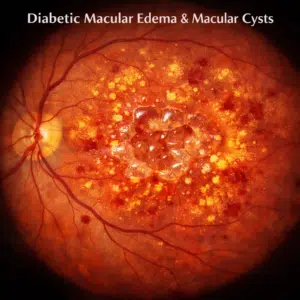
Wet AMD is time-critical. In neovascular AMD, abnormal vessels grow beneath or into the retina and leak fluid and blood. Untreated, this destroys central vision over weeks to months and ends in a fibrotic scar. Once photoreceptors are lost and fibrosis has formed, no treatment recovers that vision. Anti-VEGF therapy – injections of bevacizumab, ranibizumab, aflibercept, brolucizumab or faricimab into the eye – stabilises or improves vision in the majority of treated eyes, and it is the single most effective intervention in the history of this disease. The benefit is greatest when treatment starts early, and studies of delayed initiation consistently show worse final acuity.
That is the concrete harm at issue. A reader with new distortion who spends three months on homeopathic remedies before seeking treatment may permanently lose vision that would have been preserved. This is not a theoretical objection to alternative medicine in general; it is a specific, mechanistic consequence in this specific disease.
Seek same-week assessment if you notice: new distortion of straight lines (doorframes, window blinds, text lines bending); a new grey, dark or blank patch in central vision; sudden blurring of central vision; or a sudden change in the size or shape of objects. Check each eye separately – a good fellow eye masks early loss remarkably well, and many people discover monocular loss by accident.
A note on “nothing more can be done”. If you have been told this, it usually refers to the disease process, not to your function. Low vision rehabilitation frequently makes a substantial difference to reading and independence even when no further medical treatment is available. That is a different question with better answers, and it is worth asking specifically.
Are homeopathic remedies at least harmless?
Usually, but not always, and the caveats matter.
- Delay of effective treatment is the principal risk, and in AMD it is a serious one.
- Product quality varies. Because these products are not subject to pre-market efficacy or safety review in the US, contamination and mislabelling have occurred. The FDA has issued warnings and recalls, including for homeopathic teething products found to contain inconsistent amounts of belladonna alkaloids.
- Low-dilution products may contain active substance at pharmacologically meaningful levels, with real potential for toxicity and drug interaction.
- Anything placed in or near the eye carries risk. Non-sterile eye drops, including some marketed as homeopathic, have been associated with serious ocular infection. Do not instil any preparation into your eye that is not sterile and intended for ophthalmic use.
- Tell your ophthalmologist what you are taking. Some supplements affect bleeding risk, which is relevant before intraocular injections or surgery. Clinicians ask this question to keep you safe, not to disapprove.
On the specific remedies previously listed on this page
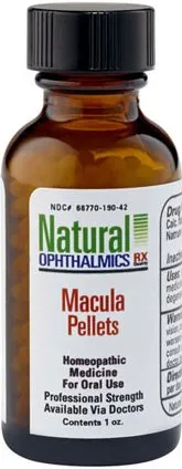
Readers arriving from searches for particular remedies deserve a direct answer about each. For all of the preparations previously described on this page – Agaricus, Apis mellifica, Arnica montana, Bovista, Secale, Sepia, Staphysagria, Carboneum sulphuratum, Hyoscyamus, Tabacum, Lycopodium, Silicea, Phosphorus and Mercurius – the position is the same: none has been evaluated in a controlled clinical trial for age-related macular degeneration using visual acuity, lesion growth, or progression to advanced disease as an endpoint. Their traditional indications derive from homeopathic provings, which are symptom records from healthy volunteers taking the substance, not from clinical outcome studies in patients with retinal disease.
A few points of specific correction, since the previous version stated these as established:
- Agaricus blazei. Some in vitro and animal work has examined immunomodulatory properties of this mushroom, but no clinical trial has shown any effect on macular degeneration. The prior claim that “a recent study has demonstrated that extracts derived from Agaricus Blazei Murill mushroom can help slow macular degeneration” was not supported by a citation, and no such study is identifiable in the ophthalmic literature.
- Bovista. The previous text conflated Bovista (a puffball genus) with Scleroderma bovista (an unrelated earthball) and described the fruiting body in detail. Neither the taxonomy nor the morphology has any bearing on retinal disease, and no clinical evidence supports use in AMD.
- Arnica montana. Has been studied for bruising and post-surgical swelling with mixed and generally unconvincing results. It has no established role in retinal disease. The prior statement that it is “an excellent natural treatment option for conditions associated with age, such as glaucoma that leads to maculae deterioration” is doubly incorrect – glaucoma does not cause macular degeneration, and arnica does not treat either.
- Tabacum. Prepared from Nicotiana tabacum. It is worth noting the irony that smoking is the strongest modifiable risk factor for AMD, roughly doubling to quadrupling risk. Whatever the merits of the homeopathic preparation, tobacco itself is among the best-established causes of the disease this page addresses.
- On “sclerosing agents”. The previous text stated that “sclerosing agents are an integral component of homeopathic treatment for macular degeneration” and that they reduce inflammation of retinal blood vessels. Sclerosing agents are pharmacological compounds used to obliterate veins in conditions such as varicose veins. They are not a homeopathic modality, they are not used in retinal disease, and the claim appears to be an error rather than a contested position.
For optimal functioning, the macula requires an abundance of blood flow. Any decrease in this flow could contribute to macular degeneration (MD). Smoking reduces this blood flow directly to retinal tissue. This is one of the few statements on the original page that holds up, and it is worth expanding: smoking is the strongest modifiable risk factor for AMD, associated with roughly two- to four-fold increased risk and earlier onset, through oxidative stress, reduced choroidal perfusion and complement activation. Risk declines over years following cessation.
What does have evidence: non-surgical measures that genuinely help
This is the section most readers of this page actually need. Everything below is supported by clinical trial or strong observational evidence, and none of it requires an injection.
1. Stop smoking – the highest-value single action
Nothing else on this list has a comparable effect size. If you smoke and have AMD, cessation is the most powerful thing within your control. Risk falls over subsequent years, and the benefit applies to both the dry and wet forms.
2. AREDS2 supplementation – if you are at the right stage
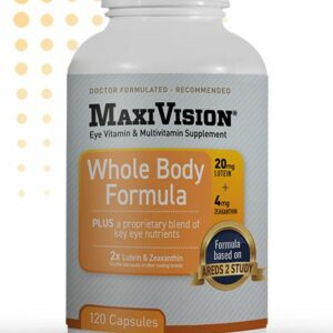
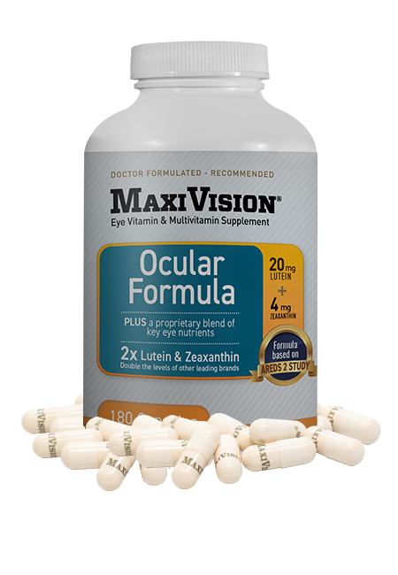
The Age-Related Eye Disease Studies are the largest randomised trials of nutritional supplementation in eye disease. The AREDS2 formulation is: 500 mg vitamin C, 400 IU vitamin E, 10 mg lutein, 2 mg zeaxanthin, 80 mg zinc (as zinc oxide), 2 mg copper (as cupric oxide). It reduces the risk of progression to advanced AMD by approximately 25%.
The qualifications matter as much as the headline:
- It is indicated for intermediate AMD (large drusen, or medium drusen with pigmentary change) or advanced AMD in one eye. It has not been shown to benefit early AMD or to prevent AMD in people who do not have it.
- It slows progression; it does not restore vision already lost.
- The original AREDS formula contained beta-carotene, which increased lung cancer risk in current and former smokers. Lutein and zeaxanthin replaced it. Any product still containing beta-carotene uses the superseded formula – check the label, particularly if you have ever smoked.
- The 80 mg zinc dose is high; copper is included specifically to prevent zinc-induced copper deficiency anaemia. Do not construct your own version from separate products.
- Adding omega-3 fatty acids to the formulation did not produce additional benefit in AREDS2.
Confirm with your eye specialist that your stage of AMD is one where AREDS2 is indicated before starting. This is a case where more is not better and where the wrong formulation carries real risk.
3. Diet
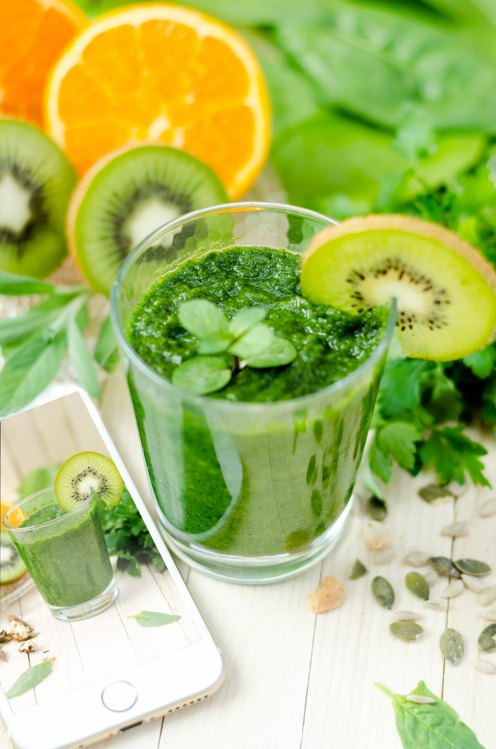
Observational evidence consistently associates a Mediterranean-pattern diet with lower risk of progression to advanced AMD. The relevant components are green leafy vegetables (the dietary source of lutein and zeaxanthin, which are concentrated in the macular pigment), oily fish, fruit, nuts, legumes, olive oil, and limited processed and high-glycaemic foods. Some evidence suggests a high dietary glycaemic index is associated with greater progression risk. This is observational rather than trial evidence, so causality is not established – but the recommendation carries essentially no downside and substantial general cardiovascular benefit.
4. Cardiovascular risk factor control
AMD shares risk factors with cardiovascular disease. Managing blood pressure, lipids and body weight, and maintaining regular physical activity, is sensible on general grounds and plausibly relevant here.
5. Monitor for conversion to wet AMD
Roughly 10–15% of AMD converts to the neovascular form, and early detection substantially changes the outcome. Use an Amsler grid daily or several times weekly, testing one eye at a time with reading glasses on. Report any new distortion, blurring or missing area immediately. Home OCT monitoring devices are becoming available for selected high-risk patients. Understand the limitation: the Amsler grid is insensitive, and a normal grid does not exclude progression – it is a supplement to scheduled examinations, not a replacement.
6. UV and blue light protection
Evidence linking light exposure to AMD progression is mixed, but sunglasses with UV protection and a wide-brimmed hat are harmless, inexpensive, and reduce glare discomfort, which is itself a common complaint in AMD.
7. Low vision rehabilitation – the most under-used option
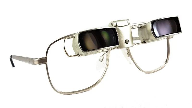
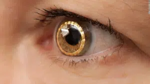
If AMD has already reduced your vision, rehabilitation addresses function rather than disease, and it works. A low vision assessment measures acuity at working distance, contrast sensitivity, and scotoma position, then builds a solution around specific goals. Interventions include magnification calculated to your actual reading requirement, high-add reading spectacles, hand and stand magnifiers, electronic and portable video magnifiers, head-mounted electronic systems, task lighting and contrast enhancement, screen readers and OCR software, and – particularly valuable in macular disease – eccentric viewing and steady-eye training, which teaches use of a preferred retinal locus outside the damaged fovea. That training alone can restore reading ability without any change in measured acuity.
8. Ask about clinical trials
If you want to pursue something beyond current standard care, a clinical trial is the evidence-based route. Trials in AMD currently include complement pathway inhibitors, gene therapies designed to produce sustained intraocular anti-VEGF expression, longer-acting drug delivery implants, and cell-based approaches. ClinicalTrials.gov lists open studies, and your retina specialist can advise on suitability. This is the appropriate channel for the entirely reasonable impulse to try something new.
How to talk to your eye specialist about complementary approaches
Tell them what you are taking. Clinicians ask about supplements to check for interactions and bleeding risk before injections or surgery, not to disapprove. Non-disclosure is the actual risk.
Ask direct questions and expect direct answers: What stage is my AMD, dry or wet? Is AREDS2 indicated for me specifically? What are the warning signs I should act on, and how quickly? What is my risk of converting to the wet form? Would a low vision assessment help me now?
Do not stop or delay prescribed treatment to try something else without discussing it first. In wet AMD in particular, a gap in anti-VEGF therapy can cost vision that cannot be recovered.
Frequently asked questions
Can homeopathy cure macular degeneration?
No. There is no reliable clinical evidence that homeopathic remedies cure, reverse or slow AMD, and no controlled trial has demonstrated benefit on visual acuity or disease progression. AMD currently has no cure in any system of medicine – but it does have treatments that meaningfully change outcomes, particularly anti-VEGF therapy for the wet form.
Is it safe to try homeopathy alongside conventional treatment?
Most homeopathic preparations at standard potencies are inert and unlikely to cause direct harm. The risk is delay or substitution. If you use them in addition to – never instead of – your prescribed treatment, and you tell your ophthalmologist, the direct risk is low. Never instil a non-sterile preparation into your eye.
What about herbal supplements such as bilberry, ginkgo or saffron?
These are herbal rather than homeopathic, and are pharmacologically distinct. Some small studies of saffron in AMD have reported modest changes in retinal function, but the trials are small, short and not yet sufficient to support a recommendation. Bilberry and ginkgo lack convincing evidence in AMD. Herbal products contain active compounds and can interact with medications – ginkgo, for instance, may affect bleeding risk, which matters before intraocular injection. Discuss any herbal supplement with your clinician.
I have dry AMD and was told there is no treatment. Is that true?
Not quite, and the phrasing matters. For intermediate dry AMD, AREDS2 supplementation reduces progression risk by about 25%. For geographic atrophy – the advanced dry form – two complement inhibitors, pegcetacoplan and avacincaptad pegol, were approved in 2023 and slow lesion growth, though they have not been shown to improve visual function and require ongoing injections; whether they are worthwhile is a genuine discussion to have with a retina specialist. Alongside this, smoking cessation, dietary measures, monitoring for conversion to wet AMD, and low vision rehabilitation all have a role. “No treatment” is more accurately “no cure”.
Why does this article take such a firm position?
Because in this specific disease, the cost of hedging is measured in permanent vision loss. On many health questions the honest answer is “the evidence is mixed”. Here it is not: there are no controlled trials showing homeopathic benefit in AMD, and there is a highly effective time-critical treatment for the wet form that must not be delayed. Presenting these as balanced alternatives would misrepresent the evidence to people making decisions about their sight.
References
1. National Health and Medical Research Council (Australia). NHMRC Statement on Homeopathy and NHMRC Information Paper: Evidence on the effectiveness of homeopathy for treating health conditions. Canberra: NHMRC; March 2015.
2. Federal Trade Commission. Enforcement Policy Statement on Marketing Claims for OTC Homeopathic Drugs. Washington DC: FTC; November 2016.
3. Federal Trade Commission. Staff Report on the Homeopathic Medicine & Advertising Workshop. ftc.gov.
4. Mathie RT, Lloyd SM, Legg LA, et al. Randomised placebo-controlled trials of individualised homeopathic treatment: systematic review and meta-analysis. Syst Rev. 2014;3:142.
5. Age-Related Eye Disease Study 2 Research Group. Lutein + zeaxanthin and omega-3 fatty acids for age-related macular degeneration: the AREDS2 randomized clinical trial. JAMA. 2013;309(19):2005–2015.
6. Chew EY, Clemons TE, Agrón E, et al. Long-term outcomes of adding lutein/zeaxanthin and omega-3 fatty acids to the AREDS supplements: AREDS2 report 28. JAMA Ophthalmol. 2022;140(7):692–698.
7. National Eye Institute. AREDS/AREDS2 clinical trials. nei.nih.gov.
8. Rosenfeld PJ, Brown DM, Heier JS, et al. Ranibizumab for neovascular age-related macular degeneration. N Engl J Med. 2006;355(14):1419–1431.
9. Heier JS, Lad EM, Holz FG, et al. Pegcetacoplan for the treatment of geographic atrophy secondary to age-related macular degeneration (OAKS and DERBY). Lancet. 2023;402(10411):1434–1448.
10. Thornton J, Edwards R, Mitchell P, et al. Smoking and age-related macular degeneration: a review of association. Eye. 2005;19(9):935–944.
11. Merle BMJ, Colijn JM, Cougnard-Grégoire A, et al. Mediterranean diet and incidence of advanced age-related macular degeneration: the EYE-RISK Consortium. Ophthalmology. 2019;126(3):381–390.
12. American Academy of Ophthalmology. Age-Related Macular Degeneration Preferred Practice Pattern. aao.org.
13. US Food and Drug Administration. Homeopathic products – consumer information and safety communications. fda.gov.





 Shop Best Low-Vision Aids with FREE Doctor Consultation.