Eyes are often the first indicator of elevated cholesterol levels, showing symptoms like yellow or fatty bumps around eyelids known as xanthomas.
An additional sign is a grey ring on the cornea known as an “arcus senilis” in adults and an “arcus juvenilis” in children.
Xanthomas
Xanthomas are yellowish skin lesions caused by fat accumulation in body tissues. They serve as an indicator of high cholesterol and an issue related to lipid metabolism; some types may even indicate diabetes and hereditary disorders like familial hypertriglyceridaemia.
There are various factors that contribute to xanthomas, and their cause varies depending on where and how it develops. Eruptive xanthomas typically appear as groups of reddish-yellow papules on symmetric areas such as trunk, buttocks, extensor surfaces of legs and arms as well as in the groin or armpits; their lesions may also form underarmpits. Their root cause involves disrupting transport of cholesterol and triglycerides through vascular systems to skin tissue where macrophages take up these lipids which then accumulate.
Lipids may become trapped within tissue structures or subcutaneous fat and eventually be consumed by macrophages, leading to atherosclerosis. They may also travel via lymphatic channels to other organs where they deposit as adipose tissue deposits – depending on its type, various treatments exist for treating this xanthoma.
Xanthelasma palpebrum is the most prevalent type of eye xanthoma and results from an imbalance in metabolism involving lipoproteins and lipids. This may result in genetic disorders like hyperlipoprotenaemia or metabolic conditions like diabetes mellitus or nephrotic syndrome. Lesions on the liver can be brought on by events such as acute pancreatitis or hereditary liver cirrhosis; however, they can also arise without any identifiable risk factor. Diagnosis typically relies upon clinical examination and blood screening tests such as serum lipid screens and glucose screening can further confirm diagnosis. Combining diet with cholesterol-reducing drugs is often enough to manage and prevent xanthelasmas from reappearing, while surgical removal should only be considered when large or multiple lesions affect cosmetic appearances; qualified dermatologists or ultra pulsed carbon dioxide lasers are usually sufficient to safely and effectively perform this procedure, as it causes minimal pain or scarring during removal.
Corneal Arcus
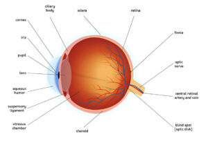
Deposition of white, grey or yellowish opaque material on the corneal margin due to deposition of lipids associated with familial hypercholesterolemia. Usually beginning below and above the sclerocorneal junction it typically progresses toward 9 and 3 o’clock or forms a full circle around the cornea. While rare at birth it becomes increasingly prevalent with age and may become widespread for individuals over 50 (called Arcus Senilis). Arcus does not impair vision nor require treatment.
Symptoms of elevated cholesterol levels can include this arcing effect. Other potential causes could be lecithin cholesterol acyl transferase deficiency or fish eye disease. If diagnosed as homozygous familial hypercholesterolemia, this arc may be the first clinical sign that leads to diagnosis; often seen alongside other xanthomas and elevated liver enzymes; treatments include ursodeoxycholic acid or immunosuppressive agents for initial stages; liver transplantation may become necessary later on.
Studies with 243 individuals aged 20-63 years found that corneal arcus is associated with dyslipidemia, one of the risk factors for coronary heart disease. Sensitivity for detection was 19% while specificity was 100%; authors of this research concluded that corneal arcus serves as an early marker for familial hypercholesterolemia and should be identified early among younger adults.
Signs and symptoms of FH include xanthomas, which are white or yellow deposits of lipid material found under the skin at joints such as the knuckles or Achilles tendon, commonly seen in children and pathognomonic of FH. Lecithin cholesterol acyl transferase deficit (also referred to as fish eye disease) is another hereditary disorder characterized by an excessive accumulation of lipids in the lens that leads to corneal opacification; its diagnosis requires genetic testing as early treatment can prevent blindness.
Age-Related Macular Degeneration
Age-related macular degeneration, or ARMD, is an eye disease in which the central portion of the retina (known as the macula) deteriorates over time. This area of our vision allows us to read, drive safely, identify faces and colors accurately, as well as perform other activities that require central vision such as reading. ARMD is the leading cause of legal blindness among Americans aged 60 or above and symptoms of this disorder include blurry or distorted vision and buildup of drusen, small deposits of fatty lipids beneath our retina which convert light signals into signals processed by our brain into images for processing by our eyesight systems and eventually processed through our visual system and processed through our visual cortex which ultimately turns light into electrical signals processed by our visual cortex which translate into sight through neurons to our visual pathways before reaching our visual cortex and processing it into visual images in our visual cortex where visual processing takes place allowing us to see.
ARMD symptoms tend to be mild to moderate; however, they can progress and worsen over time. Some individuals with dry ARMD can eventually progress into wet macular degeneration in which abnormal blood vessels grow beneath the retina and leak blood into the macula; this type of wet macular degeneration may eventually result in rapid and severe loss of central vision.
Researchers still aren’t entirely certain of what causes ARMD, though risk factors include smoking, high cholesterol levels and eating too many fat-laden calories. It tends to run in families; more women than men tend to develop it. Regular eye examinations are important in helping detect early symptoms of ARMD.
Treatment options for ARMD include special eyedrops and nutritional supplements designed to slow the progression of the condition, along with laser procedures like photodynamic therapy that stimulate new blood vessels to grow under the retina, in order to stop further vision loss. Our team at Associated Eye Physicians provide state-of-the-art macular degeneration diagnosis and treatment at our offices in Rockford and Elgin – get in touch today for more information; our friendly staff would love to be of service! We hope to hear from you soon.
Retinal Vein Occlusion
Retinal vein occlusion refers to a condition in which there is a blockage in one or more small veins that carry blood away from the retina – the layer at the back of your eye inside that converts light images into nerve signals sent directly to your brain). Retinal vein occlusion most often results from thickening or hardening of arteries (atherosclerosis), leading to thickened or hardened retinal veins (retina veins) becoming blocked with blood. Risk increases with age; risk increases more among those living with diabetes or those living with high blood pressure; smokers also increased risks as risk factors – while risk increases with age as risk also increases with age; more common among those living with diabetes or high blood pressure while smokers also increase; diabetics and those suffering from myeloma (thrombophilic disorders).
Branch retinal vein occlusion occurs when blood clots block the flow of fluid through retinal capillaries (tiny blood vessels that provide nutrients to the retina). This can result in swelling or leakage of blood onto the retina, impairing vision. Hemorrhages can either spread throughout all retinal veins affected by an occlusion, or occur locally if only one branch retinal vein was blocked off; more serious is central retinal vein occlusion which includes all three.
Occlusion of retinal veins can result in macular edema, neovascularization and neovascular glaucoma – with symptoms including macular edema, neovascularization and even glaucoma resulting from this. Although usually painless, retinal vein occlusion symptoms may include sudden blurring or loss of vision affecting one or both eyes. Diagnosing this condition typically begins with a comprehensive eye exam and follow-up exams are often necessary to monitor its progression. Digital fundus photography, optical coherence tomography and fluorescein angiography may be performed for confirmation of diagnosis. Treatment usually entails medication injections into the eye as well as laser or surgical treatments or procedures. It’s also essential that risk factors like diabetes, high blood pressure and cholesterol levels be controlled to reduce complications that could arise; follow-up exams usually serve this purpose.


