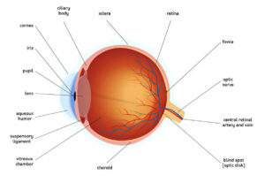
Hypertensive Retinopathy occurs when high blood pressure damages the blood vessels that supply your retina (a transparent layer at the back of your eyeball).
Eye problems can result in pain, redness of the eye or seeing halos around lights; vision may also become blurry or distorted.
Causes
Blood pressure measures the force exerted by your heart’s pumping action against arterial walls. A reading will show two numbers; one indicates your systolic blood pressure while the second number represents diastolic, or between heartbeat pressure, when resting. High blood pressure is a serious health risk that can damage blood vessels in your eyes, heart, kidneys, brain and other organs.
High blood pressure significantly increases the risk of eye disorders such as glaucoma, which results from the build-up of fluid inside the eyeball and can damage its optic nerve, leading to blurred vision.
Untreated hypertension can also result in hypertensive retinopathy, which damages the retina – the clear layer at the back of your eye that provides blood and nutrients – through changes to blood vessels that supply this essential service to our eyes. This condition should be considered an urgent medical matter and is more likely to occur among people who have longstanding high blood pressure although sudden instances may also arise.
As well as symptoms such as breathlessness or sudden, severe headache, other indicators of high blood pressure that affect the eyes include feeling breathless or sudden, severe chest pain that does not respond to physical exercise and persistent, unrelated chest pain can indicate high blood pressure as well. Frequent hard-to-stop nosebleeds as well as confusion or impaired reasoning could be further indicators.
Blood spots or subconjunctival hemorrhage is often the result of sudden fluctuations in blood pressure that strain the vessels. While these spots appear alarming at first, they will typically go away on their own after some days; gestational hypertension (preeclampsia) often experience gestational hypertension as a warning sign and require monitoring and treatment in future.
Symptoms
High blood pressure can damage the tiny blood vessels found in your retina (the light-sensitive tissue at the back of your eyeball), eventually decreasing blood flow to it and leading to its swelling, which in turn could eventually lead to glaucoma – where pressure inside the eye causes vision loss.
Untreated high blood pressure can also damage the optic nerve and cause permanent vision loss.
Your healthcare provider will conduct a physical exam and blood pressure measurement. Additional tests may be necessary depending on your risk factors for ocular hypertension and other health conditions.
High risk individuals for ocular hypertension include those with a family history of high blood pressure, those who have diabetes or other health conditions that increase the likelihood of diabetic retinopathy, as well as older adults – because their chances of ocular hypertension increase with age it’s essential that your blood pressure be regularly assessed and checked.
Signs of high blood pressure in the eyes include bright red patches on the white of each eye known as subconjunctival hemorrhages. Although they can look alarming, these spots typically disappear within two weeks as your body absorbs any remaining blood in them.
Other indicators of ocular hypertension may include lightheadedness or blurred vision. Pressure changes within your eye may disrupt nerve signals sent from eye to brain and result in dizziness or diminished vision, leading to dizziness or blurring.
Eye doctors will typically start by testing your blood pressure to assess any potential issues that need treating, such as high or low blood pressure. If this is indeed your problem, avoid caffeinated beverages and exercise to keep it under control; alternatively consult with a physician if any symptoms emerge that cannot be explained by other medical issues – chest pain or trouble breathing are just two examples.
Diagnosis
Your eye care professional can determine if high blood pressure is harming your eyes by looking into them with an ophthalmoscope and looking at changes to their structure caused by elevated pressure levels. Your physician will also take your medical history and check any associated problems, like heart or vessel conditions that could also contribute.
If the pain in the center of your eyelid persists, an eye care professional may suspect an infected sinus or orbital cellulitis. This infection usually spreads from lacrimal sac to eyelid and surrounding tissues via Staphylococcus aureus or Streptococci species and signs include pain with eye movement, restricted ocular movement restrictions, proptosis and swelling around eyelid margins. A CT scan of your brain and orbits may be ordered to help diagnose its source.
Central retinal vein occlusion (CRVO), a side effect of high blood pressure, may cause eye pain. In this condition, blood vessels in the back of your eye (retina) become damaged by blood clots forming at their origins. Your eye care provider will examine both eyes using an ophthalmoscope and possibly also use a slit lamp to examine more closely what’s happening behind each pupil.
Your doctor will look out for narrowing of blood vessels in your retina, inflammation of the macula or other areas of the retina and cotton wool spots and hard exudates that indicate damage; mild signs usually mean minimal loss of vision; however if signs progressed they could lead to some loss.
Your doctor will prescribe medication to reduce your blood pressure and work closely with both you and your primary care physician to ensure consistent follow-up. In most cases, controlling blood pressure will heal eye tissue and avoid serious complications from occurring. If your glaucoma is severe enough, however, surgery may be required; such procedures include trabeculoplasty whereby your doctor creates a new channel for draining fluid from your eye, or an iridotomy or cyclophotocoagulation to decrease fluid production.
Treatment
Subconjunctival hemorrhages typically subside within days or weeks without any cause for alarm, often within 24 hours or 48 hours if left alone. A bright red patch will eventually fade to brown or yellow and return to being transparent again; no pain or vision impairment occurs from such hemorrhages; however if multiple instances arise contact your healthcare provider as it could indicate serious vascular disorders among older adults.
Your health care provider will assess your past health history and conduct a physical exam, in addition to performing an eye exam using special lighted instruments that examine retina. This allows them to identify if bleeding originates in either side of the eye (inner/outer/back), or is concentrated around the macula (back part).
If you have glaucoma, your doctor will conduct an eye pressure test called tonometry in order to measure fluid pressure inside of your eye. Knowing your fluid pressure levels is important because high pressure can damage optic nerves and lead to blindness; if at risk for glaucoma, medications will likely be given in order to decrease eye pressure.
Glaucoma treatment options include beta blockers that reduce the production of fluid in the eye. They’re usually administered as eye drops and take several weeks before beginning their effects; side effects may include wheezing or difficulty breathing, reduced heart rate, depression fatigue or low blood pressure.
Prostaglandin analogs are medications designed to lower eye pressure by optimizing fluid drainage from the eye. They may be taken alone or combined with other medications; side effects similar to beta blockers may include dry mouth, red or irritated eyes, blurred vision, bitter metallic taste in mouth, dry throat/nose/throat congestion and fatigue as well as low blood pressure and light sensitivity.
Surgery to drain excess eye fluid may also be an option to treat glaucoma; this treatment option should only be undertaken if your symptoms are severe or vision has already been lost, and will usually only be done under local anesthetic by your physician.


