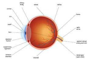
Eye doctors can regularly observe blood vessels through observation of the eyes. This makes the eyes an excellent spot to check for signs of elevated blood pressure; many studies have demonstrated its association with subclinical and clinical coronary heart disease, congestive heart failure, and stroke.
Hypertensive retinopathy affects retinal vessels and has funduscopic symptoms of arteriolar narrowing, arteriovenous crossing abnormalities, flame-shaped hemorrhages, cotton wool spots, yellow hard exudates and optic disc edema. Treatment primarily aims at controlling blood pressure with medication.
Stage 1
High blood pressure damages the vessels that deliver oxygen and nutrients to the retina, potentially leading to vision loss and other complications over time. Treatment typically involves controlling blood pressure while, if necessary, treating eyes to protect from further damage.
Hypertensive Retinopathy (HR) occurs as the result of untreated high blood pressure that damages retinal blood vessels over time, damaging their walls to thicken over time and restrict blood flow to the retina, leading to vision loss from Hypertensive Retinopathy. Over time, high blood pressure causes its walls to thicken, restricting its flow and leading to visual impairment associated with HR.
Hypertension retinopathy’s early signs include reversible vasoconstriction (widening of retinal blood vessels). More serious hypertensive retinopathy may cause permanent damage such as flame-shaped hemorrhages, cotton wool spots, yellow hard exudates and optic disk edema which can only be identified through retinal examination and funduscopic photography.
Mild hypertensive retinopathy (generalized retinal arteriolar narrowing and arteriovenous nicking) is often seen in those with high blood pressure; however, this does not always indicate cardiovascular disease or an adverse prognosis. Furthermore, older individuals who maintain normal blood pressure often develop it, and other risk factors for cardiovascular disease like left ventricular hypertrophy or renal impairment have also been found to contribute.
Moderate hypertensive retinopathy has been found to be associated with both subclinical and clinical cardiovascular conditions, including coronary heart disease and congestive heart failure. Correlation studies also demonstrate strong associations between brachial-ankle pulse wave velocity and aortic root diameter, both measured with noninvasive methods such as ultrasound and M-mode echocardiography, and brachial-ankle pulse wave velocity. Assessing the severity and relation of hypertensive retinopathy to target organ damage is therefore critical in cardiovascular risk stratification. Patients with mild or moderate hypertensive retinopathy should receive regular care, with blood pressure monitored according to established guidelines. Furthermore, those with severe hypertension should undergo careful evaluation and evaluation in order to reduce risks such as cardiovascular disease, stroke, kidney failure and vision loss.
Stage 2
Hypertensive Retinopathy typically does not present with symptoms until late in its progression and may include blurred vision, dark spots in the visual field and reduced visual acuity. Diagnosis for this disease relies upon history review, an eye exam including fluorescein angiography to obtain more detailed images of retinal blood vessels as well as treatment that involves controlling blood pressure.
An acute increase in blood pressure results in temporary vasoconstriction of retinal blood vessels; long-term exposure, on the other hand, leads to permanent damage which compromises their integrity over time. Grading systems typically assign grades 1 through 4 as indicators of retinal blood vessel damage severity. Grade 1 signs include mild generalized arteriolar narrowing and arteriovenous (AV) crossing abnormalities; Grade 2 refers to more severe generalized or focal areas of arteriolar narrowing, AV nicking, and vascular wall changes; grade 3 includes signs from grade 1, plus retinal hemorrhages and microaneurysms; while grade 4, also known as accelerated or malignant hypertensive retinopathy, comprises all four previous grades combined with optic disc swelling and macular edema.
Damage to retinal blood vessel walls begins with vasoconstriction of smaller capillaries, leading to increased resistance for fluid flow through these vessels. This resistance occurs due to deposition of vascular endothelial growth factor into capillary walls which then causes them to swell with increased pressure exerted upon endothelial cells leading to further constriction of vessels and leakage of fluid from them.
These changes also contribute to a decrease in the ratio between retinal arterioles and venules width, leading to decreased ratio. With increased pressure exerted upon cell walls due to this condition, branch retinal vein occlusion may result from pressure build up. It typically features narrowing at VPA crossings but no such constriction at VAA crossings.
Stage 3
Hypertension can compromise various body systems, including cardiovascular, renal, and cerebrovascular. Damage to retinal blood vessels may cause hypertensive retinopathy; studies indicate it as a predictor of target organ disease-related systemic morbidity and mortality; the severity and duration of hypertension is directly correlated to its onset in an individual.
Retinal blood vessels respond to chronic hypertension by going through both a vasoconstrictive phase and exudative phase, the former occurring early due to an increase in retinal vein blood pressure that causes them to dilate, creating an initial dilation which then dilates further leading to dilation and eventually dilation which forms edema within retinal blood vessels; then gradually becoming exudative due to an imbalance of fluid within them, eventually creating flame-shaped hemorrhages, cotton wool spots, yellow hard exudates in the retinal.
Exudative changes occur due to endothelial cell death and necrosis. This results in the breakdown of the blood-retinal barrier and leakage of fluids, proteins, and fats into the retinal nerve fiber layer resulting in vision loss that can be severe.
Signs of hypertensive retinopathy often manifest themselves over a long-term high blood pressure condition, though their appearance may not be as dramatic. Signs can usually be detected with funduscopic examination; computerized image analysis will often show a decreased ratio between widths of retinal arterioles to retinal venules; additionally, arteriovenous nicking or changes can also occur resulting from hypertension retinopathy.
Ocular hypertensive hypnotic retinopathy severity correlates with blood pressure levels and duration. Ocular signs can be an early warning signal of coronary heart disease or stroke. Ocular changes related to hypertensive retinopathy – including generalized and focal arteriolar narrowing, arteriovenous nicking and wall changes – tend to correlate strongly with existing levels of blood pressure but may not accurately reflect future readings.
Stage 4
Hypertensive Retinopathy is the result of chronic hypertension causing damage to retinal and choroidal blood vessels. The damage causes vasoconstrictive and exudative changes which eventually progress into complications of the sclerotic phase; occasionally such end-organ damage leads to malignant hypertension; in such instances retinopathy acts as an indicator for target-organ damage predicting morbidity and mortality rates associated with it.
Damage to retinal blood vessels typically results when retinal vascular endothelial cells become dysfunctional and leak fluids. Unfortunately, symptoms often aren’t easily visible and must be diagnosed through careful eye exams – symptoms include blurred vision, dark spots in the visual field and decreased visual acuity. Funduscopic examination by an ophthalmologist will reveal flame-shaped hemorrhages; small white superficial foci of ischemia (cotton wool spots); yellow hard exudates; optic disk edema as well as macular stars which resemble stars in silhouette.
Mild hypertensive retinopathy symptoms, including generalized retinal arterial narrowing and arteriovenous crossing abnormalities (arteriovenous nicking) have only weak associations with cardiovascular disease; while moderate hypertensive retinopathy signs such as retinal hemorrhages and cotton wool spots have strong links to both subclinical coronary heart disease as well as congestive heart failure. Therefore, in patients with mild to moderate hypertensive retinopathy it is wise to conduct an in-depth cardiovascular risk evaluation as well as considering appropriate risk-reduction therapy measures.
Fluorescein angiography allows an ophthalmologist to obtain more detailed images of retinal blood vessels, providing more information to assess and treat hypertensive retinopathy effectively. They use this data to assess its severity and plan treatment; typically this involves controlling blood pressure or treating eye issues with laser or intravitreal injections as needed; those suffering severe hypertensive retinopathy require urgently and gradually lowering it, possibly through controlled surgery, to maintain normal retinal vasculature health; while those afflicted need immediate and controlled lowering to maintain proper retinal vasculature health; potentially becoming candidates for further surgery to further reduce hypertensive retinopathy.


