Anyone experiencing new floaters, flashes of light or an opaque or cloud-like shadow in their vision should see an eye doctor immediately as these could be indications of retinal detachment – which is a very serious condition and can even lead to blindness.
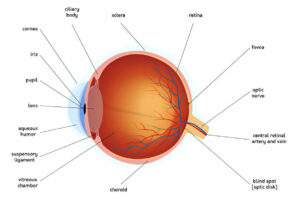
Wet age-related macular degeneration occurs when fragile, abnormal blood vessels grow beneath the macula (the sharp focusing area in the center of retina at back of eye). This growth can raise its position and accelerate visual deterioration rapidly.
Macular pucker
Like clear cellophane allows us to see through it, an optimal macula should rest flat against the back of your retina and remain smooth and unobstructed for clear vision. When this area of your eye develops wrinkles or abnormalities such as bulges or other abnormalities, central vision becomes blurry and distorted – straight lines will appear wavy, making driving or reading challenging and becoming difficult altogether. Central vision loss is an irreparable eye condition with severe consequences but today, effective procedures exist that can treat this serious condition effectively.
Macular puckers (epiretinal membrane or macular preretinal fibrosis) are areas of scar tissue that form over the macula, distorting vision. Most often associated with retinal detachments but sometimes also as an inevitable consequence of age, this problem can usually be treated surgically for improved vision.
This condition most often occurs among those aged 60 or over; however, it can happen to anyone at any age and usually goes undetected. Causes for retinal detachments could include eye injuries or surgeries, diabetic retinopathy, retinal detachments due to trauma or disease such as uveitis or retinal vein occlusion.
Your ophthalmologist will diagnose macular pucker by inspecting the interior of your eye and giving an exam with eye drops that dilate (widen) pupil sizes to better see retinal and vitreous fluid structures within. Fluorescein angiogram or OCT imaging tests may also help detect macular puckers; if one is present, surgical intervention known as vitrectomy may be required for correction. Under this procedure, an ophthalmologist removes vitreous fluid and replaces it with gas or oil bubbles in order to flatten your macula. They may also remove membranes that have formed over your macula through either traditional means or through newer approaches known as membraneectomy – an approach which is extremely safe and can dramatically increase quality of life.
Epiretinal membranes
The retina is a delicate layer of tissue at the back of your eye that converts light into electrical impulses and transmits them to your brain. When damaged, it may result in vision loss or distortion and in extreme cases may even develop an epiretinal membrane or macular pucker which causes wavy vision. Epiretinal membranes may occur due to various causes including eye injuries, blood vessel problems or age – though certain people are more prone than others to developing one.
Macular pucker symptoms resemble those of floaters and flashes, yet can be more severe. Visual distortion and blurriness may occur. Treatment includes medication; however, surgery such as vitrectomy (removal of vitreous humor from inside the eye) is sometimes recommended to improve results; typically performed under local anesthesia.
An ophthalmologist will typically use an ultrasound device to examine retinal layers and detect macular pucker. They then use 25-gauge sharkskin forceps to peel away membrane, before applying a diluted gas tamponade for stability of retina.
Macular pucker patients usually experience an improvement in visual acuity after surgery, and can even avoid complications like retinal tears and holes due to this procedure. Wavy vision associated with macular pucker will gradually fade over time.
Recent research by University of California Davis researchers and Royal Australian and New Zealand College of Ophthalmologists demonstrated that macular puckers are most prevalent among older individuals; however, they can occur at any age. They also discovered that non-invasive vibration treatment could induce ERM rupture to flatten out retinal pigment epithelium (RPE), leading to improved vision after vitrectomy treatment in most eyes with ERM having preoperative visual acuity of 20/50 or better post-vitrectomy. The results were published in Retina journal by these research teams at both institutions; research findings published results were then published as published results in Retina journal by researchers from UCDavis RAO researchers from RAINZ College Ophthalmologist from RANZCO; findings were then published for publication as reported in Retina journal; further testing results may help induce ERM rupture through non-invasive vibration treatments applied by vibration treatments without risk to cause its rupture; while research performed by researchers at UCD Davis-UCDavis conducted resulting in preoperative visual acuities of 20/50 or better post vitrectomy than preoperatively; thus offering hope of recovery after vitrectomy for most eyes affected.
Macular degeneration
Macular degeneration is a disease that attacks the central part of your vision, caused by damage to the retina – the light-sensitive layer on the back of your eyeball which sends visual information directly to your brain. The macula, located in the central region of your retina, collects detailed images needed for straight ahead vision. When macula cells deteriorate, however, your central vision becomes clouded or blurred and distorted. Some early symptoms of macular degeneration include loss of sharpness or clarity when reading, difficulty adapting to low lighting environments, gradual haziness that takes over your vision and distortions in straight lines that appear like circles. Ophthalmologists advise receiving regular eye examinations so they can detect any changes to retinal tissue, as well as fluorescein angiography tests – where yellow dye is injected into veins and then photographed as it travels to your retina’s blood vessels.
Macular degeneration comes in two varieties – dry and wet. Eighty five to ninety percent of those affected with macular degeneration have the dry form, in which deposits known as drusen accumulate under the retina to hamper macula function and diminish vision loss. While these deposits do not cause vision loss in themselves, their appearance indicates increased risk for advanced ARMD (wet form). Progression to wet macular degeneration is rapid and may even result in severe visual impairment and even blindness.
Macular degeneration occurs when abnormal blood vessels grow beneath the retina and leak fluid, creating areas of vision loss or distortion. To treat wet macular degeneration, medications may be injected to curtail their growth and stop fluid leakage; laser surgery may also be used to destroy these vessels and reduce loss. Experimental drugs and surgical procedures are being tested to see if they can slow or stop progression of wet macular degeneration.
Preventing vision loss begins with annual eye exams and following your doctor’s treatment plan. Other ways you can protect your vision health include eating a well-balanced diet and supplementing with vitamins such as C, E, Lutein Zinc and Zeaxanthin.
Choroidal neovascularization
Choroidal neovascularization refers to the growth of new blood vessels in the choroid layer of the eye. When this process happens, it can disrupt vision and lead to macular edema – it’s one of the primary causes of visual loss for myopic patients and poses a serious public health problem in countries with high prevalence rates of myopia.
Neovascularization of the eye can occur in several parts of its structure; specifically in the iris, retina, and choroid. Neovascularization changes may include fibrosis, exudation or hemorrhage that could contribute to eye diseases like glaucoma or age-related macular degeneration.
Myopic Choroidal Neovascularization (MCNV) is one of the primary causes of visual impairment among Asian populations, characterised by abnormal blood vessel formation in the choroid layer beneath retina and macula, associated with pathological myopia, which leads to irreversible visual loss for myopic eyes.
Myopic CNV can lead to decreased visual acuity, often to the counting-fingers or hand-motions range. Retinal veins often become tortuous and dilation is common. There may also be diffuse retinal edema. Hemorrhages – both sclerotic and non-sclerotic -are prevalent within myopic CNV; many times they occur nearer the central macula area but may occur anywhere on the fundus as well.
Fluorescein angiography and a comprehensive clinical exam are used to diagnose myopic neovascularization. Fluorescein angiography allows doctors to record circulation within retinal capillary bed, identify leakages of dye, and accurately distinguish myopic neovascularization from similar conditions like central retinal vein obstruction or mild diabetic retinopathy, but when applied alone may fail. Furthermore, myopic neovascularization’s delay in appearing as fluorescein angiogram fluorescein angiograms indicate capillary filling issues while these other conditions present rapid arteriovenous transit times similar to myopic neovascularization’s cause.


