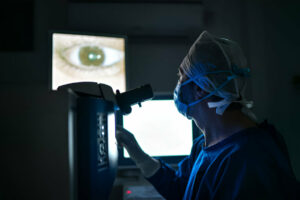
Ocular hypertension typically does not manifest with any noticeable symptoms, making diagnosis through a routine eye exam with a tonometer painless. A small plastic probe gently bounces over your cornea after using numbing drops for this test.
High Pressure
Hypertension (high blood pressure) can be an eye condition that directly or indirectly leads to eye issues like hypertensive retinopathy and choroidopathy. High blood pressure damages the tiny blood vessels supplying the retina with blood supply; this damages causes changes in vision as well as increased risks for glaucoma.
Your optometrist can detect these changes by inspecting the inside of your eyes during a comprehensive and dilated eye exam, including bends or kinks in blood vessels that provide your retina with blood, and leakage of vitreous hemorrhage into its white region – known as vitreous hemorrhage.
These changes may also cause your eyes to accumulate a build-up of fluid that puts pressure on delicate blood vessels in the back, eventually leading to glaucoma. But early warning signs can often be avoided by managing overall health and visiting an eye doctor regularly for dilated eye exams.
Over time, high blood pressure can also damage the delicate blood vessels within your eyes, potentially leading to serious eye diseases like glaucoma and macular degeneration. This occurs because elevated blood pressure weakens your vessels, making it more challenging for them to adjust when your eye pressure rises or falls.
Hypertensive Retinopathy is one of the most prominent ocular manifestations of high blood pressure, with approximately 50-80% of people who have it developing it over time. Prolonged exposure to high blood pressure damages the blood vessels in both retina and choroid making them less resilient against changes in blood flow; severity usually correlates to length of time spent having high blood pressure along with age and coexisting conditions.
Hypertensive retinopathy can be effectively managed with medications from groups including beta blockers, diuretics, calcium channel blockers and angiotensin converting enzyme inhibitors. Laser surgery may be necessary in cases involving visual-threatening retinal edema; otherwise controlling overall blood pressure through medication adherence usually suffices in healing most retinas suffering from hypertensive retinopathy.
Glaucoma
Glaucoma is an eye disease that can cause blindness over time. Typically characterized by an increase in pressure within the eye that then damages the optic nerve that transmits visual signals from eyeball to brain, and leading cause of blindness among people aged 60+. This pressure buildup usually stems from fluid buildup or drainage system issues; though all people can be susceptible; certain ethnic backgrounds, races, and age groups can be more at risk than others.
Open-angle glaucoma is one of the most prevalent types of glaucoma, occurring when fluid builds up inside of an eye due to an obstruction or blockage in its natural drainage system, leading to increased pressure that damages optic nerve cells and causes vision loss. People living with open-angle glaucoma typically show no symptoms at first; though peripheral vision loss may begin, their straight-ahead vision generally remains unaffected; untreated, this form of glaucoma can lead to irreversible vision loss over time.
Closed-angle glaucoma occurs when an eye’s drainage angle narrows slowly over time, restricting fluid drainage. This causes increased eye pressure that is more obvious than open-angle glaucoma; people suffering from it typically experience discomfort in their eye as well as blurred vision that worsens at night.
Multiple studies have concluded that individuals with high blood pressure are at a greater risk for glaucoma; although, its exact cause remains unknown. Some researchers suggest that high blood pressure may impact how fluid flows through the eye, leading to an increase in pressure inside. Researchers have also discovered that age may play a larger role than expected when it comes to developing glaucoma; older adults are at an increased risk than their younger counterparts for this eye condition. While prescription eye drops or surgical drainage improvements may reduce eye pressure enough for vision loss to be prevented by these treatments, regular eye exams should still be scheduled so as to catch any high eye pressure episodes before vision loss becomes permanent.
Subconjunctival Hemorrhage
Subconjunctival hemorrhages appear as bright red patches on the white of your eye (sclera). They result from blood vessel rupture within your conjunctiva’s clear layer, usually without pain or impacting vision; they are common among those suffering from diabetes and high blood pressure.
An increase in pressure to the blood vessels under the conjunctiva may cause small blood vessels to burst and bleed, leaving behind blood stains on the white part of your eye that resemble bruises – this is often the first symptom of this condition among elderly individuals.
People with an extended history of wearing contact lenses are at greater risk for this condition. This is due to how contact lenses can cause dryness and friction that leads to ruptured blood vessels under the conjunctiva. Furthermore, medications which disrupt clotting processes like aspirin or warfarin could increase this risk further.
Subconjunctival hemorrhages may come about for seemingly no obvious reason, although certain activities and medical situations could temporarily increase blood pressure to your eye veins, potentially causing ruptured vessels to form. Such causes include straining while coughing, sneezing or vomiting as well as using the Valsalva maneuver.
Subconjunctival blood vessels usually vanish on their own within several weeks. If they persist longer than that, however, it could be an indicator of more serious vascular conditions like atherosclerosis that require medical advice; should this occur it should be immediately addressed with medical professionals.
Nothing can be done for this condition except reassuring patients that it will resolve naturally over time, similar to a bruise. A blood test that measures coagulation factors should be conducted to rule out atherosclerosis or hypercoagulable states as possible causes for clot formation, while taking aspirin daily, controlling atherosclerotic risk factors and ceasing smoking are recommended as ways to help reduce future episodes.
Red Eye
Red eyes result from small blood vessels becoming engorged and covering the white area of the eye called the sclera, or they could appear red due to subconjunctival hemorrhage, when one or more small blood vessels under a thin clear membrane called conjunctiva rupture and fill with blood that appears like bloodshot eyes but are actually harmless, provided there are no pain or vision changes involved.
Blood vessels within the sclera are under constant strain, and may break under certain circumstances such as sudden eye strain or the use of contact lenses. Other conditions that cause red-tinged eyes include bacteria infections, herpes simplex infections, allergy, uveitis (inflammation of the middle layer of eye) or trauma from accidents or sports injuries.
If red eyes are accompanied by pain or changes in vision, it is imperative to see an eye doctor as soon as possible. Some serious ocular conditions can be treated early through medication or surgery and prevent permanent damage to the eyes.
Red eye symptoms may range in severity depending on their underlying cause. You can ascertain its intensity by inspecting both eyes and noting other associated symptoms, such as sneezing, running nose or discharge from them.
Typically, eye doctors will measure eye pressure using a tonometer and perform a physical examination of structures within the eye as well as testing visual acuity and peripheral vision. They may prescribe medicated eye drops designed to lower eye blood pressure; in certain instances they may recommend oral anti-glaucoma medication; if the cause of increased eye pressure turns out to be systemic disease (diabetes or African American ethnicity) then you will likely be referred to another physician for an in-depth medical assessment including history taking as well as laboratory tests that rule out other potential causes of elevated eye blood pressure.


