There are numerous eye diseases that could compromise your vision, with the most prevalent ones including:
Diabetic Retinopathy – High blood sugar levels can wreak havoc with the small blood vessels that supply your retina (the light-sensitive inner layer at the back of your eye), leading to swelling and leakage of fluid into it that blurs your central vision and dark spots called “floaters.”
Macular Degeneration
Macular degeneration refers to deterioration or breakdown of the macula located within your retina (light-sensitive tissue at the back of your eye). The macula provides central vision that allows for fine details like threading a needle, driving or reading – but when its function stops functioning normally your central vision becomes dim or even blurry, with distortion such as straight lines appearing bent or crooked; it is one of the primary causes of severe vision loss in people over 50.
Macular degeneration typically presents itself in either its dry or wet forms; typically the former involves yellow protein deposits called drusen slowly building up under the retina, eventually leading to its thinning and weakening over time. Ten to fifteen percent of cases progress into more serious wet form macular degeneration involving abnormal blood vessels growing under retina leaking fluid or blood, accounting for 90-100% of severe vision loss in macular degeneration cases.
There is no known cure for wet macular degeneration; however, certain vitamins and supplements such as vitamins C, E, lutein and zeaxanthin may slow its progress. Furthermore, injectable medications like Avastin or Lucentis may help counteract the growth and leakage of abnormal blood vessels; injections with Avastin or Lucentis may counteract their leakage; laser photocoagulation or photodynamic therapy may be required in some instances; regular examinations, OCT and Fluorescein angiography plus follow up injection treatment are necessary in order to optimize patient results and maximize outcomes.
Keratoconus
Keratoconus is a progressive corneal dystrophy that results in blurry vision due to an irregular shape of the cornea, often first noticed during teen or early 20 years but can develop at any age. Initial symptoms may be mild-moderate and correctable with eyeglasses or traditional contact lenses; as the condition worsens, rigid gas permeable (RGP) contacts or hybrid or scleral lenses may become necessary as more surface area of cornea is covered by rigid lenses; its causes remain unknown but could include excessive eye rubbing during long term contact lens wear as well as excessive eye rubbing due to excessive eye rubbing from excessive eye rubbing or long term contact lens wear.
The cornea is a thin transparent layer on the front of your eye that helps focus light onto the retina. When someone has keratoconus, their normally round cornea thins out and bulges into a cone-like shape, preventing light entering their eye from being focused onto their retina correctly, leading to distortion of vision.
Keratoconus can be diagnosed by performing a slit lamp examination and topographical map of the cornea. Your physician will also consider your medical history, family history and symptoms you are experiencing. Teenagers or young adults should notify their ophthalmologist of any changes to their vision such as blurriness and need for frequent eyeglass or contact lens adjustments; failure to treat may lead to more severe visual distortion that requires corneal transplant.
Retinal Detachment
The retina is a layer of light-sensitive tissue located inside your eyeball’s inner backwall and contains millions of photoreceptors that convert light entering your eye into electrical impulses that travel back to your brain and become visual images. When this process happens, vision forms; should the retina detach, this could result in severe loss.
Retinal detachments are medical emergencies that, if left untreated promptly, could lead to permanent blindness. They typically manifest as sudden flashes of light such as cobwebs or curtains moving across your vision; or as an expanding dark shadow encroaching upon central vision (similar to curtains moving).
Detachments often begin with retinal tears that go unrepaired, which allow fluid from vitreous gel to pass through and collect underneath, pulling away from its blood supply and pulling the retina away from its connection with its blood source. Detachment may then take various forms. For instance:
Rhegmatogenous Retinal Detachment – This type is the most prevalent, occurring when a hole or tear allows fluid to pass through and collect underneath the retina, detaching it from its underlying blood supply and leaving behind scar tissue beneath. It may occur naturally with age or trauma, or due to certain medical conditions like diabetic retinopathy or cataracts.
Tractional Retinal Detachment – Caused by scar tissue on the retina’s surface, this form is similar to rhegmatogenous detachment but with one important difference: scarring pulls the center of the macula away from its blood supply, potentially associated with certain inflammatory disorders and Coats’ disease.
Diabetic Retinopathy
Diabetes can damage the light-sensitive retina at the back of the eye. Damaged vessels bleed or leak fluid, blurring and distorting vision. With advanced diabetic retinopathy, abnormal new blood vessels begin growing on the retina to compensate for those that have closed or bled; eventually these new blood vessels bleed or swell, blocking vision altogether resulting in blindness – leading to detachment from its original position and blindness; diabetic retinopathy being the number one cause among American adults today – therefore regular eye exams with dilation as well as controlling blood sugar is key when living with diabetes.
Non-proliferative diabetic retinopathy (NPDR) is the initial stage of this disease. Damaged blood vessels swell and leak clear fluid and fats onto the retina, creating deposits which cause reduced or blurred vision. Treatment in this phase typically is not necessary unless vision has already become significantly impaired. When this is the case, laser scatter laser photocoagulation treatment can be conducted. Laser surgery uses laser light to destroy damaged blood vessels, which helps prevent bleeding and scarring of the retina. If bleeding becomes severe, vitrectomy surgery may need to be performed; otherwise keeping glucose levels under control, quitting smoking, and having regular dilated eye exams are the best ways to lessen risk of vision loss.
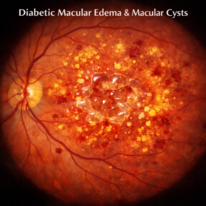
Macular Edema
Macular edema, the abnormal accumulation of fluid in the central retina that results in blurry vision, is a result of blood-retinal barrier breakdown, allowing proteins and solutes into retinal tissue and leading to fluid accumulation. Recent treatments include using vascular endothelial growth factor blockers or corticosteroids which reduce inflammatory mediators that disrupt it as ways to manage this complication.
Left untreated, macular edema can result in significant visual impairment or even blindness. Its root cause lies with damage to the macula due to increased intraocular pressure, poor blood flow or retinal ischemia; other contributing conditions include proliferative diabetic retinopathy, vitreomacular traction syndrome or taut posterior hyaloid face (TPHF).
Preventing macular edema requires annual comprehensive dilated eye exams; pregnant women with diabetes should undergo additional exams as directed by their healthcare providers.
Focal or grid laser photocoagulation is one of the most effective treatments for macular edema. This surgical technique uses highly targeted laser light to seal retinal blood vessels and decrease macular edema. Fluorescein angiography, optical coherence tomography and MRI are less invasive options that may also help; other less invasive options include fluorescein angiography, optical coherence tomography MRI and strabismus are less invasive options; vitrectomy may also be performed to address macular edema caused by epiretinal membrane or vitreomacular traction syndromes or taut posterior hyaloid fold.
Glaucoma
Glaucoma is an eye disease that affects the optic nerve that transmits visual messages to the brain, and is the second-leading cause of blindness worldwide. It develops due to increased pressure within the eye caused by fluid buildup; often without symptoms and only becoming noticeable once significant vision loss has taken place; that is why having regular eye exams is so essential.
Your eyes continuously produce fluid to keep them moist and healthy, which drains out through drainage angles in your eye. However, if the drainage angle becomes blocked or clogged up it can increase intraocular pressure (IOP) leading to an increase in eye internal pressure which leads to glaucoma; damage to optic nerve can then result in permanent vision loss.
Open-angle glaucoma is the most prevalent form of glaucoma, typically developing slowly without pain or symptoms. Over time, peripheral vision will progressively decline into tunnel vision; if this is your situation it is essential that we meet regularly so we can identify it early and provide preventative treatments.
Closed-angle glaucoma is less common but often progresses much more rapidly than its open counterpart. Here, eye drainage suddenly becomes blocked leading to an exponentially high increase in eye pressure resulting in symptoms like blurry vision, rainbow halos around lights, intense eye pain and nausea and vomiting.