Hypertension, or high blood pressure, is a prevalent health issue that can damage blood vessels throughout your body – including your eye. If left untreated, it can lead to retinal detachment, blurred vision and blindness – potentially leaving patients vulnerable for decades.
Studies demonstrate that moderate retinopathy signs such as retinal hemorrhages and microaneurysms are strongly correlated with cardiovascular diseases ranging from subclinical stroke to congestive heart failure, independent of traditional risk factors. International management guidelines now support assessment of retinopathy as part of hypertension risk stratification strategies.
Stage 1: Arterial Narrowing
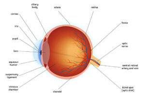
Hypertension, defined as blood pressure exceeding 140/90 mm Hg, can damage the retina (the light-sensitive layer at the back of your eye). As a result of this damage, fluid that provides nutrients to the eye seeps into its tissue, creating macular exudates; over time these deposits could damage retinal nerves and lead to permanent vision loss.
Chronically elevated blood pressure causes retinal vein walls to thicken, narrowing points where arterioles and retinal veins cross each other in their common adventitial sheath (an adventitial sheath that protects both structures). As the walls thicken further, space in this sheath becomes reduced further reducing space for compression of retinal veins within it and creating hourglass-shaped constrictions with dilatation on either side (arteriovenous nicking). This imbalance in vascular flow allows fluid seepage into retinal tissues leading to macular exudates or optic disc edemas.
Fluorescein angiography can detect fluid deposits that form early on in hypertensive retinopathy as yellow or white spots on retinal photographs, which can then be detected with special dye injected directly into retinal veins to highlight them and surrounding areas of retinal tissue. An experienced ophthalmologist administers this test through injecting dye directly into veins within the retina which then shows up as yellow spots on photos taken of your retinal pictures.
Beaver Dam Eye Study and other population studies demonstrated that generalized retinal arteriolar narrowing on photographs was strongly associated with elevated blood pressure levels, making it a reliable indicator of cardiovascular disease risk, particularly among those already taking treatment for high blood pressure.
Retinal lesions caused by long-term elevated blood pressure are reversible when blood pressure returns to normal; however, even with adequate control measures in place there may still be permanent retinal lesions present.
Hypertensive retinopathy is an excellent predictor of future cardiovascular complications in addition to traditional risk factors like age and family history. Signs of hypertensive retinopathy show strong correlation with current and past blood pressure levels, suggesting chronic damage from hypertension. Furthermore, mild signs such as generalized retinal arteriolar narrowing or arteriovenous nicking have been found strongly associated with subclinical or clinical cardiovascular diseases — such as congestive heart failure or cerebrovascular events like strokes or transient ischemic attacks — without other risk factors being present.
Stage 2: Arterial Dilation
The heart is an organ designed to pump blood around the body through an intricate network of blood vessels. Each heartbeat (cardiac cycle), it pushes blood into arteries; during diastole (resting phase), their walls relax to widen these blood vessels – this process known as vasodilation increases blood flow by decreasing resistance in its pathways and creates greater vasodilation (see above).
Hypertension damages blood vessels, leading to complications for eye health. Hypertension in particular has an adverse impact on eye vessels that supply the retina – the light-sensitive tissue at the back of your eyeball – as its blood supply comes under tremendous strain from being subjected to extreme forces that exert force upon these vital vessels delivering oxygen and nutrients directly to it. Over time this force causes these blood vessels to rupture, ultimately damaging them over time and leading to eye problems.
Damage to retinal blood vessels left untreated can quickly worsen retinopathy and vision loss. Blood vessels may narrow, close off completely or leak fluid out from within them to the outside causing blurred or darkened central vision; fluid may even enter macula area resulting in distortion or darkened central vision. Furthermore, newly growing blood vessels may rupture resulting in retinal vascular accidents or strokes which are potentially deadly and even blinding events.
Retinopathy prevention relies on blood pressure control, with lifestyle modifications like eating healthily, exercising regularly and cutting salt intake and alcohol consumption being key components. Furthermore, regular ophthalmologic exams – specifically using fluorescein angiography to provide more detailed images of retinal blood vessels – is necessary.
Studies have demonstrated that moderate retinopathy signs, such as generalized or focal arteriolar narrowing and arteriovenous nicking or increased tortuosity of the arteries – are strongly correlated to cardiovascular disease ranging from congestive heart failure and clinical stroke, through transient ischaemic attack (TIA). Furthermore, these correlations exist regardless of traditional risk factors for such conditions.
Flow-mediated dilation (FMD) is an early noninvasive marker of endothelial dysfunction and an independent predictor of coronary heart disease, but the relationship between FMD and changes seen in retina is unclear. In order to investigate how FMD relates to early retinal changes in hypertension patients, specifically by investigating how blood pressure (systolic and diastolic), arterial wall shear stress, and blood flow affect an artery with compliant walls during FMD.
Stage 3: Arterial Bifurcation
As hypertension damages retinal blood vessels, they begin to narrow and possibly bulge or form tortuosities, increasing risk for hypertensive retinopathy (HR), which could eventually lead to vision loss if untreated early. Therefore, improved computer-aided diagnosis systems for screening and grading HR are necessary; current methods have used automatic segmentation of retinal images as screening and classification tools; however more modern techniques use deep learning for more accurate results.
Studies have revealed that hypertensive retinopathy signs exhibit a graded relationship to blood pressure. While mild signs such as generalized or focal retinal arteriolar narrowing and arteriovenous nicking only have weak links to blood pressure, moderate signs like retinal hemorrhages and microaneurysms have strong connections with subclinical and clinical cardiovascular diseases such as congestive heart failure, strokes, transient ischaemic attacks and atherosclerosis – providing further support that retinopathy acts as an invaluable indicator of systemic cardiovascular health independent of traditional risk factors.
Hypertensive retinopathy most frequently develops at the carotid bifurcation, where left and right common arteries meet, thus branching off into abdominal aorta. Multiple studies have confirmed that presence of hypertensive retinopathy at this bifurcation can serve as an early predictor for coronary artery disease, renal impairment, strokes, dementias or any number of related conditions.
As with other vascular conditions, maintaining low blood pressure levels is the cornerstone of reducing hypertensive retinopathy progression risk. Regular blood pressure monitoring should be conducted alongside eating healthily and engaging in physical activities in order to ensure all vessels within your body are working efficiently. A physician can help patients manage their blood pressure by prescribing suitable medication tailored specifically to each person’s medical needs.
Stage 4: Tortuosity
Artery tortuosity syndrome occurs when blood vessel walls become elongated and stiffened, increasing stress on vessel walls, leading to more plaque formation. Shear stress leads to the formation of thrombus that blocks blood flow through an artery – potentially leading to sudden death due to lack of oxygen for vital organs. People affected may also suffer severe abdominal pain, nausea and vomiting as well as having an excavated chest (pectus excavatum). Other features may include long slender fingers and toes as well as unusually soft stretchable skin.
Tortuosity is a prevalent finding on angiography and has been linked with hypertension, atherosclerosis and aging as well as subclinical and clinical cardiovascular disease, including heart failure and stroke. Numerous hypotheses exist for its link to atherosclerosis including one wherein tortuous vessels support atherosclerotic lesion formation or enhanced shear forces may contribute to its progression.
Researchers have investigated the relationship between coronary artery tortuosity and atherosclerosis in individuals both with and without diabetes mellitus. They discovered that when tortuous arteries exist, there is an direct relationship between their tortuosity value and atherosclerosis score measured on an angiographic film.
Coronary artery tortuosity has been linked with an increased rate of early and late major adverse events following coronary stent placement, according to one study analyzing individual patient data from 6 prospective stent trials for effects of moderate to severe tortuosity on target vessel failure at 30 days and 5 years (comprising cardiac death, coronary dissection/stenosis/stenosis and myocardial infarction related events). Results demonstrated that patients with moderate to severe tortuosity experienced significantly higher rates of early major adverse events than those with low/ no tortuosity.
The international hypertension management guidelines recommend that anyone diagnosed with high blood pressure should receive regular evaluations of retinal blood vessels to detect and treat signs of hypertensive retinopathy, and ensure proper and effective blood pressure control through lifestyle modifications and prescription medication.
