Hypertensive Retinopathy (HR) is the result of prolonged elevation of blood pressure, leading to damage of retinal blood vessels and potentially leading to vision loss if not diagnosed and treated early enough. If not addressed early enough, HR could even lead to irreparable vision loss.
Studies demonstrate that mild hypertensive retinopathy signs such as retinal hemorrhages, microaneurysms and cotton wool spots are strongly associated with subclinical and clinical stroke events, other cerebrovascular outcomes, congestive heart failure and cardiovascular mortality independent of traditional risk factors.
Arterial Narrowing
Chronic hypertension causes retinal vein walls to thicken as blood pressure rises, which then narrows those points where arterioles and retinal veins cross each other in the retina, providing space within their common adventitial sheath that surrounds both structures – this narrowing reduces space within this sheath, compressing veins in turn creating an hourglass-shaped constriction with aneurysm-like dilatation on either side (arterio-venous nipping).
Narrowing is often caused by an imbalance of vascular flow as both arteries and veins try to maintain their regular diameter, leading to fluid seeping into retinal tissue and creating macular exudates or damaging the retinal nerve.
Cotton wool spots, caused by ischaemia-related dysfunction and leakage from optic nerve cells axoplasmic flow, may appear on the retina as yellow or white deposits in the peripheral part of the fundus, as seen through an examination of leaking fluid deposits on retinal photographs.
At later stages of hypertensive retinopathy, more widespread endothelial cell layer breakdown occurs within retinal veins supplying optic discs, leading to haemorrhage and cotton wool spots (macular exudates) on retina. Fluorescein angiography may detect these dense areas where there is nonperfusion and microaneurysm formation – this phenomenon is called fluorescein angiogram detection.
As hypertension damages retinal blood vessels, an individual becomes increasingly susceptible to cardiovascular conditions like heart disease and stroke. Therefore, one of the primary goals in treating hypertensive retinopathy should be effective control of blood pressure to either prevent or treat associated medical conditions.
Recent studies have demonstrated that moderate hypertensive retinopathy can serve as an invaluable indicator for cardiovascular disease independent of traditional risk factors. Therefore, its severity assessment serves as an invaluable way of stratifying cardiovascular risks. As evidenced in hypertension management guidelines such as those issued by the U.S. Joint National Committee for Prevention, Detection Evaluation and Treatment of High Blood Pressure that recommend clinical examination for hypertensive retinopathy as an indicator for risk evaluation purposes.
Arterial Bifurcation
Arterior bifurcation or narrowing is one of the earliest and most frequently occurring vascular abnormalities seen among hypertensive patients, and an excellent predictor of future systemic vascular disease and cardiovascular events. Target organ damage indicates arterial bifurcation as it requires immediate, aggressive therapy to reduce future risks; however, due to its often asymptomatic nature early detection is especially vital in this regard.
Flow patterns at arterial bifurcations are determined by both hemodynamic and non-hemodynamic factors, with their roles determined by their specific locations within the vessel wall. Local mechanical strain is predicted to play a pivotal role in atherosclerotic plaque formation and progression.
In this study, computer simulations were employed to investigate the influence of geometry and flow on atherosclerosis formation at carotid artery bifurcations sites. Three realistic geometrical models with various bifurcation angles were analysed using CFD simulations for optimal results. Results confirmed previous experimental findings regarding WSS/atheroscoleric plaque formation ratios as predicted by prior WSS studies; they also demonstrated how geometry/flow pattern have an impactful role in spiraling phenomena development around sinus outer walls of internal carotid bifurcations sites as demonstrated by previous experimental studies in relation to WSS/atheroscoleric plaque formation ratios; plus results showed good agreement with previous experimental findings regarding WSS/atherosclerotic plaque formation relation.
The carotid arteries, major blood vessels of the head and neck, provide oxygenated blood directly to the brain. If there is a blockage in one or more carotid arteries, clots in these can lead to strokes that result in temporary or permanent loss of vision, brain damage such as dementia or permanent neurodegeneration; symptoms include numbness and difficulty speaking – it could even be transient ischemic attack (TIA), which is less serious but potentially deadly.
Studies have revealed that moderate hypertensive retinopathy signs, which affect up to 10 percent of adults age 40 years and older, can significantly increase the risk of subclinical and clinical stroke, other cerebrovascular outcomes, congestive heart failure, and cardiovascular mortality independent of traditional risk factors. Thus, an evaluation of retinal blood vessels can identify patients at higher risk who require an aggressive treatment strategy.
Arterial Tortuosity
Hypertension causes changes to the retina’s blood vessels that feed it, resulting in abnormalities on fundus photos and fluorescein angiography of both. One significant sign is increased tortuosity that appears primarily in second and third order retinal blood vessels such as terminal arterioles and venules; dilation also results in visible leak points on fluorescein angiogram (cotton-wool spots).
Tortuosity occurs as the result of abnormal vasoconstriction caused by high blood pressure, leading to weakening of vascular structures that make them susceptible to dissection and sudden clot formation. When blood cannot flow freely out of the eye and out of its circulation system, retinal hemorrhages occur with either diffuse or patchy bleeding resulting from blocked flow of blood out. Leakage into retina can result in pigmentary changes called chorioretinopathy resulting from increased pressure within it’s core.
People living with high blood pressure have an increased risk of cardiovascular disease. They may develop clinical stroke, congestive heart failure or experience other vascular conditions of the kidney and brain. Damage to retinal blood vessels is an early indicator of cardiovascular disease that can be detected among the general population; mild hypertensive retinopathy signs such as generalized and focal arteriolar narrowing, arteriovenous nicking or increased tortuosity of arteries are weakly associated with current blood pressure levels but more closely tied to individual history of having high blood pressure than current levels alone.
Condition characterized by sunken chest (pectus excavatum) or protrusion of stomach through gaps in abdominal muscle structure (hernias). Other features may include long face with wide-base nose (beaked nose), long slender fingers and toes and extremely soft and stretchable skin.
Fluorescein angiography can provide more detailed images of retinal blood vessels for accurate diagnosis of hypertensive retinopathy. Treatment will often prevent or limit further damage, though effective blood pressure management with antihypertensive drugs remains key in doing so.
Arterial Swelling
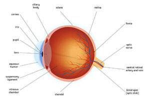
Retinopathy refers to damage done to the retina – the light-sensitive layer lining the back of your eye that converts light into visual signals that travel along the optic nerve to reach the brain. High blood pressure (hypertension) exerts excess force on blood vessels throughout your body, eventually leading to damage of this kind and leading to retinopathy.
Nonproliferative retinopathy, which occurs early on in hypertensive retinopathy, involves blood vessels closing off, leading to fluid, fat or protein leaking out from retinal blood vessels and collecting on the surface of the retina or macula, leading to blurry vision or in some cases retinal detachment if left untreated.
Hypertensive retinopathy progresses into its later stages when retinal blood vessels thicken and narrow to such an extent that they close off entirely, allowing more fluid from within them to leak out onto the retina or macula, creating blurry or dark vision.
Some new blood vessels may form hard deposits on the retinal surface that are known as cotton wool spots, copper wiring or flame haemorrhages. Over time these vessels can rupture causing retinal vascular accidents (stroke) leading to blindness.
Lowering blood pressure regularly is the main treatment for hypertensive retinopathy and may reduce the risk of stroke or other forms of vascular disease in the brain and other organs. Most retinal changes disappear as soon as blood pressure decreases; however, some permanent lesions may remain.
Studies demonstrate that moderate retinopathy signs (generalized or focal retinal arteriolar narrowing, arteriovenous nicking and macular edema) are strongly associated with cardiovascular diseases – from congestive heart failure and cerebrovascular events like stroke or transient ischaemic attack, to other forms of ischemic stroke – regardless of other traditional risk factors. This information suggests that clinical assessment of retinopathy signs is an invaluable way of identifying people at preclinical hypertension levels who might suffer target organ damage as early as preclinically. International management guidelines now support using retinopathy this way as an early warning indicator of disease progression.

