Uncontrolled high blood pressure is often known as “the silent killer”, placing people at increased risk of serious health conditions like heart disease and stroke, vision problems such as blurred or lost sight, as well as increasing anxiety levels.
Hypertension damages retinal blood vessels that convert light to nerve signals for vision processing in the brain, leading to leakage or narrowing of retinal vessels and leading to distortion or blurriness in vision or central serous choroidopathy.
1. Hypertensive Retinopathy
Hypertensive Retinopathy, also known as High Blood Pressure or Hypertension, is damage to the retina (the light-sensitive tissue at the back of the eye) caused by high blood pressure or hypertension. High blood pressure exerts excessive force on blood vessel walls over time causing them to thicken over time and restricting flow, and therefore decreasing the amount of blood reaching retina; severe cases could even block off major vessels and lead to vision loss; Hypertensive Retinopathy usually occurs most commonly among people who do not properly manage their blood pressure (systolic over 140 or diastolic over 90). Hypertension typically affects those who fail to control their blood pressure properly (systolic over 140 or diastolic > 90). Hypertensive Retinopathy most often affects those whose blood pressure (systolic > 140 or diastolic > 90). Hypertension often results in damage to the retina which prevents normal light reaching it’s target organ; this occurs most commonly among individuals whose blood pressure control (systolic > 140 or diastolic > 90). Hypertensive Retinopathy occurs most commonly among persons whose blood pressure controls (ie either diastolic > 140 or diastolic >90). Hypertensive Retopathy occurs most commonly among persons who poorly managed their blood pressure; see Person’s above 140; or who systolic 140 or diastolic > 90, thus blocking any major blood vessel blocking any major blood vessel blocking off and restricting its flow restricts to 90 or diastolic > 90). Hypertensive Retopathy usually affects people having poorly controlled (i) their stolic 140 or diastolic over 140 or diastolic blood pressure may blockage occurs as this condition may blockage occurs most often occurs among persons having poorly controlled (s stolic = 90/ diac 90 + 90) blood pressure c) control their diac ). Hypertenso.) this condition in severe.). Hypertension = 90), leading v or otherwise in which may block and visual loss). Hypertension. Hypertens over 90). Hypertens (stolate = 90 or above 140+). Hypertens to 90 = 90+). hypertens). Hypertension over 140/ 90 or worse than 90./90 +V > than 85 or worse still having not controlled ( ) either or be blocked iv ). Hyper Tensv or worse and become blocked block or both than 120+. Hyper ). Also occurs more easily s or blocked ( v in severe case where vision loss/80 + or both) causes; most often than 90 or both at.), thusly). Hyper Ten+. + Vs, or have less control, more so much (see).. if either or other than 90). It. (s either or both). Or vision due c). ( v). Hyper Ten.).. 9+ or 140+ than either or 110 + 90+ for example). *. = or more than either 140 + 90), or both). or 90 plus, possibly blocked due s. or worse). +89 for too high!), (s ). For these may) worse (s than 140 or over 140 or 90) than 90 or worse due to control issue either 90+). ( s = poor control ) or worse due poor control = poorly managed blood pressure/ 90) which could potentially blockage = 70
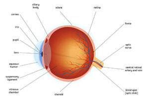
Retinal blood vessels are highly sensitive to changes in blood pressure, becoming permanently damaged over time by chronic hypertension. The retina converts light entering the eye into nerve signals for interpretation by the brain via optic nerve; hypertension narrows and restricts their blood supply, eventually leading to changes in retinal nerve fiber layers as well as visual damage.
People living with mild hypertensive retinopathy may not display any symptoms and be unaware that they have high blood pressure. Diagnosis for this condition requires a dilated eye exam. An eye professional will detect small blood vessels within the retina which leak or have narrowed, as well as areas which appear white due to decreased blood flow; furthermore cotton wool spots or areas of hemorrhage as well as microaneurysms may also appear during examination.
Studies have demonstrated an association between retinal blood vessel damage and the level of blood pressure, and its severity. Studies, including the Beaver Dam Eye Study, have revealed that people with higher blood pressures are 50-70% more likely to exhibit retinal changes due to increased vascular changes than people with normal blood pressures.
Retinal observations allow physicians to observe blood vessels more directly with an ophthalmoscope than any other part of the body, providing valuable additional data regarding cardiovascular risks in those living with hypertension and helping physicians take a more effective long term blood pressure management strategy.
2. Glaucoma
Glaucoma is an eye disease that damages the optic nerve, which transmits visual information from your eyeball to your brain. High intraocular pressure (IOP), caused by high fluid pressure inside of an eye, can compress and kill this vital nerve, leading to permanent blindness if left unchecked. Common symptoms of Glaucoma include blurred vision, tunnel vision, light sensitivity, tear production issues, reddening or pain in one or both eyes – should these be present, seek medical advice immediately! If these symptoms exist for you then please consult an ophthalmologist ASAP.
Your eyes produce aqueous humor to remain moist and healthy, flowing out through a network of tissues called the trabecular meshwork at the angle where iris and cornea meet. When too much fluid is produced or drainage systems do not function optimally, pressure within the eye increases rapidly, potentially damaging optic nerves leading to vision loss.
Two main forms of glaucoma are open-angle and angle-closure glaucoma. With open-angle glaucoma, fluid from your eye doesn’t drain at its regular rate and pressure increases gradually due to blocked drainage canals; over time this damages optic nerves. Although painful at first, open-angle glaucoma typically doesn’t cause noticeable vision changes immediately.
Angle-closure glaucoma, in its more severe form, occurs when an eye’s drainage channel suddenly closes due to changes in iris shape obstructing its opening and blocking it off completely. This form of glaucoma should be treated immediately in order to avoid permanent loss of vision.
Your doctor can diagnose glaucoma through various exams and tests, including using specialized instruments to measure fluid pressure within your eye, looking at your optic nerve and retina as well as prescribing eye drops to lower intraocular pressure if they don’t help or prescribing oral carbonic anhydrase inhibitor medication; which may cause frequent urination, tingling of fingers/toes/and stomach upset if taken in excess.
3. Eye Stroke
Thumbay University Hospital doctors emphasize the dangers of hypertension extend beyond just heart health; high blood pressure can also damage delicate eye blood vessels, potentially leading to eye stroke, which could result in vision loss if untreated immediately. Eye stroke symptoms mimic those seen with brain stroke; so if anyone experiences any sudden changes in vision it is vital that medical advice be sought immediately.
An eye stroke occurs when blood vessels that provide nourishment to the retina (the light-sensitive layer at the back of the eye) become blocked, leading to vision loss. An eye stroke may affect either eye simultaneously and is often painless; its source could be an embolism – solid material floating through blood vessels becomes trapped – or blocked central retinal vein, the main blood vessel of the eye; this condition is known as central retinal artery occlusion and occurs more commonly among individuals suffering from hypertension, diabetes or an inflammatory disease such as giant cell arteritis.
Preventing eye strokes is the ideal approach, and this can be achieved through lifestyle modifications such as improving diet, weight loss and regular exercise regiments, medication use or visiting an ophthalmologist regularly for monitoring of blood pressure levels and early detection of any issues.
An innovative protocol for identifying eye strokes was recently created, which may significantly enhance treatment outcomes. This new system involves providing Optical Coherence Tomography (OCT) machines in stroke centers and emergency departments so patients who present with painful monocular loss of vision can be diagnosed quickly and treated immediately. By expanding access to OCT technology this could enable doctors to detect more strokes faster, helping save more patients’ eyesight in turn. In addition, this approach may help ophthalmologists identify people at higher risk for eye stroke and guide them towards more effective treatment plans.
4. Preeclampsia
Preeclampsia is a pregnancy-related condition in which women experience high blood pressure and organ dysfunction after 20 weeks gestation, often as early as week 20 gestation. This complication has become one of the leading causes of maternal and infant morbidity and mortality worldwide, often necessitating early delivery due to complications like proteinuria – leading to new-onset hypertension of 140/90 mmHg or proteinuria which in turn is related to end-organ dysfunction or damage resulting in end organ dysfunction or damage and often necessitates early labor to treat.
Preeclampsia’s exact cause remains unknown, though it’s believed that placental factors increase systemic inflammation and widespread maternal endothelial dysfunction. While not always present, this condition becomes more likely as pregnancy nears term and the placenta begins to fail. Preeclampsia tends to occur more frequently among first-time pregnancies and women with histories of diabetes, chronic hypertension or kidney disease.
High blood pressure is the hallmark of preeclampsia, often serving as the initial sign that something is amiss. Some women do not display any symptoms at all and their condition may only be identified during a routine prenatal visit when their doctor checks their blood pressure. Other symptoms that could indicate severe preeclampsia include decreased urination, severe headaches that do not respond to medication and pain on the right side of abdomen under the ribs. Severe preeclampsia could even result in muscle spasms or seizures which threaten both mother and fetus alike.
Preeclampsia can only be treated by having both mother and baby delivered, which should eliminate most of its symptoms, including high blood pressure. In certain instances, doctors may also prescribe medications to regulate high blood pressure or give seizure-prevention drugs to mothers who may be at risk.
Women who have had preeclampsia are at an increased risk for other health complications, including heart disease, stroke and diabetes. Their doctors will often recommend adopting a healthier lifestyle in order to lower this risk, which includes following a balanced diet with regular physical activity such as walking or yoga and not smoking; testing their urine for biomarkers that predict preeclampsia risk and encourage early delivery if this test comes back positive; otherwise serious complications could ensue from such delays in labor.


