Eye diseases range from minor, such as pink eye, to severe conditions like glaucoma that could potentially blind. Early diagnosis is key.
Leukocoria, or white pupils in infants and babies, could be an indicator that tumors have formed behind their eye, leading to retinal cancer or retinoblastoma.
Glaucoma
Glaucoma is one of the primary causes of blindness in America. This chronic eye condition damages the optic nerve, which transmits visual messages between eyes and brain, due to high eye pressure levels. A good optic nerve carries over one million nerve fibers that carry information about color, contrast sensitivity, side vision and depth perception directly to brain. Glaucoma blindness rates continue to climb in America as more individuals are diagnosed.
There are multiple types of glaucoma. Primary open-angle glaucoma is the most prevalent form, in which individuals experience higher than usual inner eye pressure without showing symptoms. Closed-angle glaucoma occurs less frequently; when its drainage area becomes blocked it quickly raises eye pressure to an alarmingly high level resulting in vision loss within minutes requiring medical assistance to resolve.
Acute angle-closure glaucoma occurs when part of the iris blocks the drainage angle and rapidly increases eye pressure, necessitating immediate treatment to restore fluid flow within the eye and maintain normal vision. This medical emergency requires immediate treatment in order to restore fluid drainage within it.
Age increases your risk for glaucoma, so it is wise to visit an eye doctor regularly and be aware of its signs. Other risk factors for glaucoma may include family history of it as well as diabetes or being of African or Hispanic heritage.
An eye exam is the best way to detect glaucoma. Your eye doctor will place numbing drops in your eyes, test corneal thickness using a pachymeter, take photographs of the inside of the eye with a Pentacam and examine your pupils using an OCT scan or camera. They may use laser technology if narrow-angle glaucoma is present to create a hole in the iris to allow fluid drainage from drain angles more quickly. Prescription medication may help alleviate pressure; however they only work if taken consistently over time; discuss this with your ophthalmologist regarding changing or discontinuing them with them while giving him/her all information regarding all medicines you take regularly with him/her along with their lists from each physician who prescribe them to ensure full effectiveness of care for best possible treatment and effectiveness of each.
Red Eye
Red eyes occur when the small blood vessels in the white part of the eye (known as the sclera) dilate, creating the appearance of bloodshot eyes. They could be an indicator of various conditions ranging from allergies and infections to head trauma or infections; oftentimes sudden redness accompanied by pain or changes in vision is indicative of some sort of issue requiring medical intervention; to properly diagnose its source. A comprehensive eye exam should be performed by your healthcare provider in order to ascertain why redness exists in your case.
Most cases of red eye are caused by viral or bacterial conjunctivitis, also known as conjunctivitis. While viral conjunctivitis typically self-limits itself with antiviral or antibiotic treatment, bacterial conjunctivitis can become much more serious; corneal ulcers or scarring may result from bacteria belonging to Staphylococcus species or Streptococcus group bacteria; less frequently it can be caused by herpes virus infection or Chlamydia infections.
When patients present with red eyes and symptoms that suggest serious eye injury or infection, immediate referral to an ophthalmologist should be made. This includes patients experiencing pain that is unresponsive to topical anesthetics; purulent or mucous-like discharge; decrease in visual acuity or having had contact lens wear history, recent eye surgery procedures, trauma injuries or herpes infections in the past.
After an initial comprehensive medical history and physical exam, patients should also undergo evaluation using a slit lamp to closely inspect pupils, eyelids, lashes, sclera and deeper eye structures. An ophthalmologist should be consulted if a patient has diabetes, cancer, is taking steroids drops or medication or has systemic inflammation disease of skin or organ systems; acute glaucoma is difficult to diagnose without professional help and must be confirmed by an ophthalmologist in order to be confirmed as sight threatening complications associated with certain eye diseases; therefore it is vital that regular eye exams be scheduled with everyone in order to assess them properly.
Retina
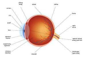
The retina is a thin and fragile layer within the eye that converts light into images visible to us. It resides firmly adhered to its walls (the sclera), with three main layers comprising its tissue: nuclear/cell; white “plexiforn; and ganglion cells, responsible for relaying information from retina to brain.
Retinal detachment is typically caused by natural aging processes; however, it may also result from certain eye diseases or injuries. Signs of retinal detachment include sudden vision changes such as floating spots (tiny specks or spots in your vision) and curtain-like shadows that start in one corner and progress across your visual field – which if left untreated could eventually lead to permanent blindness.
An detached retina requires surgical repair in order to restore vision. During this process, fluid under the retina must be extracted before scar tissue forms around its base in order to reattach it back onto the back wall of the eye. A silicone rubber band called a scleral buckle may also be sutured on to help push it against its surroundings and push the retina down against it against the sclera; an alternative treatment option could involve lasering or freezing probeing as methods of producing scar tissue at its source.
Some individuals are at an increased risk for retinal detachment than others, typically those who are nearsighted, have a family history of eye problems, or who have sustained severe eye injuries. Age related macular degeneration also puts individuals at an increased risk; this disease reduces central vision (macula), leading to reduced fine detail vision needed for reading, driving a car safely or recognising faces and colors.
Before recently, retinal detachments were often irreversible and blinding if left untreated. But with modern vitreoretinal surgery techniques now in use, over 90% of retinal detachments are being successfully reattached – an impressive accomplishment considering this disease was once thought untreatable and irreversible.
Strabismus
Strabismus is the medical term for misalignment of the eyes. Commonly referred to as cross-eyedness, wall eyes or squint, it occurs when two or both eyes don’t aim at the same point – whether this be turning inwards (esotropia), outwards (exotropia) or up and down (hypertropia or hypotropia). Strabismus may be constant or intermittent – while in children it often diminishes over time as their brain learns to use both eyes together as one three dimensional image; however in adults it may linger or worsen.
Six external eye muscles control each eye’s movement and positioning, and must work perfectly to enable binocular vision to work optimally. Any anatomical abnormality, neurological condition or problems in the region of the brain controlling these muscles could result in strabismus resulting from improper coordination or functioning issues affecting these muscles causing misalignments between eyes.
Strabismus can occur as a result of refractive errors or eye conditions such as nearsightedness, farsightedness and astigmatism. Other risk factors that contribute to its development include head injuries, general health conditions such as Down syndrome or cerebral palsy and neurological diseases like stroke or thyroid disease; family history can also play an influential role; individuals who come from families where someone had or currently has strabismus may be more prone to it themselves.
Strabismus may be present at birth – known as congenital strabismus or esotropia – making it one of the primary causes of infantile squinting. It may also arise later on due to abnormalities in either eye muscle structure or function or nerve control systems that regulate them, particularly those responsible for controlling them during infancy and childhood.
Strabismus can often correct itself naturally as the brain adapts to using both eyes together. Surgery and vision therapy may also help, with vision therapy helping improve eye teaming and focusing skills to restore normal binocular vision; optical correction with glasses/contact lenses/occlusive patching providing another effective strategy to force weaker eye to focus and increase depth perception.


