Eye diseases often progress without showing symptoms, making regular dilated eye exams essential to your overall eye health. Speak with your eye doctor about the best course of treatment and make an appointment today.
Nearsightedness, more commonly referred to as myopia, affects people of all ages and can be corrected using eyeglasses or contact lenses. Other common eye conditions include dry eyes and conditions that cause floaters.
Diabetes-related Retinopathy
Over time, diabetes can damage blood vessels in the retina. When these blood vessels leak fluid and deposits into it, which may cause macular edema – an eye disease which if left untreated can lead to blindness. People living with either Type 1 or 2 diabetes are at a greater risk than ever of developing this eye disease – those living for long periods with poor control over their diabetes as well as coexisting conditions like high blood pressure or cholesterol are particularly prone.
Diabetic retinopathy can range in severity from mild to severe. Early on, those suffering may not notice any changes to their vision; as the disease advances further, retinal tissues can begin leaking blood which leads to vision loss or in extreme cases blindness.
Your ophthalmologist (eye doctor) can detect diabetic retinopathy during a comprehensive eye exam by looking out for telltale signs, including macular edema, microaneurysms and vitreous hemorrhages.
Optometrists can treat diabetic retinopathy by injecting patients with prescription medication called anti-vascular endothelial growth factor (anti-VEGF) agents and steroid injections, which help reduce retinal blood vessel swelling. Laser treatments may also be effective, targeting either specific areas or panretinal photocoagulation to combat irregular growth of retinal blood vessels associated with diabetic retinopathy.
Age-related Macular Degeneration
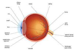
Age-related macular degeneration (AMD) is a progressive eye condition that gradually compromises your central vision, leading to severe visual loss among adults over 50. It’s the leading cause of severe vision loss among these adults and is caused by damage to your macula in the retina at the back of your eye – responsible for reading, driving and seeing fine details clearly – in its central region of retina that allows focusing close objects. With AMD it may still be possible for you to drive and walk normally but your central vision will become blurry while peripheral (side vision) vision will remain normal compared with peripheral (side vision).
Early symptoms of macular degeneration include distortions in straight lines and dark spots in the center of your vision, both of which do not require medical treatment; however, patients should monitor their vision closely and report any waviness or missing areas to their physician immediately. About 15% of people living with ARMD can develop wet macular degeneration which is more serious because it can lead to rapid vision loss; wet ARMD occurs when abnormal blood vessels form beneath retina and macula and leak blood and fluid, potentially blurring or even blinding central vision completely.
Wet ARMD can be treated using medications known as anti-VEGF therapy that will block new blood vessel formation, seal existing ones from leaking and reverse existing leakage and damage. Studies have demonstrated this can improve vision in those suffering from wet macular degeneration; however it should take some time before any improvements appear in your vision.
Glaucoma
The optic nerve provides visual information to the vision centers in our brain via electrical impulses. Comprised of 1.2 million nerve fibers that carry this data, this nerve is responsible for our color vision, contrast sensitivity and peripheral (peripheral) vision; damage to it leads to blindness. Glaucoma is one of the leading causes of irreversible blindness worldwide and damages the optic nerve by increasing intraocular pressure (IOP). An accumulation of fluid within the eye increases this pressure, pushing against optic nerve fibers that are delicate and can die under continued high pressure; everyone should get their eyes checked regularly, particularly those over 60, in order to detect any possible glaucoma cases.
Open-angle glaucoma is one of the most prevalent forms of glaucoma, in which drainage canals of the eye become gradually blocked due to too close proximity between an eye’s colored part (iris) and clear front window (cornea). As natural channels drain away their contents, eye pressure increases gradually or quickly over time.
Conventional surgery for glaucoma involves inserting a small piece of tissue into the anterior chamber to drain away fluid and lower intraocular pressure; this procedure is known as trabeculectomy. After this procedure is performed, doctors numb and remove tissue while placing medication to protect from infection and inflammation in the eyes – drugs like beta-blockers, adrenergic agonists and prostaglandin analogs may be used to manage eye pressure; however these do not treat or cure glaucoma while laser surgery for glaucoma offers another alternative that improves flow by decreasing resistance in trabecular meshwork networks – so laser surgery for glaucoma is another viable treatment option to consider for eyes suffering from high intraocular pressure than its conventional counterpart.
Trichiasis
Trichiasis is an eye condition in which eyelashes rub against the cornea and cause irritation, pain and vision loss. In extreme cases it can even lead to infections of the eyes.
Trichiasis can be treated by surgery that restores normal alignment of eyelid margin and rotates away eyelashes from cornea. Trichiasis should only become permanent in severe cases; otherwise it can result in blindness. Trichiasis surgery restores alignment by realigning eyelid margin and rotating away from cornea for permanent relief.
Trichiasis can be caused by various conditions, including trachoma, glaucoma and eyelid laxity. Furthermore, some of these conditions can also contribute to entropion (the inward rolling of lid margin allowing eyelashes to rub against cornea) so efforts should be made first to treat any existing entropion prior to treating trichiasis.
Other causes of trichiasis may include epiblepharon (a congenital condition in which folds of skin and muscle roll upward and push the eyelashes vertically toward the cornea) and distichiasis, in which an aberrant second row of lashes appears behind the normal lash line due to chronic meibomian gland inflammation, such as occurs with Ocular Pemphigoid or Stevens-Johnson Syndrome or due to conditions that cause cicatricial entropion such as trachoma.
Recurrent trichiasis after surgery is an increasingly prevalent and potentially sight-threatening issue, particularly among people living in remote regions where access to treatments may be limited. According to a community randomised trial in The Gambia, village-based trachomatous trichiasis surgery proved more successful than health centre-based procedures due to being both less costly and more convenient for patients.
Prevalence of Ocular Disease among Different Races and Ethnicities
Refractive errors, cataracts, glaucoma, macular degeneration, retinal disorders and conjunctivitis are among the most prevalent eye diseases or disorders that cause blurred vision or blindness – most are preventable, so annual eye exams should be undertaken to safeguard good eye health and vision.
Optic disorders most prevalent among older Americans include cataracts, macular degeneration, glaucoma and diabetic retinopathy. Furthermore, nearsightedness (myopia), which affects distance vision but provides good close-up vision is another prevalent eye disease among adults. Keratitis may occur due to extended contact lens wear or scratched contact lenses while styes – red bumps on the inner eyelid – may also appear frequently.
Eye disease diagnoses in the US tend to be higher among whites than blacks or Hispanics, possibly reflecting disparities in healthcare utilization or medical conditions that affect these groups differently. Eye disease prevalence also varies across geographic regions and income groups – though this may reflect differences in disease prevalence rather than any regional or socioeconomic disparities.
American Indian and Alaska Native individuals tend to experience higher rates of diabetes and other ophthalmic conditions compared with non-American Indian or Alaska Native populations, and have worse overall health as a result of less accessible health services and reduced eye examination rates. This could be the result of several factors; such as limited health care resources being readily accessible.
Ophthalmic genetics is an emerging field that studies inherited eye conditions like Usher Syndrome and thyroid eye disease. At University of Kentucky Advanced Eye Care team we have one of the few practitioners specializing in this subspecialty – Dr. Ramiro Maldonado who joined in 2017. Dr. Maldonado has expertise in treating many rare inherited conditions as he strives to find treatments for them.
