Many eye diseases and conditions that affect vision can be avoided or managed with regular eye exams. By keeping up-to-date with routine exams, regular screening can prevent loss of vision while detecting diseases in their early stages and providing treatment plans accordingly.
Glaucoma, peripheral vision loss and macular degeneration resulting in central vision loss can all be treated effectively with medication and laser surgery.
Herpes eye disease
Herpes simplex virus (HSV) is the primary cause of herpetic keratitis (HSK), an eye infection which leads to scarring and vision loss, often occurring recurrently. Most often, HSV enters through the front part of cornea and travels to nerve cells in trigeminal ganglion where it latently rests until later reactivated; so patients who experience HSK should take oral antiviral medications like Acyclovir as prevention measures against further outbreaks.
Herpes viruses come in two main varieties, HSV-1 and HSV-2. HSV-1 typically causes cold sores and blisters in the mouth or genital area while HSV-2 causes shingles on torso and face. Herpes can also appear in eye conditions causing redness, pain and teariness from its effect on cornea – this condition known as Herpes Keratitis can be primary, recurrent, or chronic in nature depending on which herpes virus it comes from.
Herpetic keratitis often develops following LASIK or PRK surgery, while the herpes virus in the cornea can spread into deeper layers, sometimes occurring days later or during daylight. Reactivated by intense light exposure or by being released back into neurons for replication and back to the cornea again OHSU researchers discovered that the herpes virus spreads into deeper layers by traveling into neurons where it replicates before returning back out again into neurons to replicate before returning out onto its target organ – the cornea. By creating a mutant herpes virus without US9, which promotes its movement into neuronal axons; instead it did not move into either the cornea nor did it remain latent within neurons after being released back from its host cell for replication within neurons allowing more light exposure from hitting it, and herpes virus created no latent infection by making rounds trips into neurons to replicate and return back out from neurons when returning back out again to surface layer of cornea or become latent within cells where it had lain latent during day time!
Glaucoma
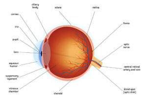
Glaucoma is a progressive eye disease that can lead to vision loss and blindness, harming a nerve in the back of the eye that connects light signals from vision sensors to your brain. It is one of the main causes of blindness worldwide and usually results from increased intraocular pressure due to improper production or drainage of fluid that slowly kills off optic nerve cells – this results in blind spots appearing around your peripheral vision before eventually leaving you completely sightless without treatment. Therefore it is imperative that regular eye exams be scheduled, since glaucoma often forms without warning until it is too late!
Open-angle glaucoma is one of the two major types of glaucoma; open-angle is by far the more prevalent variety and most prevalent in America, yet deadly as well. When drains in your eye become blocked and not enough liquid drains out, pressure in the eye rises rapidly resulting in optic nerve death; its symptoms often include gradual peripheral vision loss that often goes undetected until too late, when much vision has already been lost.
Glaucoma treatment options aim to lower intraocular pressure. These may include prescription eye drops and oral medications, surgery or laser therapy; surgical options like trabeculectomy create an opening in your sclera that allows fluid to pass freely and thus lowers pressure; as well as laser peripheral iridotomy and selective cyclophotocoagulation are also available surgical options.
Macular degeneration
Macular degeneration, also known as age-related macular degeneration (AMD), is a progressive condition which gradually impairs your central vision, due to damage to the macula–a small area located at the back of your eye that allows you to clearly view fine details such as reading, driving and recognising faces. While macular degeneration occurs gradually over time, your doctor can monitor its progression with regular exams and the Amsler grid chart to detect changes in your vision.
Macular degeneration often takes the form of dry macular degeneration. Drusen deposits grow under the retina and cause it to thin and dry out, while wet macular degeneration occurs less commonly but more quickly: abnormal blood vessels form under your retina and leak fluid or even blood into your macula, blurring or distorting central vision and possibly leading to rapid loss of vision.
Treating wet macular degeneration may help slow its progress and improve your visual acuity. Most commonly, medications to block the hormone known as vascular endothelial growth factor – believed to contribute to leakage and bleeding – are administered. Laser therapy may also be effective at eliminating abnormal blood vessels found in certain cases of wet macular degeneration. However, therapy cannot restore vision that has already been lost to this condition. *The table below presents estimates of prevalence rates derived from published examination-based population studies; variations likely reflect differences in study designs, definitions of disease and population characteristics.
Cataracts
Cataracts are an eye disease that is widely prevalent, often leading to vision impairment. Cataracts occur when the lens of the eye becomes cloudy, leading to blurry or hazy vision and often progressing more slowly in one eye than the other. Cataracts are one of the leading causes of blindness worldwide and significantly diminish quality of life for patients affected.
Lenses in our eyes should be clear and help to focus light onto our retina – the light-sensitive tissue at the back of each eye. When someone has cataracts, their lens becomes opaque and dense leading to blurred vision and progressive blindness which may require surgery treatment in severe cases.
symptoms of cataracts include blurry or hazy vision; poor night vision; glare or halos around lights; the need for brighter lighting for reading or other activities; and frequently needing new prescription glasses or contact lenses. While these symptoms may only interfere with daily activities slightly, it’s important to visit your physician if they become bothersome or interfere with daily life activities.
Age is often the cause of cataracts, though other factors may contribute to their formation as well. Smoking, diabetes, high blood pressure, prolonged sunlight exposure, eye injuries and an unhealthy diet all increase your risk. Unfortunately there is no cure for cataracts; however healthy diet, exercise, quitting smoking cessation and wearing sunglasses can all slow their progression and protect vision if symptoms arise. For any concerns related to your vision please schedule an appointment with your physician immediately.
Strabismus
Strabismus, or eye misalignment, occurs when eyes do not line up correctly with each other. Eyes may turn inward toward the nose (crossed eyes, convergent), outward away from it (walleyes, divergent), or one eye may be higher than another (hypertropia or hypotropia). Strabismus is classified by its direction of deviation, age of onset and cause. Hereditary and congenital forms often develop in infants and children while other causes include large refractive errors, droopy eyelids, cataracts as well as other ocular or orbital conditions.
Amblyopia accounts for half of all cases of strabismus, occurring when one eye does not produce an image that is clearly perceived by the brain. Left untreated, amblyopia may result in permanent loss of binocular vision. Thus it is crucial that treatment for strabismus be initiated early by addressing its source before amblyopia occurs.
Treatment options depend on the type of strabismus present; common treatments for esotropia, or crossed eyes, include prescription glasses, drops and vision therapy. Vision therapy helps strengthen coordination and strengthen muscles in affected eye; often helping people with refractive errors related strabismus. Eye patching may also encourage weaker eye to take on more visual work to align better with its counterpart eye.
Some patients suffering from strabismus who cannot undergo vision therapy may require eye muscle surgery as an alternative form of treatment. This may be beneficial if their strabismus was brought about by health conditions like stroke or thyroid disease; to determine this possibility a thorough assessment must be undertaken regarding both eye movement patterns and medical history of each individual being considered for surgery.


