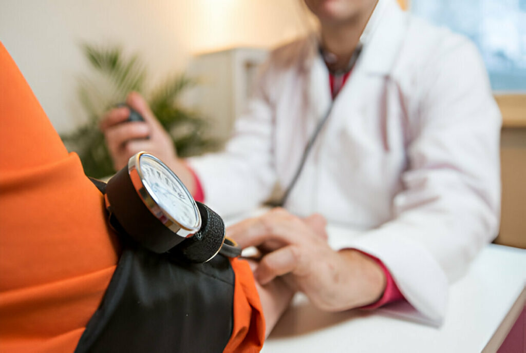


At an annual eye exam, your doctor will check your intraocular pressure to detect elevated intraocular pressure that could lead to glaucoma. Be sure to notify them about any medications, such as steroids.
Uncontrolled high blood pressure damages retinal blood vessels, leading to hypertensive retinopathy. Symptoms may include hemorrhages and areas of white spots on the retina.
Subconjunctival Hemorrhage
Subconjunctival hemorrhages, or bright red patches in the white part of your eye, are known as subconjunctival hemorrhages. They occur when blood vessels underlying the conjunctiva break and bleed, filling a space beneath your white of eye (sclera). Though easily visible, subconjunctival hemorrhages do not interfere with vision and do not hurt; in most cases they resolve themselves within days or weeks on their own unless repeated episodes occur indicating some health conditions which require treatment.
Subconjunctival hemorrhage can often be traced back to increased vein pressure leading to an internal blood vessel rupture below the conjunctiva. This may occur after you sneeze, cough, strain, rub your eyes or have an injury to the eye; or it could even be brought on by medications like aspirin and warfarin that thin blood or cause it. A subconjunctival hemorrhage could also indicate clotting disorders affecting circulation in your veins.
Chorionic hemorrhages that appear at the corners of eyes are called chorionic hemorrhage and are relatively uncommon. More likely to affect older individuals, they could signal serious vascular disease or be caused by medications like birth control pills.
If you notice a bright red spot in the white of your eye, contact your health care provider immediately. They will conduct a physical exam and check your blood pressure before looking at your eye and possibly suggesting over-the-counter artificial tears to reduce discomfort. If it turns out you have a condition that leads to hemorrhages, they will treat that first before prescribing medication that could trigger them again; or alter dosage or switch medications altogether as appropriate; in certain instances a blood test can even help determine your risk for more serious conditions than anticipated.
Red Eye
Red eye may occur when blood vessels in your eye’s white part, the sclera, expand and obscure its visibility, leading to pink or bloodshot eyes. While most cases resolve themselves within several hours or days without medical intervention, persistent red eyes could indicate more serious health conditions that require medical treatment.
Red eyes can often be caused by allergies or irritation, and to decrease your risk you should wash your hands frequently and avoid smoke and pollen as potential triggers. You should also wear wraparound sunglasses to protect against harmful UV rays; get plenty of restful sleep, eat well balanced meals and make lifestyle changes to reduce eye irritation.
Some cases of red eye require immediate medical intervention, namely those associated with pain or an obvious decrease in visual acuity. Should this occur to you, seek the advice of an ophthalmologist as soon as possible for diagnosis and treatment.
Red eye can be caused by many things, from bacteria, viral, and parasitic infections to foreign bodies, herpes outbreaks, contact lens use and other eye issues. If you’re uncertain what’s causing yours, it is essential that a thorough history is gathered as well as examination. This will enable a proper diagnosis to be made as well as determine an effective course of treatment.
Depending on the severity of your symptoms, eyedrops and cooling compresses may suffice as home treatments for mild cases. You could also try using non-irritating saline solutions to rinse away debris. In more serious instances, antibiotics or even surgery may be required. If you notice sudden redness and blurring of vision, seek medical treatment immediately as this could be an indicator of acute angle-closure glaucoma – a medical emergency requiring prompt referral to an ophthalmologist for evaluation and possible diagnosis using flourescein staining to confirm diagnosis.
Glaucoma
Glaucoma occurs when fluid pressure in the eye increases to levels that could damage the optic nerve, leading to loss of vision and potentially blindness. Unfortunately, symptoms are often subtle and not noticed until vision has already been lost – hence why this condition is known as “sneak thief of sight.” The most prevalent form of glaucoma occurs when eye drainage canals become blocked over time causing intraocular pressure (IOP) levels to rise and damage nerve tissue located behind it – once this damage has taken place it cannot be restored!
Closed-angle glaucoma, while less prevalent than open-angle glaucoma, occurs when an eye’s drainage area quickly becomes blocked due to distortion in its peripheral iris, often making symptoms such as blurred vision or rainbow halos around lights difficult to detect until later symptoms arise. Closed-angle glaucoma should be diagnosed and treated immediately in order to avoid serious pain and visual field loss.
Some individuals with elevated eye pressure but without other signs or symptoms of glaucoma such as optic nerve damage or blank spots in peripheral vision on tests (ocular hypertension) are at an increased risk for developing glaucoma and should be regularly evaluated by an eye care provider. These individuals should be closely monitored by an ophthalmologist.
Eye pressure can be reduced using prescription eye drops, medication, laser treatments or surgery; measuring it with a handheld tonometer is part of a complete eye exam.
Diabetic Eye Disease
Diabetes increases blood glucose levels, which over time can damage tiny blood vessels that feed the retina at the back of the eye, known as diabetic retinopathy. It is the leading cause of blindness among Americans and leads to blood vessels swelling up, leaking fluid or growing abnormal new vessels which over time may blur vision, cause glaucoma or retinal detachments; for this reason it’s vital that those living with diabetes visit an eye doctor annually in order to detect and treat problems early. It is advised that at least once annually they visit an eye doctor who can diagnose any developing issues before any worsened symptoms emerges – an annual exam should help identify and treat potential issues before any further complications develops causing vision impairment or blindness occurs.
Diabetic retinopathy typically progresses gradually without producing symptoms in its early stages. If leakage from blood vessels occurs, macular edema may result from it, leading to blurred central vision as well as dark spots or sudden appearance of floaters (known as vitreous halos). Other symptoms may include feeling of scratchy or watery eyes; loss of peripheral vision; and changes in light reflection off eyes.
Left untreated, diabetic retinopathy can rapidly progress to proliferative retinopathy – the most dangerous stage – leading to bleeding and scar tissue growth in the retina that could eventually result in its detached condition. Treatment includes laser surgery aimed at shrinking abnormal blood vessels or sealing any leaks; alternatively vitreous surgery might be performed, replacing its cloudy jelly-like substance with saline solution instead; chemicals may even be injected directly into eye to slow their growth and possibly slow proliferative retinopathy further.
To reduce diabetic retinopathy, it’s essential that your blood pressure, cholesterol and sugar are within a specified target range. Also be sure to get regular eye exams with dilation and follow your physician’s advice regarding eyecare – our online search tool makes this easier than ever by helping you locate one near your location! To do so.


