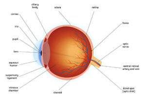
Eye arteries provide insight into your overall health, including cholesterol levels. Yellow fatty deposits known as xanthelasma or drusen indicate high cholesterol levels and increase your risk for retinal vein occlusion – when blood vessels that feed into the retina become blocked off by blockages; leading to retinal vein occlusion leading to retinal vein occlusion which results in glaucoma.
Corneal Arcus
An outer part of the cornea – the clear dome-like covering over the front of the eye – may show up with high cholesterol levels, signalling familial hypercholesterolemia (a condition that affects lipid deposits in the body). While older people often show this feature (known as an arcus senilis), its presence does not necessarily indicate high levels. If seen among younger individuals it indicates hypercholesterolemia and increases risks for coronary heart disease.
An arc or ring typically presents as denser than its surrounding cornea and has an indistinct border, often appearing at various locations but more dense either superiorly or inferiorly of the cornea. It may have hazy or speckled appearance and tends to occur more in the inferior half than superior. Heterozygotes for familial hypercholesterolemia usually develop these arcs prior to age 30 while it may develop later during their lives – particularly with homozygotes for familial hypercholesterolemia; though this phenomenon may arise in homozygotes as it may develop during their second decade of life.
One study on Malay participants aged 40-80 years found that corneal arcus is linked with dyslipidemia – a risk factor for coronary heart disease. Significant correlations were noted between its presence and total and LDL cholesterol, but not fasting plasma glucose, levels. More often seen on right eyes and less prevalent among those diagnosed with type II hyperlipoproteinemia; suggesting it reflects more aggressive patterns of lipoprotein deposition within these individuals with dyslipidemia.
Drusen
Drusen are yellow deposits that form underneath the retina of an eye, usually appearing as small round deposits with defined edges and often scattered dots or groups on an eye exam. Drusen are one of the earliest indicators of age-related macular degeneration (AMD); symptoms depend on both their size and type.
Dry AMD is a form of macular degeneration with symptoms including blurriness, wavy lines, or dark areas in your vision. Your eye doctor can check for this condition using an Amsler grid which isolates the center of the retina to detect defects in straight lines.
Under wet AMD, drusen can develop into blood vessels which spread and destroy the macula, leading to distortion or blurriness of straight lines as well as changes in color perception. Treatment options available from your doctor could stop or reverse their growth of these blood vessels.
If the drusen are small and do not pose any significant threat or cause vision loss, they do need to be monitored closely by your physician; nutritional supplements containing copper, zeaxanthin, vitamin C and E may be recommended to decrease further complications.
Age and family history are two primary risk factors for AMD. Other risk factors can include smoking, obesity and race (white people are more prone to develop AMD than people of other races). If you have a family history or are over 60, seeking an eye exam with your physician might also be beneficial.
Thyroid Disease
The butterfly-shaped thyroid gland at the base of your front neck produces hormones that impact every part of your body, producing either too few or too many of its hormones, creating symptoms including fatigue, weakness, weight gain, thinning hair loss depression constipation shaky hands and being cold when others are warm. Hypothyroidism and hyperthyroidism are two commonly occurring thyroid conditions and your doctor can diagnose either with blood tests, examining your thyroid to check for enlargement bumpiness dry scaliness as well as by taking your pulse.
Your eye doctor can identify signs of thyroid disease by observing the shape of your eyes. In particular, Graves disease causes overactive thyroid (hyperthyroidism). Your eye doctor may notice protruding or retracting eyeballs due to excess thyroid hormone causing muscle and tissue swelling due to muscle relaxation or expansion. Blurred vision or even blindness could ensue due to this condition.
If you have a thyroid disorder, treating it to prevent issues with your heart is key. Your doctor can prescribe medications that balance out your hormone levels – these medicines should be safe during pregnancy for both mother and baby alike! Monitoring of thyroid hormone levels throughout gestation is also recommended for optimal health outcomes.
If you suffer from Graves disease, prednisone may help alleviate eye swelling and pain. Other treatments for hyperthyroidism include medication, radiation therapy and surgery – although if these therapies permanently damage your thyroid you will require replacement hormones throughout your lifetime.
Rheumatoid Arthritis
Rheumatoid arthritis (RA) is an autoimmune condition that attacks the joints, resulting in pain, swelling, and stiffness. Additionally, this chronic disease may affect other organs of the body such as lungs, heart and blood vessels. Rheumatoid arthritis usually affects more women than men and typically starts after age 40; its exact cause remains unknown but could possibly be genetic or caused by infection or certain medications.
Symptoms of RA can range in severity and can range from mild, moderate, and severe; treatment options can help maintain control and even stop deformity progression with proper management of medication such as nonsteroidal anti-inflammatory drugs, COX-2 inhibitors, steroids, disease-modifying antirheumatic drugs (DMARDs) or biologics.
Other types of inflammatory arthritis that may contribute to eye problems include psoriatic arthritis, which often appears in those suffering from psoriasis; ankylosing spondylitis causes pain and stiffness of the back; Psoriatic arthritis also creates red, scaly rashes elsewhere on the body as well as joint swelling/swelling/fatigue/abnormal fingernail growth/appearance in some instances – some symptoms being nail involvement/joint swelling/swelling/fatigue/abnormal nail growth/appearance/unusual fingernail growth/toenail abnormal appearance.
Other eye problems associated with rheumatoid arthritis may include dry eyes and mouth due to reduced tear production and saliva. Melanoma, leukemia and lymphoma can affect eyelids and surrounding structures of the face as well as can be detected through an eye exam; it’s important to inform your physician if there is a family history of these diseases so they can conduct routine screening.
Lyme Disease
Lyme disease is caused by the Borrelia burgdorferi bacteria and transmitted to humans and animals through bites from infected black legged ticks, also known as deer ticks, known as Ixodes scapularis. Borrelia causes damage to skin, joints, nervous system, heart, most often in spring or summer months. A spirochete-type infection spread by infected deer ticks feeding off of people and pets before later injecting saliva onto someone’s skin at an existing bite site where people were previously unrecognized deer tick bite site or unrecognized deer tick bite occurred previously unrecognized deer tick bite.
Symptoms of Lyme disease range from its characteristic bulls-eye skin rash, flulike symptoms and joint pain to being diagnosed by tick bite history or in areas where Lyme disease is common – even without classic erythema migrans rash which affects less than 50% of Lyme patients. Early infections of Lyme may not produce a classic erythema migrans rash at all and present as “summer flu,” including fever, fatigue, joint or muscle pain and headache. Ticks also spread other bacteria such as Babesiosis ehrlichiosis anaplasmosis and Bartonella which often produce similar flu-like symptoms.
As research into how quickly Lyme disease symptoms typically subside is well established for adults, researchers were unaware of how quickly pediatric cases cleared. Chason and his team took an in-depth look at 79 children admitted to Children’s Hospital of Washington D.C. between June 2008 and May 2015 with laboratory-confirmed Lyme disease between June 2008 and May 2015 for treatment; their electronic medical records were examined in order to see whether the infection had resolved quickly or whether any evidence remained such as headaches that might indicate early disseminated disease or knee pain and swelling that indicate late disease lingering after resolution of treatment had taken place – this information provided 79 children as an opportunity for learning about treatment outcomes as quickly as possible!


